Duplex vein scan. Duplex scanning of the veins of the lower extremities, what is it? All about diagnostics
duplex scanning remains one of the most informative methods for examining blood flow and the condition of veins and vessels. Some do not distinguish it from or Doppler, but all three of these diagnostic methods have a number of differences.
Painless and safe duplex scanning allows you to identify vascular and venous diseases on early stages and to identify the cause of their occurrence.
Ultrasound of the veins of the lower extremities: what is this examination?
Duplex scanning is also called UZDS (ultrasonic duplex scanning). This method combines ultrasound and dopplerography, which makes it one of the most informative in the study of veins and blood vessels.
How is a duplex vein scan performed? lower extremities what it is and where it can be done, not everyone knows. For the patient according to the sensations and external features procedure there is no difference between ultrasound, ultrasound and dopplerography.
Conventional ultrasound will not allow you to assess the state of the vessels, since it is not designed to examine the moving blood flow. Ultrasound examines only immobile organs. With ultrasound, the ability is used ultrasonic wave coincide with the direction of moving particles, namely the uniform units of blood in the bloodstream. This allows you to evaluate the movement of blood through the vessels and veins.
The possibilities of ultrasound are used in cardiology, phlebology, and neurology.With the help of ultrasound, it is possible to assess not only the patency of blood vessels, but also the speed of blood flow.Unlike Doppler ultrasound, ultrasound allows not only to determine plaques and blood clots in the vessels, but also to identify the cause of their occurrence, to determine the cause of the recurrence of the disease.
With the help of ultrasound, you can see even small blood clots in the lumen of blood vessels.
The ultrasound shows the blood flow in the "online" mode, that is, at the moment. The picture is displayed in the light and allows you to determine the fullness of the vessel, its patency.If Dopplerography can only determine that the patency is impaired, ultrasound determines why it is impaired: is there an obstruction to blood flow, is the lumen narrowed, how much is it narrowed and what caused it.
Duplex scanning helps not only to make a diagnosis, but also to determine the stage of the disease.Duplex scanning is prescribed both for certain complaints and as a preventive measure for representatives of "sedentary" and "standing" professions: waiters, hairdressers, office workers.
Advantages and disadvantages of the method

Ultrasound is safe, non-invasive and informative method diagnostics
Duplex scanning has many advantages. Since the method is based on ultrasound, it is considered safe for pregnant women.
Simplicity and lack of complex preparation make ultrasound popular and affordable. The price for the examination depends on the medical center, but in comparison with the cost is much lower.
Advantages of ultrasound:
- Safety. Ultrasound is an absolutely safe method of examination. It is based on ultrasound examination, which does not provide any negative impact on the body, has no harmful radiation and does not lead to negative consequences. Ultrasound is safe for the fetus during pregnancy and for child's body. side effects and there are practically no contraindications. None are used chemicals causing allergies.
- High information content. Ultrasound is a survey method with a high degree of information content. The accuracy of the study is very high, it allows the doctor to correctly diagnose. If the doctor has doubts about the diagnosis, he will prescribe another ultrasound in another clinic to clarify the diagnosis.
- Painless and non-invasive. During the procedure, the patient does not experience any pain. No injections or incisions are made skin. The only one possible discomfort- cold from the gel and sensor. The doctor makes gentle pressure with the sensor, which does not cause pain.
- Quick and easy inspection. The procedure itself lasts no more than 30-45 minutes. During this time, the doctor manages to assess the condition of the veins and vessels of the lower extremities, the speed of blood flow, record all the necessary data and give the patient the result on hand. No complex manipulations with the patient are carried out. He fulfills the doctor's requests for a change of position.
No particular shortcomings of this method were identified. It can be noted that the difficulty similar procedure in villages where there is a lack of equipment, as well as the price of the examination, which, however, remains below the cost of CT and MRI.
As a rule, duplex scanning of the veins of the lower extremities is prescribed by a doctor (therapist, phlebologist). As a preventive measure, this procedure is not often carried out, since patients rarely go to doctors in the absence of complaints.
With the help of ultrasound, many diseases can be detected: thrombosis, endarteritis. The earlier the disease is detected, the higher the effectiveness of treatment.
It is necessary to consult a doctor for examination when the first symptoms of diseases of the veins and blood vessels appear:
- Pain in the legs. At chronic pain in the legs duplex scanning of the veins is shown. In vascular and venous diseases, the pain is usually aching in nature, it occurs at the end of the day, but can also appear with complete rest.
- Edema. If noticeable swelling appears on the legs in the morning or towards the end of the day, the shoes begin to press heavily, you should undergo a duplex scan of the veins and check the functioning of the kidneys.
- Heaviness and fatigue. Heaviness in the legs is a sign of incipient varicose veins. Often it appears at the end of the working day, but it can also occur in the absence physical activity, for example, when sitting for a long time. Fatigue in the legs also does not always depend on the load.
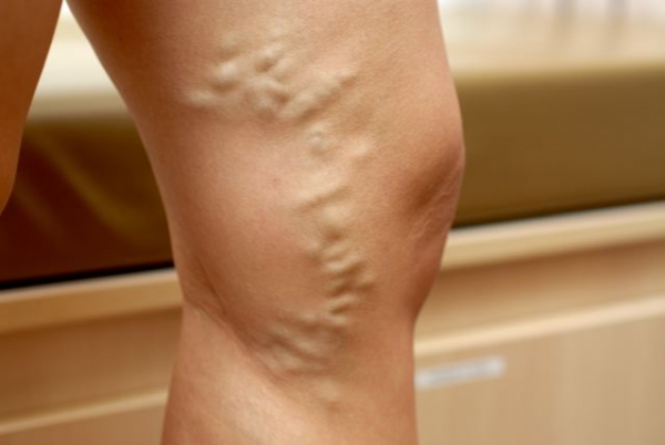
- The appearance of blueness and spider veins on foot. Appearances blue spots, spider veins is a sign of nascent varicose disease. Even small varicose asterisks should not be ignored.
- Change in the color of the skin of the legs. If the color of the skin on the legs turned blue, darkened, turned white, pigmentation appeared, you should immediately contact a phlebologist and undergo an examination.
- Palpation of seals. During palpation of the extremities, the doctor may detect seals in the veins and advise you to undergo a duplex scan.
None absolute contraindications ultrasound is not available, but in some cases the examination can be difficult. For example, if the patient has a high degree of obesity or painful ulcers that do not allow you to touch the sensor.
Preparation and procedure

Preparation before the ultrasound procedure is required only during the examination internal organs. In this case, you need to adhere to a certain diet for 2-3 days, take medications for flatulence.
Duplex scanning of the veins of the lower extremities does not require any preparation. The patient can lead habitual image life and not change the diet. All that is required of him is to come with a coupon at the appointed time to the ultrasound room.
The procedure itself is quite simple and does not require complex manipulations from the patient:
- The patient enters the office and undresses to the waist. Underwear can be left, the trousers must be removed, the skirt and dress can simply be lifted up.
- During the examination, you will have to change position several times. The examination is carried out in a standing position, lying on the back and on the stomach (with the exception of pregnant women).
- The doctor applies a special gel to the skin to improve the ultrasound signal and moves the transducer over the skin with light pressure. There is little pressure to be felt. If there are pain or discomfort should be reported to the doctor.
- If there are splints on the legs or elastic bandages must be removed prior to examination.
- During the procedure, 3 scanning modes are used. First, a two-dimensional mode is applied, which provides information about the diameter of the veins and blood vessels, the elasticity of its walls. It is possible to determine the presence of plaques and disorders already at this stage of the examination.
- Doppler mode allows you to evaluate the blood flow itself, the movement of blood through the veins and vessels.
- The color mode shows the patency of blood vessels and veins in color, that is, it allows you to determine the presence of pathologies, eddies and flows in the bloodstream.
As a rule, the whole procedure takes no more than half an hour. During the examination, the doctor records certain indicators and enters them into the protocol. After the end of the procedure, the gel is removed from the skin with napkins, the patient receives the result in his hands, which will need to be shown to his doctor.
Ultrasound results: norm and pathology

During the survey, many indicators are evaluated. The doctor should deal with the interpretation of the result.
An ultrasound specialist only assesses the condition of the vessels and blood flow, records the data, but the diagnosis is made taking into account the entire examination and all indicators:
- echogenicity. The walls of blood vessels have increased echogenicity, the lumen should be anechoic. That is, it should not have any dense formations. If the lumen of the vessel has increased echogenicity, this indicates the presence of a plaque or thrombus.
- Vessel walls. Normally, the walls of blood vessels are smooth and thin. Their thickness should not exceed 2 mm. If they are thickened (more than 4 mm), this indicates vein thrombosis.
- Diameter of arteries and veins. The diameter of the vein is usually 2 times the diameter of the artery. If the veins are narrower than arteries or more than 2 times wider, this indicates a pathology.
- Color uniformity. With the color mode, a healthy vein is stained completely and evenly. If gray spots are found on the monitor, the presence of blood clots and plaques can be suspected.
- Blood flow. Spontaneous blood flow should be visible in all veins and vessels. The absence of blood flow is allowed only in very small veins.
- Synchronization of breathing and blood flow. In a healthy person, blood flow is synchronized with respiratory function. With thrombosis, this synchronization is disrupted.
You can learn more about USDS from the video:
As a result of ultrasound, it is possible to identify such dangerous disease, how . With this pathology, cholesterol plaques block the lumen of blood vessels and veins, arteries, disrupting normal blood flow. Organs and tissues begin to suffer from oxygen starvation. Plaque in vital arteries can be fatal.
Also, ultrasound is often used to examine varicose veins. leads to bulging of the walls of the veins. On the final stages of this disease are formed trophic ulcers, the skin becomes abnormally black, and the pain in the legs becomes very severe.
Noticed an error? Select it and click Ctrl+Enter to let us know.
Duplex scanning of the veins of the lower extremities: price in Moscow, St. Petersburg, Perm. Why do a scan, what are the types and how to decipher the results. Problems with the veins of the lower extremities often violate normal course life. In order to normalize the condition, you need to undergo a course of treatment in a timely manner. The doctor can prescribe it only after setting accurate diagnosis. This is helped by modern research methods that help to see what is inaccessible. human eye. One of modern options diagnosis is duplex scanning of the veins and arteries of the lower extremities.
 For the prevention and treatment of varicose veins in the legs! Phlebologists recommend using anti-varicose gel VARIUS
against varicose veins. fast and effective disposal, acts on the cause of the disease, relieves pain, relieves swelling in the legs, relieves blue veins, restores blood outflow, strengthens the walls of blood vessels, prevents blood clots and convulsions.
For the prevention and treatment of varicose veins in the legs! Phlebologists recommend using anti-varicose gel VARIUS
against varicose veins. fast and effective disposal, acts on the cause of the disease, relieves pain, relieves swelling in the legs, relieves blue veins, restores blood outflow, strengthens the walls of blood vessels, prevents blood clots and convulsions.
Reviews of doctors and users... »
Duplex scanning of the veins of the lower extremities represents one of the varieties ultrasound diagnostics. The method allows to estimate the blood flow. Duplex scanning gives a picture of the patency of blood vessels and the stage of development of varicose veins.
This method of research has a number of clear advantages:
- The ability to assess the condition of previously inaccessible vessels;
- Identification of obvious differences between the vessels and ducts of other organs;
- The ability to diagnose even small blood clots and plaques;
- Assessment of blood flow in real time.
All this helps to accurately determine the disease, its cause and prescribe treatment.
Why do a vascular scan: the basics and capabilities of the method
Vessel scanning is a fairly informative study. It allows you to assess the condition of veins and arteries in places inaccessible to conventional ultrasound. Scanning helps to find out about blood flow disorders in the brain, which was not previously available. duplex study gives a visual representation of the vessels and their exact location.
Diagnostics is widely used. Duplex scanning helps to assess the condition thyroid gland by setting the nature of the nodes. When diagnosing neck vessels, it is possible to find out about the presence of blood clots and plaques.
Elena Malysheva says: |
| Varicose veins pass quickly, a new invention from ZDOROV in medicine. Decrease varicose mesh, improving well-being, reducing fatigue at the end of the working day, getting rid of pain in the legs. New remedy bee cream Wax is HEALTHY for varicose veins. |
Interview with Elena Malysheva and user comments... » |
Duplex scanning of the veins of the lower extremities helps to assess the condition of the organs abdominal cavity and timely diagnosis of tumors. A study using this method of the kidneys provides information about renal arteries, veins and determines the quality of their functioning.
Scan gives full information about the condition of the uterus and fallopian tubes. Thanks to the technique, doctors are able to diagnose the presence of polyps, tumors and cysts in time.
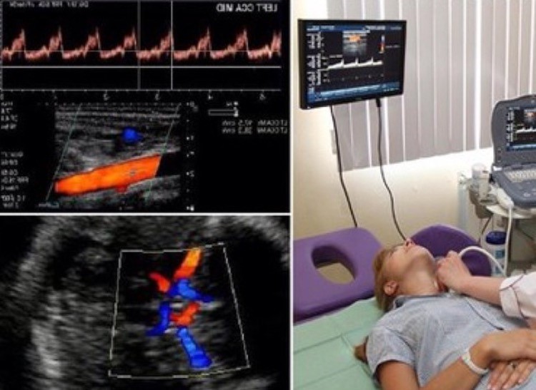
This diagnosis involves identifying problems with main vessels brain. During the procedure, the condition is assessed carotid artery, vertebral and left subclavian branches. Manipulations are made in the area of the collarbone and neck.
Scanning helps to identify the following problems:
- Thrombi and plaques.
- Vascular stenosis.
- Aneurysms.
- Underdevelopment of blood vessels.
- Elongation.
- Anomalous tortuosity.
This study is prescribed for frequent headaches, tinnitus, drowsiness, memory problems, low or high blood pressure. In addition, scanning allows you to monitor the condition of patients with vegetative-vascular dystonia, blood diseases, neck tumors, stroke survivors and those who are being prepared for heart surgery.
After the procedure, the doctor receives the results with a transcript. They describe in detail the dimensions of all scanned arteries. Judging by the study, the doctor can make an accurate diagnosis and prescribe an effective treatment.
Doppler scanning gives a less clear picture current state patient. This happens because there is no visual representation of the vessels. As a result of the scan, the doctor receives data only on the patency of blood through the vessels.
Thus, the study helps to determine the presence of pathology, but does not indicate the causes of their occurrence.
In turn, duplex scanning of the veins of the lower extremities allows you to clearly see vessels and evaluate their blood flow and patency. Already after the completion of the procedure, a person has the opportunity to learn about the condition of the walls, the shape of the vessels and the presence cholesterol plaques. That is, the technique helps to simultaneously assess the blood flow velocity and appearance arteries.
Types and features
The state of the vessels can be judged after they detailed study. Specifically for this, there are the following scanning options:
- Doppelerography (ultrasound);
- duplex scanning;
- Color duplex scanning.
A more complete picture of the state of the veins of the lower extremities is given by two latest method. They are often prescribed by phlebologists. Often referred to as triplex color scanning, it visualizes arteries, veins, and blood flow.
This allows you to better see blood clots and plaques, if any. The presented studies can be carried out in many hospitals.
Scanning of vessels is quite informative diagnostics. Thanks to the study, the doctor is able to assess the state of blood vessels and arteries in real time. Scanning can be performed on patients of any age.
The procedure is painless as the tissues are not damaged in any way. After the diagnosis, the doctor can accurately determine pathologies such as varicose veins, thrombosis, thrombophlebitis and prescribe the appropriate treatment.
Who is assigned the procedure
This method has direct indications for its purpose. As a rule, duplex scanning of the veins of the lower extremities is performed in the following cases:
- Constant pain in the legs;
- Edema and heaviness in the lower extremities;
- The formation of spider veins;
- Frequent convulsions;
- Problems with probing the pulse on the arteries of the lower extremities;
- Appearance dark spots or trophic ulcers;
- Seals in the area of dilated veins.
In order for the physician to select the right course of treatment, it assigns diagnostics that are distinguished by their accuracy.
The process of preparing and conducting
This technique is distinguished by its safety and high accuracy. In this case, no special preparation is required before using duplex scanning.
Diagnosing is quite simple. The patient lies on the couch, and the doctor applies a special gel to the scanned areas. After that, using a special sensor, the vessels are scanned, and all the information is displayed online on the monitor. As a rule, duplex scanning takes no more than 40 minutes.
Contraindications
Duplex scanning of the veins of the lower extremities is considered quite safe and exact method. It is available to people of all ages. But it will have to be abandoned in the following cases:
- In the presence of open wounds, ulcers or rashes.
- during an exacerbation of asthma.
- During an epidemic of infectious diseases.
After getting rid of all these problems, duplex scanning can be performed.
During pregnancy
Pregnancy is a rather responsible period in the life of every woman. Now there are modern techniques, which help to determine the condition of the mother and fetus and at the time to find out about the presence of any pathology.
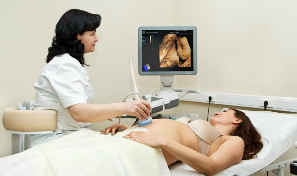
A Doppler study helps to assess the state of blood flow, in particular its speed, and determine the width of the lumen and pressure inside the vessels. Also, thanks to the diagnosis, you can learn about the development of the child's heart and understand how well the fetus is supplied with blood. Doppler helps to learn about the entanglement of the umbilical cord and problems with the placenta.
The study is safe and mandatory. Doppler ultrasound is prescribed for all expectant mothers in the third trimester of pregnancy. As a rule, it is recommended to carry out diagnostics after 30 weeks, that is, closer to childbirth, it is possible to assess the position of the child and the condition of all his organs.
Early detection listed problems helps to cope with them in time and avoid a threat to the mother and child. Already, obstetrician-gynecologists know how to deal with a particular problem that can be detected by a doppler. That is why more and more healthy children began to be born.
Results and data interpretation

The patient receives the results of duplex scanning immediately after the completion of the study. There are indicators by which one can judge the condition of the veins and arteries. are considered normal following results research:
- The walls are smooth, thin, elastic.
- Echogenic lumen with valve leaflets.
- The size deep vein does not exceed more than 2 times the size of the deep artery.
- The vein has no gray areas when stained.
- The blood flow coincides with breathing.
- When holding the breath, the inferior vena cava increases in size up to 15%.
- The vessel, when clamped by a special sensor, collapses.
doctor says about venous thrombosis in the following cases:
- The venous wall is thicker than 4 mm;
- Blood flow and breathing are not synchronized;
- Change in the diameter of the vein in which the thrombus is located;
- Increased echogenicity of the valve leaflets.
In any case, on the basis of the results of the tests, a person cannot judge his condition. Final Diagnosis should be put by a phlebologist who will select the appropriate course of treatment.
Method safety
Duplex scanning is considered to be quite safe methods diagnostics. Radiation can only adversely affect the retina, but this applies to outdated devices. Modern technology is absolutely safe, since strong filters signal at the maximum permissible level.
Duplex scanning of the veins of the lower extremities: price in Moscow, St. Petersburg, Perm
Duplex scanning of the veins of the lower extremities is a modern and very common study. In the metropolitan areas, many hospitals and private clinics provide these services. Scanning should only be done with the direction of a doctor.
Often people are interested in the cost of such a study. In Moscow, you can find many offers. Depending on the clinic, the price varies from 1500 to 4000 thousand rubles. This price is for vein scanning only, arterial examination is paid extra.
In Saint Petersburg you can find a large number of offers from clinics that perform duplex scanning of the veins of the lower extremities. Prices in St. Petersburg are in the range of 1300-2500 thousand rubles.
Choosing a clinic need to navigate not on the cost of the study, but on the reputation of the doctor and the clinic. This is very important points, since the quality of the procedure performed and the effectiveness of the treatment subsequently prescribed depend on it.
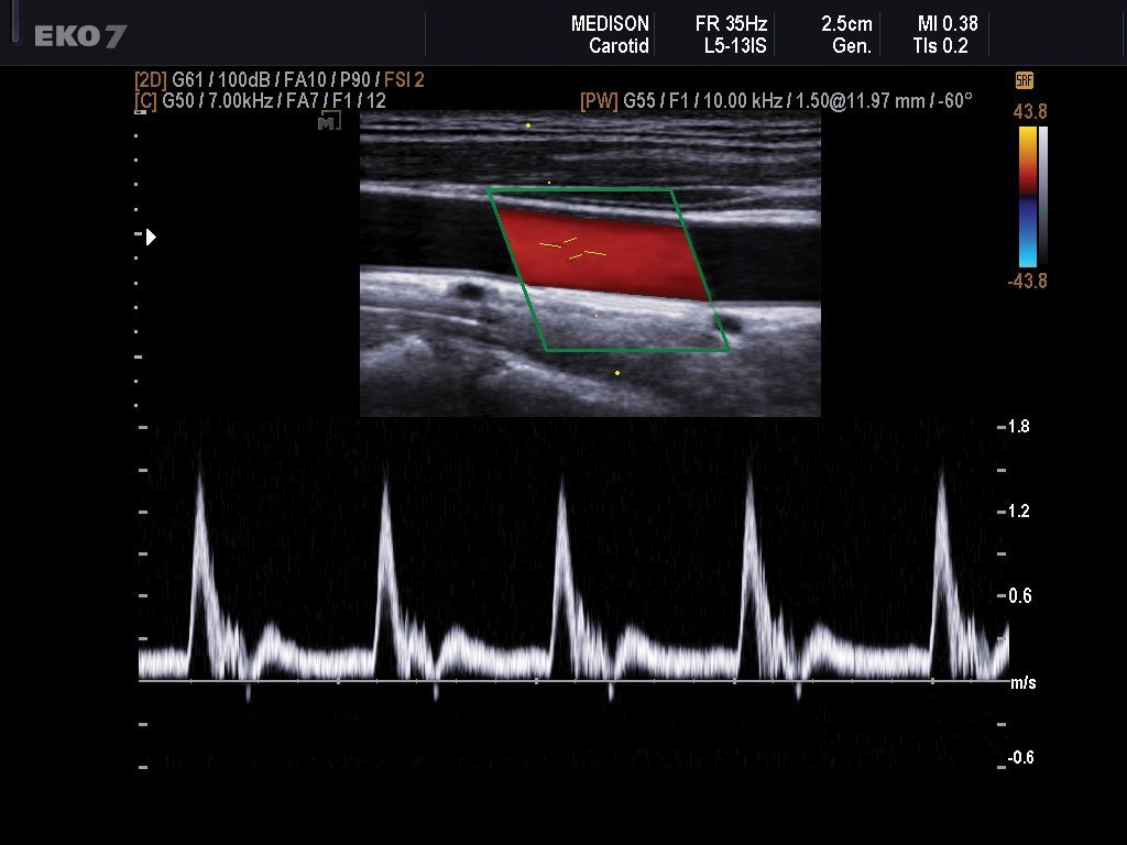
Duplex scanning - instrumental method study of veins and blood vessels, combining two-dimensional scanning and Dopplerography. In other words, this technique allows not only to see the state of the vascular system, but also to assess the blood flow, its direction and speed.
The technique of the procedure through the eyes of the patient is no different from conventional ultrasound: the specialist receives an image on the monitor through a special sensor.
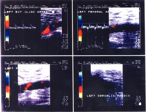
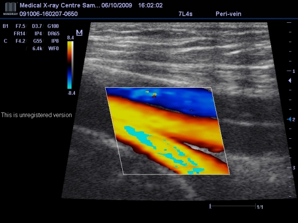
With duplex scanning, 2D real-time vascular imaging and Doppler information: blood flow velocity, its direction through a Dopplerogram (graphics) or color doppler - color overlays on the image (triplex scanning).
The condition of the vessels is examined in different duplex scanning modes:
- B-mode- two-dimensional image. Allows you to see the contours of the vessel, the thickness of its walls, gaps, valve flaps.
- Doppler curve- wavy chart. It carries information about the type of vessel, its condition.
- CDC- color overlay on the image. Blue color shows the movement of blood from the sensor, red - the flow of blood to the sensor, light colors indicate high blood flow rates, bright colors indicate low ones.
The study is carried out with a sensor with an operating frequency from 5 to 10 MHz, their frequency may vary depending on the object, for example, for the diagnosis of intradermal veins, sensors with a frequency of 12 MHz.
Indications
 Ultrasound of the lower extremities is the leading diagnostic method. Deep and superficial veins both legs.
Indications for research are following states:
- , with the available signs venous insufficiency- swelling, pain and a feeling of fullness in the legs. It is carried out in order to identify pathological refluxes (reverse blood flow) and complications in the form of blood clots and phlebitis (inflammation of the vascular wall), assess the condition of the veins - the presence valvular insufficiency, patency of veins.
- - a condition characterized by the formation of blood clots in deep veins, and as a result, impaired blood flow. The size of blood clots and their location are assessed.
- – pathological condition developing in patients who have had thrombophlebitis or phlebothrombosis. It is characterized by pronounced swelling of the legs, the formation of trophic ulcers.
Duplex scanning of vessels of the lower extremities necessarily carried out in groups of patients with increased risk development of varicose veins if they have clinical manifestations varicose veins are pregnant women, obese people, patients after surgical phlebological operations, patients with diabetes mellitus.
Contraindications
Duplex scanning of the arteries of the lower extremities has no contraindications. Even in disabled patients with severe complications of varicose veins, its use is necessary.
Important! With this research method, the Valsava test is often used - straining the patient at the time of holding the breath. The test is used to detect possible valvular insufficiency. In patients with deep vein thrombosis, this test is prohibited.
Duplex scanning during pregnancy will in no way harm the fetus. For comparison, radiation from mobile phone several times higher than that of ultrasound and dopplerography.
Where to do a duplex scan?
 Not all municipal polyclinics have trained specialists ultrasound diagnostics, respectively, services for duplex scanning of the veins of the lower extremities are not available in all medical centers.
As a rule, duplex scanning can be done in specialized phlebological centers, in urban and district hospitals that have a branch radiodiagnosis in private clinics. The study is paid and is somewhat more expensive than ultrasound of other organs and systems.
Modern research methods
Many ultrasound devices are equipped with additional scan modes that allow you to obtain advanced diagnostic data. Various ways imaging of blood vessels have a generic name - ultrasound angiography, which uses the following modes:
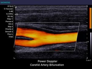
- color doppler mapping- displays the speed of movement of red blood cells and their flow in color mode, where Blue colour– direction of blood flow away from the sensor, red – movement towards the sensor. Saturated colors indicate a slowdown in blood flow, pale color indicates high speed erythrocytes.
- Power Doppler Imaging- allows you to visualize small vessels and red blood cell density, but does not provide data on blood flow velocity.
- 3D echography– allows you to get a three-dimensional image (3D mode).
- Contrast in ultrasound – intravenous administration contrast agents in the study. Enhances visualization small vessels and sensitivity of EDC and CDI methods.
The difference between duplex and triplex scanning. Research that uses standard methods duplex scanning and color mapping is called triplex scanning. Undoubtedly, this method makes it possible to obtain an extended diagnostic picture of the state of the vascular system, in contrast to duplex scanning.
Triplex scanning uses 3 modes:
- B-mode, in which the diameter of the veins, wall thickness and the presence of valves are assessed.
- TsDK - reveals the speed and direction of blood flow.
- Dopplerography - determines the phase of blood flow.
special diagnostic value triplex scanning has in the study of the inferior pudendal vein to detect thrombosis. With regard to the diagnosis of superficial veins, there is not much difference between the choice of duplex or triplex scanning.
Duplex scanning of the veins of the lower extremities
 Special preparation before the study is not required. The procedure is carried out in an office equipped with technical equipment and an ottoman. Diagnosis is performed in a patient in a standing or lying position, if the patient's condition is severe, then the study is carried out only in horizontal position.
The patient's legs should be freed from clothing, with a plastered limb, the plaster is cut before the procedure.
Technique
How is it carried out? On the patient's skin, over the examined vein a special acoustic gel is applied. Throughout the study, the sensor is moved from top to bottom, from the inferior vena cava to the foot. Scanning is carried out first in a horizontal position of the patient, then in a vertical position.
The study of the anterior superficial veins is carried out with the patient lying on his back, his legs should be slightly bent and spread apart. Scanning of the popliteal vein is performed in the supine position, a small roller is placed under the patient's feet.
Scanning scrotal vessels performed in the patient in the supine position, the gel is applied directly to the scrotum. Study pelvic organs in women, it is performed by two types of sensors: transvaginal - in the position of a woman lying on her back with legs bent at the knees, a condom is put on the sensor and inserted into the vagina, and abdominal - the condition of the veins is assessed through the area above the pubic joint. Read more about varicose veins of the pelvis.
Deciphering the results
 Normally, the veins have smooth contours, thin walls, venous valves are visualized, their leaflets fluctuate synchronously with breathing. In the color mode, healthy veins are evenly colored, blood flow increases on exhalation and decreases on inspiration.
Deciphering duplex scanning of the arteries of the lower extremities:
- Varicose disease. When removing the B-image, an enlarged vein is noted with the formation of cavities and loops, which are compressed when pressure is applied to them by the sensor. With insufficiency of the venous valve, retrograde blood flow is recorded when coughing or deep breath patient.
- Thrombosis. When pressed by the sensor, the thrombosed vein does not shrink, the blood flow in the color mode is not recorded. The thickness of the venous wall more than 2 mm indicates the transferred thrombophlebitis. The migrating thrombus is identified on the B-image, it fluctuates in time with breathing. If the vessel is somewhat wider than the thrombus itself, then given state points to high risk development of thromboembolism. The parietal thrombus is displayed as an echogenic mass, does not shrink when pressed with a sensor. With a parietal thrombus, the blood flow is synchronized with respiration, with a thromboembolic-dangerous state, the blood flow between the thrombus and the vascular wall is determined.
- Postthrombophlebic disease- insolvent perforating veins with a diameter of 5 to 10 mm are determined. Collateral (bypass) blood flow is detected.
At ultrasound scanning vascular system of the patient, it is necessary to assess the patency of the veins and the state of their valvular apparatus, diagnose or exclude possible development thrombosis, its form, localization and extent.
Useful video
Familiarize yourself visually with the principle of conducting a Duplex study:
Price
 Price for duplex scanning veins varies to a small extent from 900 to 1300 rubles, moreover, in Moscow, the price of a study is somewhat higher than in other cities of Russia. Prices for triplex scanning are higher by 200 - 300 rubles and are determined in the range from 1200 to 1500 rubles.
Duplex scanning is the "gold standard" of research venous system. It helps to trace the course of a venous disease in dynamics, determine further tactics for treating patients, and identify indications for the installation of cava filters.
If the legs often hurt and swell, dilated veins are visible, a feeling of heaviness in the legs has become habitual, that is, there is a reason to think about problems with blood supply. Quite a large part of patients' visits to hospitals is associated with vascular diseases. Diseases of the circulatory sphere are insidious: they are dangerous because they can initially occur without any symptoms at all. The doctor will help to make an accurate diagnosis and prescribe effective treatment. In this case, duplex scanning of the veins of the lower extremities is used.
This examination technique helps to find out exactly where the problem is hiding. It allows you to see the parts of the vessels changed by the disease, to find out how close the blood flow in the veins is to the norm. It is possible to identify places where the walls of the vessels have become thinner or, conversely, to detect thickenings in them. It does not matter whether the vessel is located close to the surface of the skin or is located in the deep layers of tissues. The degree of performance of the vessels is also assessed.
The procedure for scanning the veins of the lower extremities helps to reveal hidden pathological processes
Many are concerned about the symptoms of circulatory disorders in the legs. To establish the cause of the pathological condition and diagnose the disease, the doctor prescribes certain examination methods that allow you to visually examine the vessels. Few of the patients know what duplex scanning of the veins of the lower extremities is. But every patient heard about ultrasound and dopplerography. So, it's the same thing. Let's take a closer look at this issue.
When examining a patient, the doctor can only see external changes, but in order to determine the true process of the development of the disease, you need to know how the vessels function. This can be done using duplex scanning. Such a study is especially relevant when it comes to the need for surgical intervention.
Duplex scanning of the veins of the lower extremities is modern method vascular studies, gives an accurate clinical picture and greatly facilitates the process of pathology detection
Duplex vascular scanning is a modern research method that includes a standard examination of blood vessels and blood flow features, as well as Dopplerography. This diagnostic method gives an accurate clinical picture and greatly facilitates the process of detecting pathology.
In addition, it is very easy to use, can be carried out by any person without age restrictions and even allowed during pregnancy.
This method is often used for healthy people that are at risk of developing venous diseases, as it is informative even in the early stages, when the patient is not bothered by any symptoms.
The procedure itself takes about 45 minutes and allows the specialist to assess the condition of the walls of the veins, their valves and the blood flow through them. You can also see the presence of blood clots, the length of the affected area, seals, the diameter of the lumen. All these data make it possible to accurately establish the diagnosis.
For determining pathological phenomena The following types of vascular scans are used:
- Doppler ultrasound.
- Ultrasonic duplex scanning.
- Color duplex scanning.
The last two methods are used in phlebological practice most often.
Features of duplex scanning
Using this diagnostic method, it is possible to establish all kinds of parameters of the vessels, which are decisive in the diagnosis of pathology. This is especially important for varicose veins, thrombophlebitis, thrombosis.
By performing ultrasound duplex scanning of the veins of the legs, the doctor has the opportunity to see the state of the vessels in real time, to evaluate all the necessary characteristics. An undeniable advantage can be considered the fact that this method of diagnosis is allowed to be used by all patients, without age restrictions.
It is well tolerated by patients, does not require pre-training does not cause pain or discomfort does not damage tissue. Due to the lack of penetration, there is no need for the use of anesthetic drugs, in connection with this, the occurrence of allergic reactions on them.
In this way, it is possible to determine the cause of the recurrence of the disease after suffering surgical treatment. It is also possible to examine the size of formations in the vessels during thrombotic processes.
This examination is well tolerated by patients, does not require preliminary preparation, does not cause pain or discomfort.
Color duplex scanning of the veins of the lower extremities is a newer method and differs from ultrasound in that it gives a color image. A specialist can literally see a real image and evaluate all the necessary characteristics of both vessels and blood flow. This method is especially useful when it comes to emergency cases requiring a quick decision and treatment appointment.
Duplex scanning does not pose any danger to human health, so it can be used as long as necessary to make an accurate diagnosis and appointment. effective treatment patient.
Indications

It is worth going through a duplex scan of the veins of the lower extremities if you have developed the following symptoms:
- Swelling of the lower extremities, even if this symptom occurs only in evening time, and by morning it disappears on its own.
- Feeling of heaviness in the legs.
- Cramps, periodic muscle contractions.
- Pain or discomfort that can develop not only after exertion, but also in a state of complete rest.
- On the legs it is not possible to feel the pulsation of the arteries.
- Availability age spots on the skin or it has become dense, dark, pale, there is redness.
- Visually, broken capillaries - spider veins are visible on the skin of the lower extremities.
- Trophic ulcers.
Even the development of one of the above symptoms is a reason to consult a specialist, as it indicates damage to the vessels of the legs. Treat similar diseases needed immediately to prevent progression pathological process and serious consequences.
In addition, duplex scanning can also be prescribed for the development of signs that, it would seem, are in no way related to the condition of the leg veins. However, you should seek qualified medical care if you are concerned:
- Insomnia.
- Spontaneous loss of consciousness.
- Frequently recurring neck and headaches.
- Migraine attacks.
- Traumatic injuries of the neck.
- Dizziness.
- Memory deterioration.
- Decreased attention.
- There are suspicions of the development of vegetovascular dystonia.
- Intracranial pathological processes.
- Chronic diseases (hypertension, diabetes, atherosclerosis, etc.).
This connection is explained by the fact that the vessels pass throughout the body and blood is constantly moving through them, and therefore the pathological state of one area can spread to other areas.
Duplex scanning is quite effective after surgical treatment leg veins. It makes it possible to control the healing process and, if necessary, adjust the course of treatment in a timely manner.
Also this method studies should be periodically carried out by patients at risk of developing vascular diseases. These are people whose work requires a constant stay in a sitting or standing position, moving heavy objects, with a hereditary predisposition. The procedure is also shown to women taking oral contraceptives a long period, patients with overweight body. Pregnant women who had problems with blood vessels before conception.
Training
Before carrying out the manipulation, it is not necessary to adhere to any rules. It is enough just to observe hygiene standards.
Manipulation
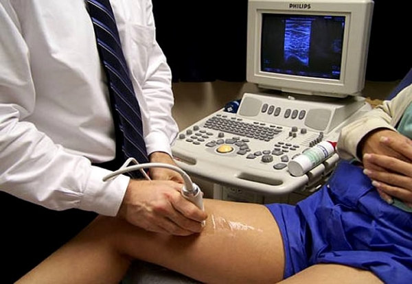
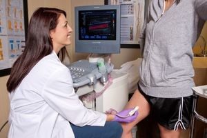
Now let's look at how duplex scanning of the vessels of the lower extremities is performed. It is carried out without prior hospitalization in a polyclinic. The methodology is as follows. The patient must lie down on the couch and provide the doctor with access to the lower limb, so the trousers will have to be removed, and the skirt should simply be raised to the level of the hips.
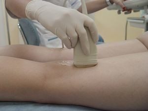
Before the scan, a special gel is applied to the skin. It is distributed over the surface by an ultrasound sensor and provides a better perception of data by the device. In order to obtain maximum information about the state of the vessels, the quality of blood flow during the examination, the patient is asked to change the position of the body several times. First, the patient is laid on his back, then he is asked to take vertical position and lie on your stomach.
To examine the large superficial and main veins of the lower limb, the patient lies on his back. In order for a specialist to assess the condition of the popliteal vessels and those that pass in the upper part of the lower leg, it is necessary to lie on the stomach. This position is not recommended during pregnancy.
During the examination, the doctor conducts special tests that allow you to more accurately assess the quality of blood flow and the condition of the valves of the veins. Sometimes the patient is asked to hold their breath or change their body position slightly.
Duplex scanning of the veins of the lower extremities is carried out without prior hospitalization in a polyclinic
This technique is very popular and is widely used in medical practice. The cost of duplex scanning of the vessels of the lower extremities varies in different price ranges, but it is quite affordable for any person with an average income. This difference depends on the prestige of the clinic, doctor, conditions.
It is used both in private clinics and in ordinary district medical institutions.
Result interpretation
Only a specialist can distinguish the norm from the pathology. However, we will give an example of what should be the result of a duplex scan in a healthy person.

normal thickness vascular walls does not exceed 2 mm, they are thin, even, without seals, the lumen is anechoic. The valves are functioning properly. The rhythm of blood ejection is synchronized with respiratory movements. The diameter of the deep veins of the extremities is twice that of superficial arteries. With color duplex scanning, vessels without signs of a pathological process are stained in one tone, gray areas are absent.
Any deviation from the norm is considered as possible violation blood flow or dysfunction of the vessels.
Duplex scanning of the vessels of the legs can provide answers to all questions regarding the state of the walls of the veins and blood flow in them. With its help, it is possible to fully examine vascular system and this greatly simplifies the diagnosis. In addition, this study is available to any person, regardless of the financial situation and the scale of the settlement.
