Dimensions and weight of a normal thyroid gland. Functional disorders of the thyroid gland
I talked about why it is useful to conduct a regular examination of the thyroid gland using ultrasound. After that, a lot of letters came to the mail with questions about what the norms of the thyroid gland should be.
Therefore, I decided to write a separate article so that everyone can get acquainted with the information.
The thyroid gland is an organ located in the neck, in front, under the larynx. It has the shape of a butterfly and consists of two symmetrical lobes and an isthmus. Since the gland is located directly under the skin, deviations in its structure or structure can be detected even during the initial examination by an endocrinologist by palpation.
The thyroid gland of normal size in most cases is not palpable, except in cases where excessive thinness or anatomical structure the patient's neck allows this.
However, with a noticeable increase in the size of the gland during palpation, it is easy to determine:
- the shape of the organ, the size and symmetry of its lobes, the total volume;
- mobility and localization of the gland;
- density and consistency of gland tissue;
- the presence of nodes and volumetric formations.
Unfortunately, the manipulation does not allow to detect formations while maintaining or reducing the normal size of the organ, therefore, the main method for reliable diagnosis of the state of the thyroid gland is ultrasound.
On ultrasound thyroid is defined as a rounded organ, vaguely resembling a butterfly in shape, with symmetrical lobes and a homogeneous structure.
- The volume of the gland: in women - from 15 to 20 cm3, in men - from 18 to 25 cm3.
- Dimensions of the lobes of the gland: length - 2.5-6 cm, width - 1.0-1.8 cm, thickness - 1.5-2.0 cm.
- Isthmus thickness: 4 to 8 mm.
- Parathyroid glands with a diameter of 2–8 mm, from 2 to 8 units.
In different medical sources, the boundaries of normal indicators of the size of the lobes and the volume of the organ differ. Studies among the population have shown that the average values of the norm are relative - for example, the population of regions with a constant iodine deficiency is different general change the size of the thyroid gland in a big way, and this is not a pathology.
Often there is asymmetry of the body - right lobe usually more than the left, but it also happens vice versa - as an individual feature of the organism. There have been cases where healthy people one of the lobes was underdeveloped or completely absent.
The difference in the volume of the thyroid gland in men and women is not associated with gender, but with the difference in the physical and physiological parameters of the body.
Normal thyroid size
Although during the menstrual cycle in women there are some fluctuations in the data of ultrasound of the thyroid gland, nevertheless, specialists during the examination take into account, first of all, the age and weight of the patient. In adults, the normal size of the thyroid gland can vary within:
- weight up to 40 kg - up to 12.3 cm3;
- 41–50 kg - up to 15.5 cm3;
- 51–60 kg - up to 18.7 cm3;
- 61–70 kg - up to 22 cm3;
- 71–80 kg - up to 25 cm3;
- 81–90 kg - up to 28.4 cm3;
- 91–100 kg - up to 32 cm3;
- 101–110 kg - up to 35 cm3.
As the data of the list show, the concept of the norm in a healthy person is very relative and often goes beyond the average indicators. In addition, it is allowed to exceed these norms by 1 cm3 or more, provided that the function of the thyroid gland is not impaired.
There are cases of individual underdevelopment (hypoplasia) of the organ with the preservation of its full functionality.
In about 1/6 of the population, the thyroid gland has a pyramidal lobe - an additional structural unit with a base in the middle of the isthmus - which is also one of the options individual norm. Specialists of diagnostic rooms periodically observe the absence of an isthmus between the lobes of the organ in some patients.
To identify pathological changes a comprehensive analysis of data from ultrasound examination of the thyroid gland is necessary:
- Contours of the gland - healthy organ has clear, even contours, the change of which indicates the development of the inflammatory process.
- Structure - homogeneous glandular tissue is an indicator of the norm and has a characteristic granularity. With the development of immune inflammatory diseases- autoimmune thyroiditis, diffuse toxic goiter– the structure becomes heterogeneous. Sometimes the heterogeneous structure of the glandular tissue is also found in healthy older people. age groups at increased production antibodies to certain enzymes of thyroid cells.
- Echogenicity is a certain value of the general acoustic response characteristic of the tissue under study. Echogenicity should be normal, i.e. meet the standards for that body. If echogenicity is reduced, the doctor may suspect the development of an inflammatory process. An increase in echogenicity may indicate acute inflammation or the development of pathological changes.
- Foci of changes are areas characterized by a decrease (hypoechogenicity), absence (anechoicity) or an increase (hyperechogenicity) of the acoustic response of ultrasound. Such formations should not normally be, although the presence of small, up to 4 mm, anechoic areas is allowed - single enlarged follicles of the glandular tissue. Pathological foci, identified in the structure of the tissue, are nodes of the thyroid gland. Nodes can be single or multiple. Solitary small nodules (1-3 mm) are usually not treatable and often disappear on their own over time. Formations larger than 3 mm, as a rule, require clarification of the diagnosis.
- The state of the lymph nodes - the latter should have clear, even contours, the absence of cysts and normal size(not enlarged).
What does a thyroid ultrasound show?
colloid nodes- formations, which are overgrown follicles. These are benign lesions that almost never degenerate into malignant tumors.
Adenoma – benign tumor subject to surgical removal. The presence of a fibrous capsule allows it to be differentiated from other pathologies. It develops with age, mainly in women.
Cyst- fluid-filled formation. Usually observable.
thyroid cancer- a dangerous single node that does not have clear boundaries and a shell. It is characterized by rapid growth, is subject to immediate removal along with the lymph nodes.
When a neoplasm is detected, the patient undergoes additional research- Dopplerography or elastography, to assess changes in the intensity of blood flow in the vessels of an organ, and the cellular and tissue structure of existing formations. If necessary, a needle biopsy is performed to histological analysis under ultrasound supervision.
Diffuse toxic goiter- a disease manifested by an increase in the volume of the gland and the heterogeneity of its structure due to the formation of multiple nodes.
Inflammatory diseases (thyroiditis)- distinguish between acute and subacute thyroiditis of infectious and viral origin, arising as complications after tonsillitis, bronchitis, pneumonia, SARS; fibrous thyroiditis - inflammation of the tissue as a result of the abundant growth of its fibrous component; autoimmune chronic thyroiditis- a feature of the body to perceive thyroid cells as foreign, as a result of which an inflammatory process occurs.
Goiter of the thyroid gland- an increase in volume due to tissue growth. Euthyroid goiter does not affect the function of the organ, hypo- and hyperthyroid goiters are associated with corresponding dysfunctions. Possible development endemic goiter among the population of areas with reduced content iodine in environment, as well as some hypertrophy of the thyroid gland during pregnancy.
Hypoplasia of the thyroid gland- congenital underdevelopment of the organ due to endocrine disorders during pregnancy of the mother or insufficient intake of iodine in the body.
Thyroid atrophy- a decrease in its size as a result of the gradual replacement of the glandular tissue with connective tissue, combined with the development of hypothyroidism, requiring constant replacement therapy.
Thus, when setting accurate diagnosis endocrinologist results ultrasound(ultrasound) are analyzed in combination with other indicators of the patient's health. The combination of complaints, individual symptoms, general well-being, blood tests and functional diagnostic data allows the doctor to determine the individual boundaries of the norm and pathology and choose the best means of treating the patient.
Dear readers, if you have any questions, then ask them in the comments, I will try to answer them in detail.
Introduction
The thyroid gland, an endocrine gland, shaped like a butterfly, is a unique organ.
Ancient philosophers associated it with fire, thereby emphasizing its significance for the body. Very small in size, no more than 18 ml in women and 25 ml in men, it is involved in almost all life processes. Without it, the functioning of the human body is impossible. Growth and development, metabolic processes, respiration, digestion... Thyroid dysfunction creates many problems in the work of all body systems.
In recent years, the number of people with identified disorders in the thyroid gland has sharply increased: diffuse and nodular goiter, Graves' disease, autoimmune thyroiditis, and oncological diseases. There are enough reasons for disappointing statistics: environmental degradation, reduced immune defense of the human body, lack of iodine, lack of planned medical prevention, unbalanced nutrition, stress as a provoking factor. Currently, thyroid diseases are leaders in the list of diseases of the endocrine system.
Quite a lot has been written about the treatment and prevention of thyroid diseases; on the Internet, you can find tips and tricks to combat the disease. However, it should be remembered that the treatment, selection and prescription of drugs should be handled by a specialist - an endocrinologist. And before you start using any method of treatment, you need to consult a doctor.
In this book, we will talk about the structural features of the thyroid gland, its functions, diseases of this vital organ, as well as give useful advice and talk about methods for examining and treating thyroid diseases.
Chapter 1 Thyroid Gland
"Butterfly" flies on iodine, without it it does not fly!
The thyroid gland and its functions
The thyroid gland is a gland of the endocrine system that stores iodine and produces iodine-containing hormones: thyroxine and triiodothyronine, which are involved in the regulation of metabolism and the growth of individual cells, as well as the body as a whole.
The gland, along with other organs of the endocrine system, performs its main function: it maintains the constancy of the internal environment of the body, which is necessary for its normal functioning.
The thyroid gland is located under the thyroid cartilage and has the shape of a butterfly (see Fig. 1).
Rice. 1. The shape of the thyroid gland can be compared with the letter "H" or with a butterfly
Interesting fact:
A brief morphological description of the thyroid gland as early as the 2nd century BC. BC e. given by Galen. He considered it part of the vocal apparatus.
Continued the study of the thyroid gland Vesalius.
And the name of this organ was given by Barton in 1656. He proceeded from its shape and purpose: it, like a shield, protects the organs located on the neck.
The concept of the function of internal secretion, which is carried out by the thyroid gland, was formulated by King.
Karling later described cretinism in people without a thyroid gland.
The gland consists of two lobes and an isthmus. The isthmus is a part of the thyroid tissue that connects the right and left lobe. It is located at the level of the second or third tracheal ring.
Lateral lobes cover the trachea and attach to it connective tissue.
An additional, pyramidal lobe may depart from the isthmus or one of the lobes. It is a long process that reaches the top of thyroid cartilage or hyoid bone.
The additional proportion is not considered a deviation, rather it is an individual feature of the organism (see Fig. 2).
The thyroid gland is located in middle third neck. Run your hand over the neck and you will find dense cartilage that shifts when you swallow. This is the thyroid cartilage. In men, it is larger than in women, and is called the Adam's apple.

Rice. 2. The lower parts of the thyroid gland are short and wide, while the upper ones are high, narrow and slightly divergent.
The thyroid cartilage somewhat covers the thyroid gland, its upper pole reaches it. It got its name from its functions: it serves as a shield, covers important organs lying on the neck.
The main characteristics of the gland: weight, height and width of shares, volume.
The thyroid gland of an adult human weighs on average 20–40 g, while in a newborn it is only 2–3 g.
Normally, the height and width of the lobes of the thyroid gland are 3–4 and 1–2 cm, respectively, and the width is 7–11 cm.
In order to understand whether the thyroid gland is enlarged, the doctor palpates (probes) it and compares the size of each lobe with the size of the terminal nail phalanx of the thumb on the patient's hand. Normally, their sizes should be the same.
Look at your fingers and you'll see how big your thyroid should be (see Figure 3).

Rice. 3. Nail phalanx of the thumb
The World Health Organization (WHO) distinguishes three degrees of thyroid size, which the doctor evaluates during examination and palpation (table 1).
Table 1
Degrees of thyroid size

If a goiter is detected, you should understand what the volume of the thyroid gland is. This is important for further treatment planning and follow-up.
Volume is the main indicator of the size of the thyroid gland.
Normally, it is up to 18 ml in women and up to 25 ml in men.
The volume of the thyroid gland is calculated using a special formula during an ultrasound examination (ultrasound).
The thyroid gland is made up of follicles. Follicles are communities of thyrocytes (thyroid cells), these are closed hollow formations of various shapes. Their walls are formed by cells that produce colloid - a thick yellowish mucous liquid.
The smallest follicles have a diameter of 0.03 to 0.1 mm, and their average size is 0.15 mm. The largest follicles can be seen with the naked eye on a transverse section of the thyroid gland.
Thyroid hormones
The thyroid gland is an endocrine gland. Its main function is the production of hormones, which include iodine, without which the normal functioning of the body is impossible (Fig. 4).
Thyroid hormones control metabolism, the processes of maturation of tissues and organs, and activate mental activity. They are necessary for active growth, the formation of bones of the skeleton, in women - for the development of the mammary glands.
The term "hormone" in Greek - "I excite", "I encourage". It was introduced into medical practice by Bayliss and Starling. Thyroxin was discovered by the American E. Kendall in 1914, and in 1927 C. Harrington synthesized it for the first time. With a decrease in the production of thyroid hormones in childhood, the growth of the body stops. In this case, you should immediately consult a doctor!
As already mentioned, the thyroid gland produces thyroid hormones: thyroxine and triiodothyronine.
In another way, thyroxine is called T4, as it carries four iodine atoms. In the blood and tissues of the human body, the T4 hormone is converted into the T3 hormone - triiodothyronine, which carries three iodine atoms.
Initially, the thyroid gland produces 70% T4 and 30% T3, but the main amount of T3 is formed during the breakdown of T4 in the body.
The biological effect of hormones is realized as follows: the hormone attaches to the receptor and, connecting with it, starts a series of reactions already in the cell of the organ.
Since thyroid hormones are responsible for the development of the body, proper metabolism and energy, receptors are everywhere: in the brain and in all tissues of the human body.
The functions of thyroid hormones are as follows:
Increase the intensity of oxidative reactions in cells;

Rice. 4. The main function of the thyroid gland is the production of hormones, without which the normal functioning of the body is impossible.
Influence the processes occurring in mitochondria, the cell membrane;
Maintain hormonal excitability of the main nerve centers;
Participate in normal functioning heart muscle;
Ensure the functioning of the immune system: stimulate the formation of T-lymphocytes responsible for fighting infection.
The thyroid gland is actively supplied with blood, it has a lot of blood vessels.
Active blood supply is carried out by four main arteries. The two superior thyroid arteries arise from
external carotid, and the two lower ones - from the thyroid cervical region subclavian arteries.
The outflow of blood from the gland occurs through paired veins. It is 4-6 ml / min / g and slightly exceeds the blood flow in the kidneys and brain.
Previously, active blood supply to the thyroid gland created difficulties during surgery on this organ. Surgeon Theodor Kocher developed safe approaches to thyroid surgery, for which he received the Nobel Prize. And it was the knowledge of the characteristics of the blood supply to the thyroid gland that helped him develop a certain tactic of surgical intervention.
It consists of two lobes and an isthmus and is located in front of the larynx. The mass of the thyroid gland is 30 g.
The main structural and functional unit of the gland are follicles - rounded cavities, the wall of which is formed by one row of cuboidal epithelium cells. Follicles are filled with colloid and contain hormones thyroxine and triiodothyronine associated with the protein thyroglobulin. In the interfollicular space are C-cells that produce the hormone thyrocalcitonin. The gland is richly supplied with blood and lymph vessels. The amount flowing through the thyroid gland in 1 min is 3-7 times higher than the mass of the gland itself.
Biosynthesis of thyroxine and triiodothyronine It is carried out due to iodination of the amino acid tyrosine, therefore, active absorption of iodine occurs in the thyroid gland. The content of iodine in the follicles is 30 times higher than its concentration in the blood, and with hyperfunction of the thyroid gland, this ratio becomes even greater. Absorption of iodine is carried out due to active transport. After the combination of tyrosine, which is part of thyroglobulin, with atomic iodine, monoiodotyrosine and diiodotyrosine are formed. Due to the combination of two diiodotyrosine molecules, tetraiodothyronine, or thyroxine, is formed; condensation of mono- and diiodotyrosine leads to the formation of triiodothyronine. Subsequently, as a result of the action of proteases that break down thyroglobulin, active hormones are released into the blood.
The activity of thyroxin is several times less than that of triiodothyronine, however, the content of thyroxin in the blood is about 20 times greater than that of triiodothyronine. Thyroxine can be deiodinated to triiodothyronine. Based on these facts, it is assumed that the main thyroid hormone is triiodothyronine, and thyroxine functions as its precursor.
The synthesis of hormones is inextricably linked with the intake of iodine in the body. If there is a deficiency of iodine in the region of residence in water and soil, it is also scarce in food products of plant and animal origin. In this case, in order to ensure sufficient synthesis of the hormone, the thyroid gland of children and adults increases in size, sometimes very significantly, i.e. goiter occurs. An increase can be not only compensatory, but also pathological, it is called endemic goiter. The lack of iodine in the diet is best compensated by seaweed and other seafood, iodized salt, table mineral water containing iodine, bakery products with iodine supplements. However, excessive intake of iodine in the body creates a load on the thyroid gland and can lead to serious consequences.
Thyroid hormones

Effects of thyroxine and triiodothyronine
Basic:
- activate the genetic apparatus of the cell, stimulate metabolism, oxygen consumption and the intensity of oxidative processes
Metabolic:
- protein metabolism: stimulate protein synthesis, but in the case when the level of hormones exceeds the norm, catabolism prevails;
- fat metabolism: stimulate lipolysis;
- carbohydrate metabolism: with hyperproduction, glycogenolysis is stimulated, the blood glucose level rises, its entry into cells is activated, and liver insulinase is activated
Functional:
- provide development and differentiation of tissues, especially nervous;
- enhance the effects of the sympathetic nervous system by increasing the number of adrenoreceptors and inhibiting monoamine oxidase;
- prosympathetic effects are manifested in an increase in heart rate, systolic volume, blood pressure, respiratory rate, intestinal peristalsis, CNS excitability, increased body temperature
Manifestations of changes in the production of thyroxine and triiodothyronine
Comparative characteristics of insufficient production of somatotropin and thyroxine
The effect of thyroid hormones on body functions
The characteristic action of thyroid hormones (thyroxine and triiodothyronine) is an increase in energy metabolism. The introduction is always accompanied by an increase in oxygen consumption, and the removal of the thyroid gland is accompanied by its decrease. With the introduction of the hormone, the metabolism increases, the amount of released energy increases, and the body temperature rises.
Thyroxine increases the expenditure. There is weight loss and intensive consumption of glucose from the blood by tissues. The decrease in glucose from the blood is compensated by its replenishment due to the increased breakdown of glycogen in the liver and muscles. The reserves of lipids in the liver decrease, the amount of cholesterol in the blood decreases. The excretion of water, calcium and phosphorus from the body increases.
Thyroid hormones cause increased excitability, irritability, insomnia, emotional imbalance.
Thyroxine increases the minute volume of blood and heart rate. Thyroid hormone is necessary for ovulation, it helps to maintain pregnancy, regulates the function of the mammary glands.
The growth and development of the body is also regulated by the thyroid gland: a decrease in its function causes growth to stop. Thyroid hormone stimulates hematopoiesis, increases the secretion of the stomach, intestines and secretion of milk.
In addition to iodine-containing hormones, the thyroid gland produces thyrocalcitonin, reducing the amount of calcium in the blood. Thyrocalcitonin is a parathyroid hormone antagonist. Thyrocalcitonin acts on bone tissue, enhances the activity of osteoblasts and the process of mineralization. In the kidneys and intestines, the hormone inhibits calcium reabsorption and stimulates phosphate reabsorption. The implementation of these effects leads to hypocalcemia.
Hyper- and hypofunction of the gland
hyperfunction (hyperthyroidism) causes a disease called Graves' disease. The main symptoms of the disease: goiter, bulging eyes, increased metabolism, heart rate, increased sweating, motor activity(fussiness), irritability (capriciousness, rapid mood swings, emotional instability), fatigue. Goiter is formed due to diffuse enlargement of the thyroid gland. Now the methods of treatment are so effective that severe cases of the disease are quite rare.
Hypofunction (hypothyroidism) thyroid gland that occurs at an early age, up to 3-4 years, causes the development of symptoms cretinism. Children suffering from cretinism lag behind in physical and mental development. Symptoms of the disease: dwarf growth and a violation of the proportions of the body, a wide, deeply sunken bridge of the nose, widely spaced eyes, an open mouth and a constantly protruding tongue, as it does not get in the mouth, short and curved limbs, a dull expression. The life expectancy of such people usually does not exceed 30-40 years. In the first 2-3 months of life, you can achieve the subsequent normal mental development. If treatment begins at the age of one, then 40% of children who have undergone this disease remain at a very low level of mental development.
Hypothyroidism in adults leads to a disease called myxedema, or mucous edema. With this disease, the intensity decreases metabolic processes(by 15-40%), body temperature, pulse becomes less frequent, blood pressure decreases, puffiness appears, hair falls out, nails break, the face becomes pale, lifeless, mask-like. Patients are characterized by slowness, drowsiness, bad memory. Myxedema is a slowly progressive disease that, if left untreated, leads to complete disability.
Regulation of thyroid function
The specific regulator of the activity of the thyroid gland is iodine, the thyroid hormone itself and TSH ( Thyroid-stimulating hormone). Iodine in small doses increases the secretion of TSH, and in large doses oppresses her. The thyroid gland is under the control of the central nervous system. Such food products, like cabbage, rutabaga, turnip, inhibit thyroid function. The production of thyroxine and triiodothyronine increases sharply in conditions of prolonged emotional arousal. It is also noted that the secretion of these hormones accelerates with a decrease in body temperature.
Manifestations of disorders of the endocrine function of the thyroid gland
With an increase functional activity thyroid gland and excessive production of thyroid hormones, a condition occurs hyperthyroidism (hyperthyroidism)), characterized by an increase in the level of thyroid hormones in the blood. The manifestations of this condition are explained by the effects of thyroid hormones in elevated concentrations. So, due to an increase in basal metabolism (hypermetabolism), patients experience slight increase body temperature (hyperthermia). Decrease in body weight despite the saved or increased appetite. This condition is manifested by an increase in oxygen demand, tachycardia, an increase in myocardial contractility, an increase in systolic blood pressure, and an increase in lung ventilation. The activity of ATP increases, the number of p-adrenergic receptors increases, sweating, heat intolerance develop. Increased excitability and emotional lability, tremor of the limbs and other changes in the body may appear.
Increased formation and secretion of thyroid hormones can cause a number of factors, the correct identification of which determines the choice of a method for correcting thyroid function. Among them are factors that cause hyperfunction of follicular cells of the thyroid gland (tumors of the gland, mutation of G-proteins) and an increase in the formation and secretion of thyroid hormones. Hyperfunction of thyrocytes is observed with excessive stimulation of thyrotropin receptors by an increased content of TSH, for example, in pituitary tumors, or with reduced sensitivity of thyroid hormone receptors in thyrotrophs of the adenohypophysis. A common cause of hyperfunction of thyrocytes, an increase in the size of the gland is the stimulation of TSH receptors by antibodies produced against them during autoimmune disease, called Graves' disease - Basedow (Fig. 1). A temporary increase in the level of thyroid hormones in the blood can develop with the destruction of thyrocytes due to inflammatory processes in the gland (toxic Hashimoto's thyroiditis), taking excessive amounts of thyroid hormones and iodine preparations.
Elevated levels of thyroid hormones may be thyrotoxicosis; in this case, one speaks of hyperthyroidism with thyrotoxicosis. But thyrotoxicosis can develop when an excessive amount of thyroid hormones is introduced into the body, in the absence of hyperthyroidism. The development of thyrotoxicosis due to increased sensitivity of cell receptors to thyroid hormones has been described. There are also opposite cases when the sensitivity of cells to thyroid hormones is reduced and a state of resistance to thyroid hormones develops.
Decreased formation and secretion of thyroid hormones can be caused by many reasons, some of which are the result of a violation of the mechanisms of regulation of thyroid function. So, hypothyroidism (hypothyroidism) can develop with a decrease in the formation of TRH in the hypothalamus (tumors, cysts, radiation, encephalitis in the hypothalamus, etc.). This hypothyroidism is called tertiary. Secondary hypothyroidism develops due to insufficient production of THG by the pituitary gland (tumors, cysts, radiation, surgical removal parts of the pituitary gland, encephalitis, etc.). Primary hypothyroidism can develop as a result of autoimmune inflammation of the gland, with a deficiency of iodine, selenium, excessive intake of goitrogenic products - goitrogens (some varieties of cabbage), after irradiation of the gland, long-term use a number of drugs (iodine, lithium, antithyroid drugs), etc.

Rice. 1. Diffuse enlargement of the thyroid gland in a 12-year-old girl with autoimmune thyroiditis (T. Foley, 2002)
Insufficient production of thyroid hormones leads to a decrease in the intensity of metabolism, oxygen consumption, ventilation, myocardial contractility and minute blood volume. In severe hypothyroidism, a condition called myxedema — mucous edema. It develops due to the accumulation (possibly under the influence of elevated TSH levels) of mucopolysaccharides and water in the basal layers of the skin, which leads to facial puffiness and pasty skin, as well as weight gain, despite a decrease in appetite. Patients with myxedema may develop mental and motor retardation, drowsiness, chilliness, decreased intelligence, tone sympathetic department ANS and other changes.
In the implementation of complex processes of the formation of thyroid hormones, ion pumps are involved that ensure the supply of iodine, a number of enzymes of a protein nature, among which thyroperoxidase plays a key role. In some cases, a person may have a genetic defect leading to a violation of their structure and function, which is accompanied by a violation of the synthesis of thyroid hormones. May be observed genetic defects thyroglobulin structures. Autoantibodies are often produced against thyroperoxidase and thyroglobulin, which is also accompanied by a violation of the synthesis of thyroid hormones. The activity of the processes of iodine uptake and its incorporation into thyroglobulin can be influenced by a number of pharmacological agents by regulating hormone synthesis. Their synthesis can be influenced by taking iodine preparations.
The development of hypothyroidism in the fetus and newborn can lead to the appearance cretinism - physical (short stature, violation of body proportions), sexual and mental underdevelopment. These changes can be prevented by adequate thyroid hormone replacement therapy in the first months after the birth of a child.
The structure of the thyroid gland
It is the largest endocrine organ in terms of mass and size. It usually consists of two lobes, connected by an isthmus, and is located on the anterior surface of the neck, being fixed to the anterior and lateral surfaces of the trachea and larynx by connective tissue. Average weight normal thyroid gland in adults ranges from 15-30 g, but its size, shape and topography of the location vary widely.
A functionally active thyroid gland is the first endocrine glands appears during embryogenesis. The laying of the thyroid gland in the human fetus is formed on the 16-17th day of intrauterine development in the form of an accumulation of endodermal cells at the root of the tongue.
On the early stages development (6-8 weeks), the rudiment of the gland is a layer of intensively proliferating epithelial cells. During this period, the gland grows rapidly, but hormones are not yet formed in it. The first signs of their secretion are detected at 10-11 weeks (in fetuses about 7 cm in size), when the gland cells are already able to absorb iodine, form a colloid and synthesize thyroxine.
Under the capsule appear single follicles in which follicular cells are formed.
Parafollicular (near-follicular), or C-cells grow into the thyroid rudiment from the 5th pair of gill pockets. By the 12-14th week of fetal development, the entire right lobe of the thyroid gland acquires a follicular structure, and the left one two weeks later. By the 16-17th week, the fetal thyroid gland is already fully differentiated. The thyroid glands of fetuses of 21-32 weeks of age are characterized by high functional activity, which continues to grow up to 33-35 weeks.

Three types of cells are distinguished in the parenchyma of the gland: A, B and C. The bulk of the parenchyma cells are thyrocytes (follicular, or A-cells). They line the wall of the follicles, in the cavities of which the colloid is located. Each follicle is surrounded by a dense network of capillaries, into the lumen of which thyroxine and triiodothyronine secreted by the thyroid gland are absorbed.
In the unchanged thyroid gland, the follicles are evenly distributed throughout the parenchyma. With a low functional activity of the gland, thyrocytes are usually flat, with a high one they are cylindrical (the height of the cells is proportional to the degree of activity of the processes carried out in them). The colloid filling the gaps of the follicles is a homogeneous viscous liquid. The bulk of the colloid is thyroglobulin secreted by thyrocytes into the lumen of the follicle.
B cells (Ashkenazi-Gurtl cells) are larger than thyrocytes, have eosinophilic cytoplasm and a rounded centrally located nucleus. Biogenic amines, including serotonin, were found in the cytoplasm of these cells. For the first time B-cells appear at the age of 14-16 years. AT in large numbers they occur in people aged 50-60 years.
Parafollicular, or C-cells (in the Russian transcription of K-cells), differ from thyrocytes in their lack of ability to absorb iodine. They provide the synthesis of calcitonin, a hormone involved in the regulation of calcium metabolism in the body. C-cells are larger than thyrocytes, they are located, as a rule, singly in the composition of follicles. Their morphology is typical for cells synthesizing protein for export (there is a rough endoplasmic reticulum, the Golgi complex, secretory granules, mitochondria). On histological preparations, the cytoplasm of C-cells looks lighter than the cytoplasm of thyrocytes, hence their name - light cells.
If at the tissue level the main structural and functional unit of the thyroid gland is follicles surrounded by basement membranes, then one of the proposed organ units of the thyroid gland can be microlobules, which include follicles, C-cells, hemocapillaries, tissue basophils. The composition of the microlobule includes 4-6 follicles surrounded by a membrane of fibroblasts.
By the time of birth, the thyroid gland is functionally active and structurally completely differentiated. In newborns, the follicles are small (60-70 microns in diameter), as the child's body develops, their size increases and reaches 250 microns in adults. In the first two weeks after birth, the follicles develop intensively, by 6 months they are well developed throughout the gland, and by the year they reach a diameter of 100 microns. During puberty, there is an increase in the growth of the parenchyma and stroma of the gland, an increase in its functional activity, manifested by an increase in the height of thyrocytes, an increase in the activity of enzymes in them.
In an adult, the thyroid gland is adjacent to the larynx and the upper part of the trachea in such a way that the isthmus is located at the level of the II-IV tracheal semirings.
The mass and size of the thyroid gland change throughout life. In a healthy newborn, the mass of the gland varies from 1.5 to 2 g. By the end of the first year of life, the mass doubles and slowly increases by puberty up to 10–14 g. The increase in mass is especially noticeable at the age of 5–7 years. The mass of the thyroid gland at the age of 20-60 years ranges from 17 to 40 g.
The thyroid gland has an exceptionally abundant blood supply compared to other organs. The volumetric rate of blood flow in the thyroid gland is about 5 ml/g per minute.
The thyroid gland is supplied with blood by the paired superior and inferior thyroid arteries. Sometimes the unpaired, most inferior artery(a. thyroideaima).
The outflow of venous blood from the thyroid gland is carried out through the veins that form plexuses in the circumference of the lateral lobes and isthmus. The thyroid gland has an extensive network of lymphatic vessels, through which the lymph takes care of the deep cervical The lymph nodes, then into the supraclavicular and lateral cervical deep lymph nodes. Take-out lymphatic vessels lateral cervical deep lymph nodes form a jugular trunk on each side of the neck, which flows into the thoracic duct on the left, and on the right into the right lymphatic duct.
The thyroid gland is innervated by postganglionic fibers of the sympathetic nervous system from the upper, middle (mainly) and lower cervical nodes. sympathetic trunk. The thyroid nerves form plexuses around the vessels that go to the gland. It is believed that these nerves perform a vasomotor function. The vagus nerve is also involved in the innervation of the thyroid gland, carrying parasympathetic fibers to the gland as part of the upper and lower laryngeal nerves. The synthesis of iodine-containing thyroid hormones T 3 and T 4 is carried out by follicular A-cells - thyrocytes. Hormones T 3 and T 4 are iodinated.
Hormones T 4 and T 3 are iodinated derivatives of the amino acid L-tyrosine. Iodine, which is part of their structure, makes up 59-65% of the mass of the hormone molecule. The need for iodine for the normal synthesis of thyroid hormones is presented in Table. 1. The sequence of synthesis processes is simplified as follows. Iodine in the form of iodide is taken from the blood with the help of an ion pump, accumulates in thyrocytes, is oxidized and included in the phenolic ring of tyrosine as part of thyroglobulin (iodine organization). Thyroglobulin iodination with the formation of mono- and diiodotyrosines occurs at the border between thyrocyte and colloid. Next, the connection (condensation) of two diiodotyrosine molecules is carried out with the formation of T 4 or diiodotyrosine and monoiodotyrosine with the formation of T 3 . Part of thyroxin undergoes deiodination in the thyroid gland with the formation of triiodothyronine.
Table 1. Norms of iodine consumption (WHO, 2005. by I. Dedov et al. 2007)
Iodized thyroglobulin, together with T4 and T3 attached to it, is accumulated and stored in the follicles as a colloid, acting as depot thyroid hormones. The release of hormones occurs as a result of pinocytosis of the follicular colloid and subsequent hydrolysis of thyroglobulin in phagolysosomes. The released T 4 and T 3 are secreted into the blood.
Basal daily secretion by the thyroid gland is about 80 μg T 4 and 4 μg T 3 At the same time, thyrocytes of the thyroid gland follicles are the only source of endogenous T 4 formation. Unlike T 4 , T 3 is formed in thyrocytes in a small amount, and the main formation of this active form of the hormone is carried out in the cells of all tissues of the body by deiodination of about 80% of T 4 .
Thus, in addition to the glandular depot of thyroid hormones, the body has a second - extra-glandular depot of thyroid hormones, represented by hormones associated with blood transport proteins. The role of these depots is to prevent rapid decline the level of thyroid hormones in the body, which could occur with a short-term decrease in their synthesis, for example, with a short decrease in the intake of iodine in the body. The bound form of hormones in the blood prevents their rapid excretion from the body through the kidneys, protects cells from uncontrolled intake of hormones. The cells enter free hormones in quantities commensurate with their functional needs.
Thyroxin entering the cells undergoes deiodination under the action of deiodinase enzymes, and when one iodine atom is cleaved, more than active hormone- triiodothyronine. In this case, depending on the deiodination pathways, both active T 3 and inactive reverse T 3 (3,3,5 "-triiodine-L-thyronine - pT 3) can be formed from T 4 . These hormones are converted by successive deiodination into metabolites T 2 , then T 1 and T 0 , which are conjugated with glucuronic acid or sulfate in the liver and excreted in the bile and through the kidneys from the body. Not only T3, but also other thyroxin metabolites can also exhibit biological activity.
The mechanism of action of thyroid hormones is primarily due to their interaction with nuclear receptors, which are non-histone proteins located directly in the cell nucleus. There are three main subtypes of thyroid hormone receptors: TPβ-2, TPβ-1 and TPa-1. As a result of interaction with T3, the receptor is activated, the hormone-receptor complex interacts with the hormone-sensitive DNA region and regulates the transcriptional activity of genes.
A number of non-genomic effects of thyroid hormones in mitochondria, the plasma membrane of cells, have been revealed. In particular, thyroid hormones can change the permeability of mitochondrial membranes for hydrogen protons and, by uncoupling the processes of respiration and phosphorylation, reduce ATP synthesis and increase the formation of heat in the body. They change permeability plasma membranes for Ca 2+ ions and affect many intracellular processes carried out with the participation of calcium.
Main effects and role of thyroid hormones
The normal functioning of all organs and tissues of the body without exception is possible with a normal level of thyroid hormones, since they affect the growth and maturation of tissues, energy metabolism and the metabolism of proteins, lipids, carbohydrates, nucleic acids, vitamins and other substances. Allocate metabolic and other physiological effects thyroid hormones.
Metabolic effects:
- activation of oxidative processes and an increase in basal metabolism, increased oxygen uptake by tissues, increased heat generation and body temperature;
- stimulation of protein synthesis (anabolic action) in physiological concentrations;
- increased oxidation fatty acids and a decrease in their level in the blood;
- hyperglycemia due to the activation of glycogenolysis in the liver.
Physiological effects:
- ensuring normal processes of growth, development, differentiation of cells, tissues and organs, including the central nervous system (myelination nerve fibers, differentiation of neurons), as well as processes physiological regeneration fabrics;
- strengthening the effects of SNS through increased sensitivity of adrenergic receptors to the action of Adr and NA;
- increased excitability of the central nervous system and activation of mental processes;
- participation in providing reproductive function(contribute to the synthesis of GH, FSH, LH and the implementation of the effects of insulin-like growth factor - IGF);
- participation in the formation of adaptive reactions of the body to adverse effects, in particular, cold;
- participation in the development of the muscular system, increasing the strength and speed of muscle contractions.
The formation, secretion, and transformation of thyroid hormones are regulated by complex hormonal, nervous, and other mechanisms. Their knowledge allows diagnosing the causes of a decrease or increase in the secretion of thyroid hormones.
The hormones of the hypothalamic-pituitary-thyroid axis play a key role in the regulation of thyroid hormone secretion (Fig. 2). Basal secretion of thyroid hormones and its changes under various influences are regulated by the level of TRH of the hypothalamus and TSH of the pituitary gland. TRH stimulates the production of TSH, which has a stimulating effect on almost all processes in the thyroid gland and the secretion of T 4 and T 3 . Under normal physiological conditions, the formation of TRH and TSH is controlled by the level of free T 4 and T in the blood based on the mechanisms of negative feedback. At the same time, the secretion of TRH and TSH is inhibited by a high level of thyroid hormones in the blood, and at their low concentration it increases.

Rice. Fig. 2. Schematic representation of the regulation of the formation and secretion of hormones in the axis of the hypothalamus - pituitary gland - thyroid gland
Important in the mechanisms of regulation of hormones of the hypothalamic-pituitary-thyroid axis is the state of sensitivity of receptors to the action of hormones on various levels axes. Changes in the structure of these receptors or their stimulation by autoantibodies may be the cause of impaired thyroid hormone production.
The formation of hormones in the gland itself depends on the entry into it from the blood enough iodide - 1-2 mcg per 1 kg of body weight (see Fig. 2).
With insufficient intake of iodine in the body, adaptation processes develop in it, which are aimed at the most careful and efficient use of the iodine present in it. They consist in increased blood flow through the gland, more efficient capture of iodine by the thyroid gland from the blood, changes in the processes of hormone synthesis and secretion of Tu. Adaptive reactions are triggered and regulated by thyrotropin, the level of which increases with iodine deficiency. If the daily intake of iodine in the body is less than 20 micrograms for a long time, then prolonged stimulation of thyroid cells leads to the growth of its tissue and the development of goiter.
Self-regulatory mechanisms of the gland in conditions of iodine deficiency provide for its greater capture by thyrocytes at a lower level of iodine in the blood and more efficient recycling. If about 50 mcg of iodine is delivered to the body per day, then by increasing the rate of its absorption by thyrocytes from the blood (iodine of food origin and reutilizable iodine from metabolic products), about 100 mcg of iodine per day enters the thyroid gland.
The intake of 50 micrograms of iodine per day from the gastrointestinal tract is the threshold at which the long-term ability of the thyroid gland to accumulate it (including reutilized iodine) is still preserved in quantities when the content of inorganic iodine in the gland remains at the lower limit of the norm (about 10 mg). Below this threshold intake of iodine in the body per day, the effectiveness increased speed the capture of iodine by the thyroid gland is insufficient, the absorption of iodine and its content in the gland are reduced. In these cases, the development of thyroid dysfunction becomes more likely.
Simultaneously with the inclusion of the adaptive mechanisms of the thyroid gland in iodine deficiency, a decrease in its excretion from the body with urine is observed. As a result, adaptive excretory mechanisms ensure the excretion of iodine from the body per day in amounts equivalent to its lower daily intake from the gastrointestinal tract.
The intake of subthreshold iodine concentrations (less than 50 mcg per day) leads to an increase in TSH secretion and its stimulating effect on the thyroid gland. This is accompanied by an acceleration of iodination of tyrosyl residues of thyroglobulin, an increase in the content of monoiodotyrosines (MIT) and a decrease in diiodotyrosines (DIT). The ratio of MIT/DIT increases, and, as a result, the synthesis of T 4 decreases and the synthesis of T 3 increases. The ratio of T 3 /T 4 increases in the gland and blood.
With severe iodine deficiency, there is a decrease in serum T 4 levels, an increase in TSH levels and normal, or increased content T 3 . The mechanisms of these changes are not exactly elucidated, but most likely, this is the result of an increase in the rate of formation and secretion of T 3, an increase in the ratio of T 3 T 4 and an increase in the conversion of T 4 to T 3 in peripheral tissues.
An increase in the formation of T 3 in conditions of iodine deficiency is justified from the point of view of achieving the greatest final metabolic effects of TG with the smallest of their "iodine" capacity. It is known that the effect on the metabolism of T 3 is approximately 3-8 times stronger than T 4, but since T 3 contains only 3 iodine atoms in its structure (and not 4 like T 4), then for the synthesis of one T 3 molecule only 75% of iodine costs are needed, compared with the synthesis of T 4 .
With a very significant iodine deficiency and a decrease in thyroid function against the background of a high level of TSH, the levels of T 4 and T 3 decrease. More thyroglobulin appears in the blood serum, the level of which correlates with the level of TSH.
Iodine deficiency in children has a stronger effect than in adults on metabolic processes in the thyrocytes of the thyroid gland. In iodine-deficient areas of residence, thyroid dysfunction in newborns and children is much more common and more pronounced than in adults.
When a small excess of iodine enters the human body, the degree of iodide organization, the synthesis of triglycerides and their secretion increase. There is an increase in the level of TSH, a slight decrease in the level of free T 4 in serum, while increasing the content of thyroglobulin in it. Longer excess iodine intake can block TG synthesis by inhibiting the activity of enzymes involved in biosynthetic processes. By the end of the first month, an increase in the size of the thyroid gland is noted. With chronic excess intake of excess iodine in the body, hypothyroidism may develop, but if the intake of iodine in the body has returned to normal, then the size and function of the thyroid gland may return to its original values.
Sources of iodine that can cause excess intake of iodine are often iodized salt, complex multivitamin preparations containing mineral supplements, foods, and certain iodine-containing drugs.
The thyroid gland has an internal regulatory mechanism that allows you to effectively cope with excess iodine intake. Although the intake of iodine in the body may fluctuate, the concentration of TG and TSH in the blood serum may remain unchanged.
It's believed that maximum amount iodine, which, when taken into the body, does not yet cause a change in thyroid function, is about 500 mcg per day for adults, but there is an increase in the level of secretion of TSH in response to the action of thyrotropin-releasing hormone.
The intake of iodine in amounts of 1.5-4.5 mg per day leads to a significant decrease in serum levels, both total and free T 4 , an increase in the level of TSH (the level of T 3 remains unchanged).
The effect of suppressing the function of the thyroid gland by an excess of iodine also takes place in thyrotoxicosis, when, by taking an excess amount of iodine (in relation to the natural daily requirement) eliminate the symptoms of thyrotoxicosis and lower the serum level of triglycerides. However, with prolonged intake of excess iodine into the body, the manifestations of thyrotoxicosis return again. It is believed that a temporary decrease in the level of TG in the blood with an excessive intake of iodine is primarily due to the inhibition of hormone secretion.
The intake of small excess amounts of iodine into the body leads to a proportional increase in its uptake by the thyroid gland, up to a certain saturating value of absorbed iodine. When this value is reached, the uptake of iodine by the gland may decrease despite its intake into the body in large quantities. Under these conditions, under the influence of pituitary TSH, the activity of the thyroid gland can vary widely.
Since when an excess of iodine enters the body TSH level increases, then one would expect not the initial suppression, but the activation of the thyroid function. However, it has been established that iodine inhibits an increase in the activity of adenylate cyclase, inhibits the synthesis of thyroperoxidase, inhibits the formation of hydrogen peroxide in response to the action of TSH, although the binding of TSH to the thyrocyte cell membrane receptor is not disturbed.
It has already been noted that the suppression of thyroid function by excess iodine is temporary and function is soon restored despite the continued intake of excess amounts of iodine into the body. There comes an adaptation or escape of the thyroid gland from the influence of iodine. One of the main mechanisms of this adaptation is a decrease in the efficiency of iodine uptake and transport into the thyrocyte. Since it is believed that the transport of iodine across the thyrocyte basement membrane is associated with the function of Na+/K+ ATPase, it can be expected that an excess of iodine may affect its properties.
Despite the existence of mechanisms for the adaptation of the thyroid gland to insufficient or excessive intake of iodine to maintain its normal function iodine balance must be maintained in the body. With a normal level of iodine in soil and water per day, up to 500 μg of iodine in the form of iodide or iodate, which are converted into iodides in the stomach, can enter the human body with plant foods and, to a lesser extent, with water. Iodides are rapidly absorbed from the gastrointestinal tract and distributed into the extracellular fluid of the body. The concentration of iodide in the extracellular spaces remains low, since part of the iodide is quickly captured from the extracellular fluid by the thyroid gland, and the rest is excreted from the body at night. The rate of iodine uptake by the thyroid gland is inversely proportional to the rate of its excretion by the kidneys. Iodine can be excreted by salivary and other glands digestive tract, but then again reabsorbed from the intestine into the blood. About 1-2% of iodine is excreted sweat glands, and with increased sweating, the proportion of iodine excreted with iodine can reach 10%.
Of the 500 μg of iodine absorbed from the upper intestine into the blood, about 115 μg is captured by the thyroid gland and about 75 μg of iodine is used per day for the synthesis of triglycerides, 40 μg is returned back to the extracellular fluid. Synthesized T 4 and T 3 are subsequently destroyed in the liver and other tissues, the iodine released in the amount of 60 μg enters the blood and extracellular fluid, and about 15 μg of iodine conjugated in the liver with glucuronides or sulfates are excreted in the bile.
In the total volume, blood is an extracellular fluid, which in an adult makes up about 35% of body weight (or about 25 liters), in which about 150 micrograms of iodine are dissolved. Iodide is freely filtered in the glomeruli and approximately 70% passively reabsorbed in the tubules. During the day, about 485 micrograms of iodine is excreted from the body with urine and about 15 micrograms with feces. The average concentration of iodine in the blood plasma is maintained at a level of about 0.3 μg / l.
With a decrease in iodine intake in the body, its amount in body fluids decreases, excretion in the urine decreases, and the thyroid gland can increase its absorption by 80-90%. The thyroid gland is able to store iodine in the form of iodothyronines and iodinated tyrosines in quantities close to the 100-day requirement of the body. Due to these iodine-sparing mechanisms and deposited iodine, TG synthesis in conditions of iodine deficiency in the body can remain undisturbed for up to two months. A longer iodine deficiency in the body leads to a decrease in the synthesis of triglycerides despite its maximum uptake by the gland from the blood. An increase in the intake of iodine in the body can accelerate the synthesis of triglycerides. However, if the daily intake of iodine exceeds 2000 mcg, the accumulation of iodine in the thyroid gland reaches a level where iodine uptake and hormone biosynthesis are inhibited. Chronic iodine intoxication occurs when its daily intake into the body is more than 20 times the daily requirement.
The iodide entering the body is excreted from it mainly with urine, therefore its total content in the volume of daily urine is the most accurate indicator of iodine intake and can be used to assess the iodine balance in the whole organism.
Thus, a sufficient intake of exogenous iodine is necessary for the synthesis of triglycerides in amounts adequate to the needs of the body. At the same time, the normal realization of the effects of TG depends on the effectiveness of their binding to the nuclear receptors of cells, which include zinc. Therefore, the intake of a sufficient amount of this microelement (15 mg/day) is also important for the manifestation of the effects of TH at the level of the cell nucleus.
The formation of active forms of TH from thyroxine in peripheral tissues occurs under the action of deiodinases, the presence of selenium is necessary for the manifestation of their activity. It has been established that the intake of selenium in the body of an adult in amounts of 55-70 μg per day is a necessary condition for the formation of a sufficient amount of T v in peripheral tissues.
The nervous mechanisms of regulation of thyroid function are carried out through the influence of the neurotransmitters ATP and PSNS. The SNS innervates the vessels of the gland and glandular tissue with its postganglionic fibers. Norepinephrine increases the level of cAMP in thyrocytes, enhances their absorption of iodine, the synthesis and secretion of thyroid hormones. PSNS fibers are also suitable for the follicles and vessels of the thyroid gland. An increase in the tone of the PSNS (or the introduction of acetylcholine) is accompanied by an increase in the level of cGMP in thyrocytes and a decrease in the secretion of thyroid hormones.
Under the control of the central nervous system is the formation and secretion of TRH by small cell neurons of the hypothalamus, and consequently, the secretion of TSH and thyroid hormones.
The level of thyroid hormones in tissue cells, their conversion into active forms and metabolites is regulated by a system of deiodinases - enzymes whose activity depends on the presence of selenocysteine in cells and the intake of selenium. There are three types of deiodinases (D1, D2, DZ), which are differently distributed in various tissues of the body and determine the pathways for the conversion of thyroxine into active T 3 or inactive pT 3 and other metabolites.
Endocrine function of parafollicular thyroid K-cells
These cells synthesize and secrete the hormone calcitonin.
Calcitonip (Thyrocalcitoin)- a peptide consisting of 32 amino acid residues, the content in the blood is 5-28 pmol / l, acts on target cells, stimulating T-TMS-membrane receptors and increasing the level of cAMP and IGF in them. It can be synthesized in the thymus, lungs, central nervous system and other organs. The role of extrathyroidal calcitonin is unknown.
The physiological role of calcitonin is the regulation of the level of calcium (Ca 2+) and phosphates (PO 3 4 -) in the blood. The function is implemented through several mechanisms:
- inhibition of the functional activity of osteoclasts and suppression of resorption bone tissue. This reduces the excretion of Ca 2+ and PO 3 4 - ions from bone tissue into the blood;
- reducing the reabsorption of Ca 2+ and PO 3 4 - ions from primary urine in the renal tubules.
Due to these effects, an increase in the level of calcitonin leads to a decrease in the content of Ca 2 and PO 3 4 ions in the blood.
Regulation of calcitonin secretion carried out with the direct participation of Ca 2 in the blood, the concentration of which is normally 2.25-2.75 mmol / l (9-11 mg%). An increase in the level of calcium in the blood (hypscalcismia) causes an active secretion of calcitonin. A decrease in calcium levels leads to a decrease in hormone secretion. Stimulate the secretion of calcitonin catecholamines, glucagon, gastrin and cholecystokinin.
An increase in the level of calcitonin (50-5000 times higher than normal) is observed in one of the forms of thyroid cancer (medullary carcinoma), which develops from parafollicular cells. At the same time, the determination of a high level of calcitonin in the blood is one of the markers of this disease.
An increase in the level of calcitonin in the blood, as well as practically complete absence calcitonin after removal of the thyroid gland may not be accompanied by a violation of calcium metabolism and the state of the skeletal system. These clinical observations suggest that physiological role calcitonin in the regulation of calcium levels remains not fully understood.
A normal and even more pathologically enlarged thyroid gland is usually easy to palpate, which makes it possible to determine its size. AT practical work the weight of the thyroid gland is judged on the basis of its size, since both in norm and in pathology there is a correspondence between the weight and size of this gland.
Palpation of a normal gland at the same time makes it possible to verify the smoothness of its surface and the absence of compaction, which, with sizes corresponding to age, indicates normal condition her.
A. V. Rumyantsev (N. A. Shereshevsky, O. L. Steppun and A. V. Rumyantsev, 1936) indicates that in an embryo with a length of 1.38 mm, the laying of the thyroid gland is already clearly visible microscopically. Consequently, in the human embryo, the rudiment of the thyroid gland appears very early. Patten (1959) and several other authors describe in detail the development of the thyroid gland in the human embryo.
After the formation of the thyroid gland, which occurs during prenatal period, this gland is characterized by those external features, namely the form and number of shares that are observed during all subsequent years.
As you know, the thyroid gland is a horseshoe-shaped organ, consisting of 2 lateral lobes (right and left), interconnected at the bottom by a narrow middle part, the isthmus (isthmus glandulae thyreoideae). Occasionally (according to some data, even in 30%) this isthmus is completely absent, which, apparently, is not associated with deviations in the function of this important gland with internal secretion.
Both lateral lobes of this horseshoe-shaped organ, located on the front of the neck, are directed upwards.
The dimensions of the lateral lobes of the thyroid gland are characterized by significant individual variability. The corresponding size data given in different guidelines differ even when they refer to the same age and the same sex with the same total weight of the examined person.
The anatomy manual Rauber-Kopsch (1911) indicates that each of the lateral lobes of this gland in an adult has a length of 5 to 8 cm and a width of 3 to 4 cm. The thickness of the middle of the gland is from 1.5 to 2.5 cm The length and width of the right and left lobes are not always the same, the right is often larger.
The size and shape of the isthmus connecting both lobes vary greatly. Its width is most often 1.5-2 cm, and its thickness is from 0.5-1.5 cm. The posterior surface of the isthmus is adjacent to the second and third tracheal rings, and sometimes to the first ring.
From isthmus up to hyoid bone the protrusion of the thyroid gland departs - the so-called pyramidal lobe (or pyramidal process). Sometimes it departs not from the middle part, but from the side, in these cases more often from the left (Rauber-Kopsch). If the isthmus is absent, then, naturally, there is no pyramidal lobe.
The average weight of the thyroid gland in a newborn is 1.9 g, in a one-year-old - 2.5 g, in a 5-year-old - 6 g, in a 10-year-old - 8.7 g, in a 15-year-old - 15.8 g adult - 20 g (according to Salzer'a).
Wohefritz (according to Neurath, 1932) indicates that the weight of the thyroid gland by the age of 5 is on average 4.39 g, by 10 years - 7.65 g, by 20 years - 18.62 g and by 30 years - 27 g. , for an organism in the period of growth, the same average weight data are given as indicated by Salzer.
The ratio of thyroid weight to body weight, according to Neurath, is as follows. In a newborn, 1:400 or even 1:243, in a three-week-old - 1:1166, in an adult - 1:1800. These data show how relatively large the weight of the thyroid gland in a newborn is. This pattern is even more pronounced in the prenatal period. In addition, all researchers emphasize that in women the weight of the thyroid gland is greater than in men. Even in the prenatal period, the weight of this gland in female embryos is greater than in male embryos (Neurath).
Wegelin (according to Neurath) indicates the following average figures for the weight of the thyroid gland in different age periods: 1 - 10 days of life - 1.9 g, 1 year - 2.4 g, 2 years - 3.73 g, 3 years - 6.1 g, 4 years old - 6.12 g, 5 years old - 8.6 g, 11-15 years old-11.2 g, 16-20 years old-22 g, 21-30 years old - 23.5 g, 31-40 years old - 24 g, 41-50 years old - 25.3 g, 51-70 years old-19-20 years old. Consequently, in old age the weight of this gland already decreases.
In tall people, the weight of the thyroid gland is somewhat larger than in people of smaller stature (according to Neurath).
Dystopia is extremely rarely observed, i.e., the displacement of a part of the thyroid rudiment to an unusual place. Sometimes one lobe or even the entire thyroid gland is displaced into the mediastinum. Occasionally, such a dystopia has been found in the area of development of a future limb. Such a germ, as well as a fully or partially formed thyroid gland in an unusual place, can continue to function, as is characteristic of the thyroid gland.
Nevertheless, an rudiment with abnormal localization can turn over one stretch or another into a part of the thyroid gland affected by cancer, with all the terrible consequences of this malignant tumor. This is revealed at different times, sometimes years and decades later.
Individual differences in weight and size of the thyroid gland are found in all age periods.
The individual functional features normal thyroid gland in all age periods.
The boundaries of normal and "still normal" in terms of size and weight are very wide. They appear to be larger than is found in all other endocrine glands.
Thyroid (glandula thyroidea) - an unpaired organ, located in the anterior region of the neck at the level of the larynx and upper division trachea. The gland consists of two lobes - the right (lobus dexter) and the left (lobus sinister), connected by a narrow isthmus. The thyroid gland lies rather superficially. In front of the gland, below the hyoid bone, there are paired muscles: sternothyroid, sternohyoid, scapular-hyoid, and only partly sternocleidomastoid, as well as superficial and pretracheal plates of the cervical fascia.
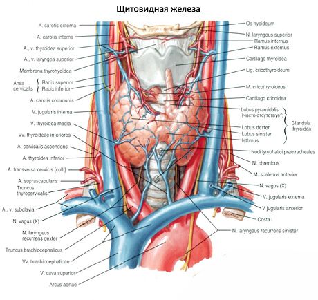

The posterior concave surface of the gland covers the front and sides of the lower sections of the larynx and upper part trachea. The isthmus of the thyroid gland (isthmus glandulae thyroidei), connecting the right and left lobes, is usually located at level II or III of the tracheal cartilage. AT rare cases the isthmus of the gland lies at the level of the I cartilage of the trachea or even the cricoid arch. Sometimes the isthmus may be absent, and then the lobes of the gland are not connected to each other at all.
The upper poles of the right and left lobes of the thyroid gland are located slightly below the upper edge of the corresponding plate of the thyroid cartilage of the larynx. The lower pole of the lobe reaches the level of the V-VI cartilage of the trachea. The posterolateral surface of each lobe of the thyroid gland is in contact with guttural part pharynx, the beginning of the esophagus and the anterior semicircle of the common carotid artery. The parathyroid glands are adjacent to the posterior surface of the right and left lobes of the thyroid gland.
From the isthmus or from one of the lobes, the pyramidal lobe (lobus pyramidalis) extends upward and is located in front of the thyroid cartilage, which occurs in about 30% of cases. This lobe with its apex sometimes reaches the body of the hyoid bone.
The transverse size of the thyroid gland in an adult reaches 50-60 mm. The longitudinal size of each share is 50-80 mm. Vertical dimension the isthmus ranges from 5 to 2.5 mm, and its thickness is 2-6 mm. The mass of the thyroid gland in adults from 20 to 60 years is on average 16.3-18.5 g. After 50-55 years, there is a slight decrease in the volume and mass of the gland. The mass and volume of the thyroid gland in women is greater than in men.
Outside, the thyroid gland is covered with a connective tissue sheath - fibrous capsule(capsula fibrosa), which is fused with the larynx and trachea. In this regard, when the larynx moves, the thyroid gland also moves. Inside the gland, connective tissue septa extend from the capsule - trabeculae, dividing the tissue of the gland into lobules, which consist of follicles. The walls of the follicles are lined from the inside with cubic-shaped epithelial follicular cells (thyrocytes), and inside the follicles there is a thick substance - a colloid. The colloid contains thyroid hormones, which consist mainly of proteins and iodine-containing amino acids.
The walls of each follicle (there are about 30 million of them) are formed by a single layer of thyrocytes located on basement membrane. The size of the follicles is 50-500 microns. The shape of thyrocytes depends on the activity of synthetic processes in them. The more active the functional state of the thyrocyte, the higher the cell. Thyrocytes have a large nucleus in the center, a significant number of ribosomes, a well-developed Golgi complex, lysosomes, mitochondria, and secretion granules in the apical part. The apical surface of thyrocytes contains microvilli immersed in a colloid located in the cavity of the follicle.
The glandular follicular epithelium of the thyroid gland, more than other tissues, has a selective ability to accumulate iodine. In the tissues of the thyroid gland, the concentration of iodine is 300 times higher than its content in the blood plasma. Thyroid hormones (thyroxine, triiodothyronine), which are complex compounds of iodinated amino acids with protein, can accumulate in the colloid of follicles and, as necessary, be released into the bloodstream and delivered to organs and tissues.
Thyroid hormones
Thyroid hormones regulate metabolism, increase heat transfer, enhance oxidative processes and the consumption of proteins, fats and carbohydrates, promote the release of water and potassium from the body, regulate growth and development processes, activate the activity of the adrenal glands, sex and mammary glands, have a stimulating effect on the activity of the central nervous system.
Between the thyrocytes on the basement membrane, as well as between the follicles, there are parafollicular cells, the tops of which reach the lumen of the follicle. Parafollicular cells have a large rounded nucleus, a large number of myofilaments in the cytoplasm, mitochondria, the Golgi complex, and a granular endoplasmic reticulum. These cells contain many granules of high electron density with a diameter of about 0.15 µm. Parafollicular cells synthesize thyrocalcitonin, which is an antagonist of parathyroid hormone - a hormone parathyroid glands. Thyrocalcitonin is involved in the exchange of calcium and phosphorus, reduces the calcium content in the blood and delays the release of calcium from the bones.
The regulation of thyroid function is provided by the nervous system and thyrotropic hormone of the anterior pituitary gland.
Thyroid embryogenesis
The thyroid gland develops from the epithelium of the foregut in the form of an unpaired median outgrowth at a level between I and II visceral arches. Up to 4 weeks embryonic development this outgrowth has a cavity, in connection with which it received the name of the thyroid duct (ductus thyroglossalis). By the end of the 4th week, this duct atrophies, and its beginning remains only in the form of a more or less deep blind hole at the border of the root and body of the tongue. The distal duct is divided into two rudiments of the future lobes of the gland. The emerging lobes of the thyroid gland are displaced caudally and take their usual position. The preserved distal part of the thyroid-lingual duct turns into a pyramidal lobe of the organ. Reducing sections of the duct can serve as the beginnings for the formation of additional thyroid glands.

Vessels and nerves of the thyroid gland
The right and left superior thyroid arteries (branches of the external carotid arteries), respectively, approach the upper poles of the right and left thyroid lobes, and the right and left inferior thyroid arteries (from the thyroid cervical trunks of the subclavian arteries) approach the lower poles of these lobes. The branches of the thyroid arteries form numerous anastomoses in the capsule of the gland and inside the organ. Sometimes the so-called inferior thyroid artery, which departs from the brachiocephalic trunk, approaches the lower pole of the thyroid gland. Deoxygenated blood from the thyroid gland flows through the superior and middle thyroid veins into the internal jugular vein, along the inferior thyroid vein - into the brachiocephalic vein (or into lower section internal jugular vein).
The lymphatic vessels of the thyroid gland flow into the thyroid, pre-laryngeal, pre- and paratracheal lymph nodes. The nerves of the thyroid gland depart from the cervical nodes of the right and left sympathetic trunks (mainly from the middle cervical node, go along the vessels), as well as from the vagus nerves.
