What are the organs of the human abdominal cavity. Ultrasound of the abdominal cavity, a comprehensive examination

Ultrasound is prescribed for diseases internal organs.
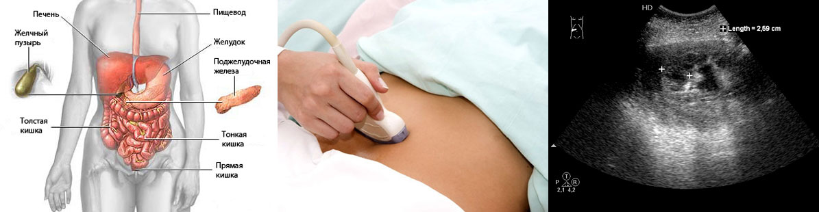
ultrasound abdominal cavity prescribed in the presence of diseases of the internal organs, to assess their condition and the effectiveness of treatment, as well as in the presence of certain complaints in the patient. Common name The "abdominal cavity" includes several organs at once: the stomach, esophagus, spleen, liver, adrenal glands, organs of the genitourinary system.
The ultrasound procedure allows you to assess the size of these organs, the structure of their tissues, location, tumors, functionality, as well as the presence of injuries and others. If the patient goes to the doctor for the first time with complaints of this kind, he is referred for an ultrasound of the abdominal cavity in the following cases:
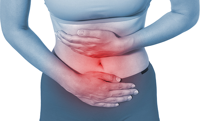
- Discomfort and pain in the stomach, a feeling of fullness after eating, the inability to eat large portions, rapid satiety, heaviness. If there is a periodic one, it is not recommended to postpone the examination.
- There is heaviness and discomfort in the right hypochondrium. That's where the liver is, so similar symptoms it is advisable not to miss. Usually, problems with the liver are also accompanied by nausea, sometimes - yellowness of the skin.
- Sharp stabbing or paroxysmal pain lower abdomen. They may be associated with the intestines, various ulcerative and inflammatory diseases one of the intestines. Similar diseases desirable to identify early stage. Sometimes ultrasound is not enough, it is used as initial method diagnosis, and then the doctor prescribes a colonoscopy or other procedures.
- , vomiting and bitterness in the mouth. Most often, these symptoms indicate problems with the liver and gallbladder, which it is desirable to examine.
- Strong, even painful gas formation, flatulence, bloating. These may be signs of bowel disease that need to be treated as soon as possible.
Indications for abdominal ultrasound are also any pain in the abdomen or lower back and test results (urine), which indicate the presence of diseases of the internal organs.
Preparation for the procedure

Before the examination, you need to go on a diet.
Ultrasound of the abdominal organs requires mandatory and thorough preparation. Many factors influence the quality and reliability of the study: bad habits, drugs taken, increased gas formation, overweight etc. Tell the doctor what medications the patient is taking. Some of them may have to be excluded for a while.
About 2-3 days before the examination, you need to sit on. This does not depend on which organ is suspicious: the liver, stomach, intestines. All foods that cause increased gas formation should be excluded (soda, beans, nuts, cabbage, chips, etc.). Food should be low-fat, balanced and fractional. It is advisable to divide meals into 4-5 times and eat small portions every 3-4 hours.
You can eat eggs, cereals, low-fat cheese, boiled meat, baked low-fat fish, drink only unsweetened tea and still water. Avoid coffee and juices. There is no need to limit yourself to liquids. It promotes normal and timely bowel movements. You need to drink about 2 liters of fluid per day. There is a list of foods that should generally be excluded from the diet completely at the time of preparation: black bread, alcohol, milk and dairy products, sweet pastries, dairy products, fresh fruits and vegetables, coffee, fatty and fried meat, lard.
As a rule, do not eat or drink on the day of the examination if the procedure is scheduled for the morning. If the ultrasound is performed in the afternoon, then you can have breakfast until 10 am. Before the procedure itself, the doctor may advise you to drink, eliminating excessive gas formation and bloating, such as Espumizan. In some cases, you will have to do a cleansing enema.
Also, on the evening before the examination, you need to drink a special laxative drug that the doctor will prescribe. Preparations should not contain lactulose (like Normase or), as it increases the formation of gases and bloating. It is advisable not to smoke at least a day before the procedure. If this is not possible, you should refrain from smoking for at least 2-3 hours immediately before the ultrasound.
You will learn about what preparation is needed for an ultrasound from the video material:
How is an abdominal ultrasound performed?
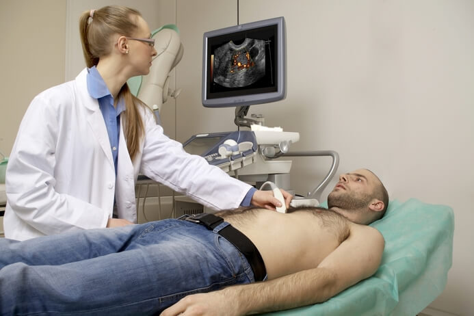
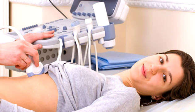
The procedure is carried out in the ultrasound room in the presence of a specialist and a nurse who will record the doctor's testimony. The patient enters the office and frees from clothing upper part torso.
If it is enough to lift the clothes, the doctor will tell you about it. Then lies down on the couch in the direction of the ultrasound machine. The doctor applies a special gel to the abdomen and moves the sensor over this area. The gel acts as a layer, helps the device to slide. It is hypoallergenic and leaves no residue, does not cause irritation.
None pain this should not be observed, however, if it constantly hurts or reacts to palpation, discomfort is possible. The subject should lie quietly and try not to move so that the doctor can see the picture. In some cases, it is necessary to press the sensor a little harder.
The liver and spleen are located behind the ribs and are covered by them. In order for the doctor to be able to examine these organs well, it should descend slightly into the peritoneal cavity. For this patient, they are asked to do deep breath and hold your breath. To get an accurate picture, patients will have to turn on their right and left sides a couple of times. The entire procedure takes no more than 20 minutes. If an acute and strong appears during the examination, the reason for this is on ultrasound, and a disease that may require the help of a surgeon. Any sensations should be reported to the doctor.
Preparation for the procedure is very important. If it was performed in bad faith, the doctor will notice this and say that some organs are poorly visible. The doctor may examine all of the abdominal organs or only some of them. Each organ is assessed as far as possible, but ultrasound is not always sufficient. For example, it can be estimated only by its size, the presence of damage. More detailed information provide blood tests.
When examining the gallbladder, stones and inflammation can be detected, and the pancreas - cysts, tumors, lesions. The cost of the procedure depends on the city, as well as the examined organs and dopplerometry (assessment of the work of blood vessels and blood circulation).
What diseases can be identified?
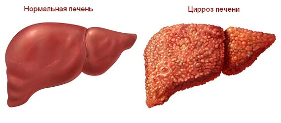
Cirrhosis of the liver - dangerous disease in which liver cells die.
Ultrasound can be used to detect a large number of disease is still at an early stage. However, some of them may require additional examination, tests and more accurate confirmation with a second ultrasound. If in doubt, you can do an ultrasound again with another. The doctor should deal with the interpretation of the ultrasound results. The study reveals the following diseases:
- . Inflammation of the gallbladder can occur abruptly or proceed slowly, turning into a chronic form. The cause can be either infection or congenital deformity bubble, which will be revealed during the examination. Cholecystitis can also be detected during tests.
- . Cirrhosis of the liver is a rather dangerous disease in which liver cells die and are replaced by connective tissue. The body cannot properly perform its functions. As a result, this can lead to the death of a person. It can affect not only people with alcohol addiction, although they have a much higher incidence rate. It is possible to determine cirrhosis by ultrasound at an early stage. The sooner treatment is started, the more likely it is to prolong a person's life.
- Tumors and cysts. Various neoplasms can appear according to the most various reasons and in all organs. They can be benign or. It is also important to identify them as early as possible, because benign neoplasms can degenerate into cancerous tumors.
- Pyelonephritis. With this disease, the kidneys, or rather the pelvis, become inflamed. The cause of this disease can be an infection, while a person has heat, nausea, headache, back pain. Sometimes the cause of pyelonephritis is also, then the pain will be quite strong. Acute pyelonephritis can flow into chronic if the cause has not been completely eliminated. Chronic form disease is accompanied by constant remissions and relapses.
- . Inflammation of the pancreas is quite common. It is connected with malnutrition and bad habits, hormonal changes and even heredity. In this disease, the pancreas begins to actively produce digestive enzymes that digest the gland itself.
Abdominal ultrasound for children and pregnant women

Preparing a child for an ultrasound has its own nuances.
The ultrasound procedure itself is no different, but the preparation of the child for the examination has its own nuances. small infants never kept on a diet or starved. It is enough for them to skip one before the procedure and put a microclyster to cleanse the intestines.
Older children (under 3 years old) should fast for approximately 4 hours before the procedure, but this will not cause any difficulties. You can drink, but not later than an hour before the ultrasound. Even older children (under 14) should fast for about 8 hours and not drink for an hour before the procedure. However, 2-3 days before the examination, you should try to remove cabbage, beans, sweets, soda, chips, etc. from the child.
Young children can be given drugs that reduce gas formation, such as Espumizan and Bobotik, as well. Even if the child is very hungry before the examination, you should not give him lollipops or chewing gum. Pregnant women with abdominal pain and suspected diseases of the internal organs can also be prescribed an ultrasound of the abdominal organs. It's safe for the child. Preparation is also needed, however, no strong laxatives are prescribed for pregnant women so as not to cause a spasm of the muscles of the uterus. An enema will be enough, for example, before the examination.
A few days before the ultrasound, it is necessary to exclude all fried, spicy, seasoned, as well as foods that increase gas formation (it is already increased in pregnant women). As prescribed by the doctor, you can drink Espumizan. However, it is not recommended for pregnant women to starve strongly. It is enough to exclude sweets, muffins and everything that causes
Ultrasound of the abdominal organs is an important additional method examinations of patients, both surgical and therapeutic. The indications for it are quite extensive. AT clinical guidelines Professor Palmer E.V. indicated following states requiring an abdominal ultrasound:
- pain in the abdomen of unknown origin;
- fever of unknown origin;
- blunt abdominal trauma;
- confirmation of ascites - the presence of fluid (transudate) in the abdominal cavity;
- the presumptive presence of volumetric processes (abscesses, hematomas, cysts, tumors).
Such different indications to the procedure can be explained large quantity vital organs in the abdominal cavity and ambiguity clinical picture that occurs when they are damaged. With many diseases of these organs, the first symptoms are extremely non-specific: pain, dyspeptic syndrome, intoxication, fever, and so on. Therefore, to assume correct diagnosis, it is necessary to evaluate the morphological structure of the formations that are in the abdominal cavity. The easiest way to do this is with an ultrasound.
Ultrasound of the abdominal organs involves the study and assessment of the condition of the following structures:
- liver and nearby vessels (portal and hepatic veins);
- gallbladder;
- bile ducts (vesical, common hepatic and common bile duct and);
- pancreas;
- walls of the stomach and intestines (extremely rare, since their visualization is almost always difficult)
- spleen;
- diaphragm;
- kidneys (despite the fact that they lie in the retroperitoneal space, clinicians evaluate their condition during an ultrasound of the abdominal cavity);
Each of these organs has its own morphological features which are studied during the ultrasound examination. With the help of them, it is possible to determine the localization of the pathological process.
Liver and blood vessels
The normal structure of the liver implies a homogeneous structure of the parenchyma throughout its entire length. Diagnostic doctors look for the presence of any formations that appear as echo-negative foci. One can estimate their size, density and suggest the nature of the focus.
The portal (portal) and hepatic veins are normally visible against the background of the liver. Walls portal vein and the branches extending from it have a high echogenicity, while the hepatic veins absolutely do not conduct ultrasound. At portal hypertension, the walls and volume of these vessels can change significantly, which is manifested by a change in signal conductivity.
gallbladder
In the absence of pathology, the gallbladder is an echo-negative formation - it does not conduct an ultrasound signal and looks like a black structure on the monitor. The presence of echogenic foci in it indicates the presence of stones. The size of the gallbladder is variable, so they are not assessed during ultrasound. Its shape is constant: on the transverse section it should be rounded, on the longitudinal - pear-shaped. The condition of the wall should be assessed, and what formations are in its cavity. Normally, the bladder cavity is filled only with bile, and the walls have a smooth and well-defined surface.
bile ducts
hepatic bile ducts and cystic duct, as a rule, do not look at ultrasound of the abdominal organs, since their visualization is difficult due to the structural features of the mucosa and the low flow rate of bile.
The common bile duct on ultrasound looks like a straight tube. Its study is often difficult due to the presence of gases in the intestines. This is due to improper preparation of the patient for the procedure. In this case, a discontinuous structure of the common bile duct can be found.
Pancreas
This organ is more echogenic than the surrounding tissues. In an unchanged state, it is visualized as a homogeneous, coarse-grained formation. Almost all diseases of the pancreas are reflected in its structure, shape and structure:
- pancreatitis - ultrasound can determine an increase in the organ, a decrease in its ability to conduct an ultrasound signal, but clear contours remain;
- pseudocyst - an echo-negative formation with limited clear contours is determined in the structure of the pancreas;
- neoplasm (most often - solid cancer) - indirect sign these are blurred borders of the pancreatic head due to diffuse echo reduction.
Doctors-diagnostics necessarily look for the presence of these signs during the ultrasound of the abdominal cavity.
Spleen
With a normal structure of the organ, you can see the parenchyma in the form of an oval formation with low echogenicity. As a rule, the vessels are not visible. Amplification of the conducted signal and visualization of blood vessels is a direct sign of splenomegaly - an increase in the size of the spleen.
The edges of the spleen are significantly different on ultrasound. The superior and lateral margins are more convex, while the inferior and medial margins have depressions of varying sizes from nearby organs. The medial surface of the spleen is always concave. Irregular outlines or blurry contours of the organ may indicate a rupture of the spleen or the formation of a volumetric process (most often cysts). Forming a double contour is an absolute sign formation of a subcapsular hematoma.
The hilum of the spleen (the place where the vessels enter and exit) has a higher echogenicity than the parenchyma.
kidneys
Although the kidneys are a retroperitoneal organ, they are often examined together with the abdominal organs. The examination plan includes an assessment of the location of the kidneys, its parenchyma, capsule, pelvis and renal veins. Normal performance the following:
- the kidneys are located between the bispinarum line (a segment connecting the anterior superior spines ilium) and diaphragm;
- clear contour of the renal capsule;
- the parenchyma is heterogeneous: the cortex is more echogenic than the medulla;
- the collecting system (pelvis) has an irregular asymmetric shape and a denser structure.
During the ultrasound, it is possible to determine an additional kidney, the presence of cystic processes, severe glomerulonephritis or long-term chronic pyelonephritis.
Assessment of the abdominal organs with ultrasound has great importance in clinical practice. With it, you can determine the affected organ, in case of non-localized or non-specific symptoms, assess the state of organs and some vessels. The pathology of each organ on ultrasound has its own echographic signs that allow you to determine the clinical diagnosis.
Abdominal ultrasound can help identify inflammatory processes and pathological condition organs in this area. This study considered informative, painless and harmless. Any age category. Used to make an accurate diagnosis. Appointed only after a preliminary consultation and at the first anxiety symptoms. Very often, Dopplerography is also used, which helps to assess blood flow in large vessels.
On ultrasound of the abdominal cavity, a specialist evaluates the condition and functioning of such organs as:
- Liver. Ultrasound can detect the presence of such abnormalities and diseases in this organ: hepatitis, cirrhosis, changes associated with diseases of cardio-vascular system, tumors, cysts and neoplasms of a different nature.
- Gallbladder. Ultrasound procedure helps to identify such abnormalities in this organ: developmental anomalies, stones, complications of cholelithiasis, cholecystitis, polyps, tumors and lesions.
- Pancreas. ultrasound this body defines: developmental anomalies, pancreatitis, abscesses, cysts, tumors different nature and age changes.
- Spleen. The study of this organ helps to identify: malformations, damage, changes in size, cyst, tumor and changes in systemic diseases blood.
Attention is also paid to the vessels - an assessment is made of the location of the main and intraorgan vessels, their size and the state of the gaps. Be sure a specialist evaluates both the retroperitoneal space and the organs that are located there.
Carrying out diagnostics
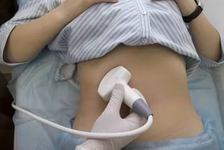 Ultrasound of the abdominal organs does not take much time, on average, the procedure lasts no more than 20 minutes. This diagnostic procedure carried out according to the following scheme:
- The patient must be in a supine position.
- The upper body must be bare.
- Then on skin patient, a special gel is applied to help the transducer slide and prevent air from entering under the transducer.
- The doctor moves the sensor along the body, while studying the necessary areas.
Ultrasound should not cause any discomfort to the patient.
What is being evaluated
With help this survey pathology and its stage can be determined. If abnormalities are found by the uzist doctor, he can refer the patient for an additional consultation with a narrow specialist.
During the ultrasound, the doctor should give Special attention such indicators:
- Dimensions.
- structure.
- Location.
- Neoplasms and inflammation.
- changes and chronic diseases.
At healthy person all indicators must be within acceptable limits. It should also be borne in mind that the norms for each age group their.
Why is an abdominal ultrasound performed?
 An organ examination is recommended if the patient has the following symptoms:
- Bitterness in the mouth.
- Heaviness in the right hypochondrium.
- Heaviness after eating.
- Pain after eating.
- Increased formation of gases in the intestines.
It is mandatory to conduct an examination of the organs if the patient has diseases or only suspicions:
- For pancreatitis and hepatitis.
- cholecystitis and cirrhosis.
- For a cyst or tumor.
- Stones.
What influences the results
Before conducting the study, the patient must carefully prepare in advance, since not proper preparation can negatively affect the results of ultrasound. The preparation itself will depend on which organs are planned to be examined.
Basically, it is recommended that the patient follow a diet for three days before the ultrasound and exclude from the diet products that contribute to the formation of gases in the intestines. As air contributes to distortion overall picture. It is also advisable not to eat fatty foods and drink carbonated drinks. Meals should be fractional and frequent.
As for the preparation of children for the examination, they have their own characteristics, which must be clarified with a specialist.
Have you ever had an abdominal ultrasound? What organs are examined during the procedure?
Modern medicine is developing rapidly and progressively, which allows us to successfully solve various problems human health. One of the most famous and popular procedures is the study of the abdominal organs using ultrasound. The method is safe and effective way for a comprehensive examination, which allows you to timely identify the symptoms of any diseases. Today, this technology is very widespread and is used in various fields medicine.

For the first time, this type of examination technology was applied in 1949. John Wild used this method to determine the thickness of intestinal tissues, and in the future the method developed very rapidly. So in 1962 the first scanner was developed composite type operating in B-mode. The end of the twentieth century was marked by increased development and revolution in this area. The development of this technology took place continuously and today the study is the most accessible, simple, but very effective method.
The use of ultrasound for diagnosing diseases of the abdomen, for example, pancreas, allows you to identify all the symptoms and manifestations of the disease. Often similar procedures ultrasound is done for a routine examination of the abdominal cavity, which confirms Very good condition health. Also, the study of organs is carried out in various difficult situations such as in the emergency department. This allows you to quickly and fairly accurately determine the condition of the abdominal organs and a set of subsequent procedures. With this method, you can put accurate diagnosis and prescribe the correct treatment.
Features of the ultrasound procedure
Diagnosis and monitoring of health status become much more effective if an examination of internal organs is carried out and ultrasound is used. Safe Method ultrasound is also completely painless. Depending on the indications, a study is made with an emphasis on various organs.
The research includes things like:
- comprehensive examination of the gallbladder;
- pancreas and assessment of its condition;
- special attention is paid to the liver;
- an ultrasound examination of the intestines and other elements is also performed.
When examining the gallbladder, its condition is determined, as well as the condition of the ducts. Their size, the presence of stones, patency and other medical indicators are recorded. An examination of the tissues of the organs and their condition is also carried out.
The study of the pancreas allows you to evaluate such parameters as size, shape, the presence of any formations, contour. Quite often it is difficult to see the body, as it partially overlaps small intestine or stomach gas. The conclusion of a specialist may contain such a diagnosis as " diffuse changes". All this suggests that the state of the abdominal cavity was affected, associated either with age-related changes, or with chronic processes inflammatory nature. In this way, this moment in the examination is very important and determines many medical indicators.
Modern research, where the liver enters allows you to get enough full information about the state of the body. Often, a lot of ailments arise due to incorrect operation liver, and that is why it is given great attention. The specialist checks the size of the organ, condition, blood flow, structure and the presence of any changes. In this process, it is defined as focal changes, and diffuse. Thus, the method is quite effective for a comprehensive study and assessment of health status.
Ultrasound is also used to examine other internal organs of the abdomen. An examination of the intestine is carried out, which determines the thickness of the walls, the presence of any formations and other indicators. Also, the method is effective for examining the kidneys, adrenal glands, spleen, which provides exact information about their work and status. The modern procedure is a safe high accuracy in determining all indicators, which makes it possible to establish an accurate diagnosis. It is important that the conclusion of an ultrasound specialist must be analyzed in conjunction with outpatient test results, clinical and anamnestic data. Thus, you can get a complete, correct and detailed picture of the health of the patient and his organs.
Preparing for ultrasound
Ultrasound is a very popular method in medicine, because it has a high permeability and allows you to get the most accurate data. The effectiveness of the result depends not only on the doctor of ultrasound diagnostics, but also on the thoroughness and correctness of the preparation of the body. This process is quite important, because otherwise the results may not be complete, or not correct. When doing an examination of the abdomen, several important rules should be considered:
- First of all, it is important to follow a three-day diet that excludes the use of fresh vegetables and fruits, black bread, legumes and carbonated drinks. These products cause gas formation and it will be very difficult to check the condition of some organs;
- to obtain a clear, high-quality image, it is necessary not to eat anything 7 hours before the organ examination procedure;
- should inform the doctor before doing an abdominal ultrasound if the patient is undergoing any course of therapy.
Simple recommendations will help you get a quality result and an accurate conclusion from a specialist. Most often, such a procedure for examining internal organs is carried out in morning time and on an empty stomach. Thus, the state of the gallbladder, pancreas and other organs will be optimal.
The popular abdominal ultrasound procedure is safe and painless. The doctor may refer the patient to a similar process with the following indications:
- if pain occurs in the upper abdomen;
- in the event that there is bitterness in the mouth, as well as unpleasant heaviness in the region of the left hypochondrium;
- increased gas formation;
- acute pain of girdle character.
Feelings like this can be a symptom various diseases such as exacerbations of chronic diseases. In no case is it recommended to postpone the visit to the doctor, but it is worth visiting a specialist on time. Thus, it is possible to avoid many unpleasant situations and keep healthy.
The study of the body by such a technique can be as paid service, as well as free. In the first case, such additional moments are provided as transferring all received data to digital media, printing an image, additional examination, interpretation and others. In the second case, the specialist issues a complete conclusion, which contains a description of the state and features of the organs. All this helps the doctor to prescribe the right course of treatment, optimal procedures and make an accurate diagnosis. If ultrasound is prescribed for children, then some preparation features should be taken into account. For example, babies under the age of one year should skip one feeding and not drink liquids for an hour before the procedure. Older children under the age of three should not eat four hours before the examination and drink one hour before. Older children should not eat food for 6-7 hours, and water - for an hour. Thus, you can get the most accurate results and make an accurate diagnosis.
Medical research, which involves the use of ultrasound, is safe and does not deliver any discomfort. At the same time, this technique is very effective, since ultrasound has a high penetrating power and transmits the most accurate data. Ultrasound includes many important elements. In some cases, before the procedure, the doctor may prescribe the use special preparations, for example, reducing gas formation. All recommendations of a specialist should be followed and this will provide an accurate conclusion and proper therapy.
