The correct size of the ovaries in women. Normal size of the ovaries in women. Changes in the uterus and ovaries during pregnancy
The female sex glands located in the pelvis and performing a generative function are the ovaries. They have a huge impact on the state of hormonal balance in the female body. The size of the ovaries indicates the possible presence of deviations in their development and functionality. These are glands of an oblong shape, whitish in color, with a heterogeneous bumpy surface. The proper production of eggs depends on the state of the ovaries and childbearing function female body. To identify violations in the health of these glands will help ultrasonography, during which the size of the ovaries in women is established, which varies depending on the age and state of health of the woman.
The aim of this study is to develop a validated model of ovarian volume in healthy women from conception throughout life to the aggregation of data from multiple sources. The approved model is a polynomial of degree 14 of the kind. With the coefficients shown in Table 1 and the relationship with the data shown in Figure 1. The model has a coefficient of determination indicating that about 69% of the lifetime variation in ovarian volume is due to age alone. Moreover, the proportions of residuals within one, two, and three standard deviations are close to the expected values of data with a Gaussian distribution.
The ovaries are formed already in the second month prenatal development fetus and continue to form until the onset of menstruation. They perform several important functions:
- generative, on which the formation of eggs depends;
- vegetative, affecting the development and formation of primary sexual characteristics, as well as the development of the mammary glands, skeletal features and hair growth related to secondary sexual characteristics;
- hormonal, due to which the female sex hormones (estrogen and progesterone) and the male sex hormone (androgen) are produced in the body of a woman.
Thanks to proper development ovaries and their functionality in a woman's body is maintained hormonal balance. Egg cells are produced, pregnancy is maintained, the necessary muscle layer and a normal fat layer is formed.
Figure 3 shows an example of a 5-fold validation process in which a model is selected that does not gain or adjust basic set data. The logarithmically unadjusted predictive normative model is shown in Figure 4. This shows the mean volume per ovary in milliliters for a healthy human population along with prediction intervals and standard deviations. Mean and guideline ranges for ovarian volume are given from birth to age 50 in Table 2. Our model shows that, on average, ovarian volume increases from 7 ml at 2 years to a peak of 7 ml at age 20 and declines throughout life up to 8 ml during menopause.
The functional activity of the ovaries begins from the moment of puberty and continues until the woman has menopause. First changes at work this body may be noticeable as early as the age of 40, when women notice a failure menstrual cycle. The purpose of the paired gonads is to prepare everything necessary for the onset of pregnancy. This period in life is the most responsible, affecting the state of the whole organism and leading to significant changes in it.
Model residual patches under 10 years of age are usually distributed normally. Model residual plots aged 10 to 30 years and over 30 years are close to ideal normal distribution. This lower peak is beyond 95% confidence interval 5-2 ml for the full peak of the model, indicating a statistically significant difference between the two peaks. We have described and validated the first normative model that describes ovarian volume in healthy women from conception to 82 years of age.
The model has a coefficient of determination indicating that 69% of the variation in ovarian volume throughout life is due to age alone. Ovarian volume increases during childhood and adolescence and is the maximum average woman at age 20, declining thereafter to menopause and beyond.
Normal size of the ovaries
A change in the size of the ovaries does not always indicate a violation of the patient's health status. Their dimensions vary depending on the phase of the menstrual cycle and the level of hormones. In addition, the size of the right ovary often differs from the parameters of the left organ, but in most cases such a discrepancy does not exceed 0.2 mm and does not cause concern.
Transvaginal ultrasound assessment has been used as a proxy for ovarian reserve in sexually mature adults active women. Our normative model now adds to this by demonstrating a steady increase in ovarian volume from birth with a modest acceleration at the onset of puberty. The main contributor to ovarian volume before puberty is likely to be stromal growth; while small antral follicles are present in the ovaries of prepubertal girls of all ages, large follicles are not found while serum gonadotropin concentrations remain low.
The normal size of the ovaries in women is:
- volume - 4-10 cm 3;
- thickness - 16-22 mm;
- width - 18-30 mm;
- length - 20-37 mm.
These parameters are determined on the fifth or seventh day of the menstrual cycle. The range is quite large, and when conducting an ultrasound examination, the specialist takes into account the individual characteristics of each woman. This is both age and the presence of children or inflammatory diseases, and disorders in the development of organs reproductive system and the woman's age.
After menarche and the onset of ovulation, the main contributor to the change in ovarian volume is likely to be the number and size of antral follicles present. Human growth during childhood is described as having three complementary and partially superimposed components: infancy, childhood, and puberty. Each component seems to be controlled by separate biological mechanisms.
Measurement of ovarian volume has been found to be useful for a wide range disorders in children and young women. Measurement of ovarian volume is an accurate diagnostic tool for adolescent girls with irregular menstruation. In most of these girls, enlarged ovaries are associated with polycystic ovary syndrome, and ovarian volume is part of the diagnostic criteria for this state. Therefore, we censored our data set to exclude all women with an ovarian volume greater than 10 ml.
During the entire menstrual cycle in a healthy woman, the size of the ovaries remains within the normal range, minor changes in certain parameters are associated with individual features body of a woman and does not require the intervention of doctors. However, if a significant deviation from the norm is detected, you should immediately consult a doctor who will establish the cause and prescribe the appropriate treatment.
Excluding these data resulted in a reduction in peak mean ovarian volume, as expected, and a slight increase in age at peak. Recent results suggest that the number of antral ray follicles has better discrimination efficiency than ovarian volume.
Girls with precocious puberty have significantly increased ovarian volumes compared to the normal population, and ovarian volume has been proposed as a useful discriminator between central precocious puberty and a precocious event.
Determination of normal parameters of the gonads is required to confirm or cancel such a diagnosis as ovarian depletion or the presence of a tumor. Often, during the examination, the doctor discovers either a cyst, which is recognized as physiological and disappears when the hormonal background changes.
Determine the level of hormones, and decide on the need for an appointment hormonal drugs Maybe experienced doctor. Such treatment can not only save the patient from these diseases, but also restore the functionality of the glands, eliminating the cause of ovarian exhaustion and the onset of early menopause.
It remains difficult to estimate ovarian reserve in adolescents and young women with cancer due to significant age-related changes in the various markers available. Our model is derived from data from several sources of measurement of ovarian volume in healthy women. This is both a strength and a weakness of the study. The strength is that measurement errors, both underestimating and overestimating ovarian volume, are likely to be negated because any bias is unlikely to always be in the same direction for each data source.
Weakness is the heterogeneity of values obtained from different sources. We cannot be sure that measurement of ovarian volume by abdominal ultrasound, which is often difficult in young children, is as accurate as measurement by transvaginal ultrasound in older women. The largest data source consists of values imputed from a very large transvaginal ultrasound data source from an ovarian cancer screening program. This study excluded patients with solid or cystic ovarian tumor detected by sonography, but not patients with polycystic ovary morphology.
The normal size of the ovaries during menopause 
The active activity of the glands continues until the age of 40-50, with the onset of this age, the production of eggs stops and female body spends accumulated in advance.
At a time when the reproductive function fades, the size of the glands also changes. Normal sizes The ovaries in menopausal women are noticeably reduced and both organs become the same size:
Our normative ovarian volume model using data obtained from multiple data sources and various methods estimate overcomes the weakness of other studies using only one imaging modality, since any potential bias in one direction is likely to be negative.
Normal size of the ovaries and uterus after childbirth
We have shown that, on average, ovarian volume increases from 7 ml at age 2 years to a peak of 7 ml at age 20 and declines later in life to about 8 ml at menopause. The study methodology used for both data collection and data analysis closely follows what is used to derive a validated normative model for anti-Millerian hormone levels found in the blood of healthy women from conception to menopause.
- the volume is from 1.5 to 4 cm 3 ;
- width is reduced to 1.2-1.5 cm;
- length - 2-2.5 cm;
- thickness becomes no more than 1-1.2 cm.
Small fluctuations in the size of the ovaries in menopause are possible due to the fact that at first in the postmenopausal period, the production of individual follicles still continues, despite the fact that menstruation is no longer present.
A written informed consent to the original work of the person that produced the tissue samples, and all data was anonymized prior to analysis. References to these identified studies were then reviewed and any other relevant research papers extracted. Papers were included if they contained ovarian volume results in healthy, normal girls with no ovarian or endocrinological abnormalities, in order to isolate data that approximates a healthy human population.
Ovarian size during pregnancy
All parameters of the reproductive system of a woman change during pregnancy. The size of the uterus and ovaries increase, and the glands can be displaced.
The reason for the enlargement of the glands is active blood flow, and the displacement is associated with the growth of the uterus and the forced rise of the glands under its pressure from the small pelvis upwards.
Abstracts of 37 studies were identified by this method. After reviewing full papers, studies were excluded if either the results consisted solely of descriptive statistics or the subjects were classified by pubertal stage rather than age. Of the remaining nine studies, there were seven data measured by trans-abdominal ultrasound and plotted - although two of them contain tabular data. Ovarian volumes were standardized to an expanded ellipsoid approximation formula because variation was used in some studies.
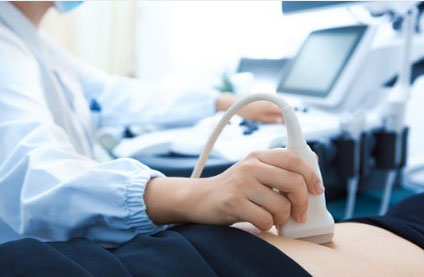
Ultrasonography
The doctor can determine the cause of the change in parameters during an ultrasound examination. It will accurately determine the onset of pregnancy or the presence of dysfunction, which also causes glandular enlargement. Changes to settings can be benign tumor or malignant neoplasm, inflammatory process, cyst corpus luteum ovary.
Zero conception volume values were added to the pooled dataset to force the models to pass through the only known volume at any age. Because variability increases with ovarian volume, we adjusted the data. Each model defines general type curve and has parameters that, when instantiated, yield a specific curve of that type. For each model, we calculated the parameter values that maximize the coefficient.
For each candidate model, the root mean square error and were calculated after removing artificial zero values at conception. The most efficient family of models were high-precision polynomials. 5-fold cross-validation: data were randomly divided into 5 subsets of the same size. For each subset, the other four subsets were used to train high-precision polynomials of degree 8 to 20, with the subset held back as test data.
The most important indicator is the volume of the gland, indicating the presence pathological process requiring urgent medical intervention. gonads may indicate the development of such pathologies as:
- cyst or;
- benign tumor;
- malignant neoplasm;
- the presence of metastases.
However, it is equally important to timely detect such a pathology as glandular depletion. Small ovaries indicate premature fading reproductive function in women aged 35-40 years. Ovarian wasting syndrome is associated with the cessation of follicle production, leading to the cessation of ovulation and a decrease in the production of female sex hormones. Ultrasound examination will also allow you to notice such changes, during which the doctor not only measures the size of the glands, but also studies their shape and location.
What can you learn about the condition of the ovaries by ultrasound?
The standard error of the test data was calculated and compared with the standard error of the training data for the same model. In other words, the estimated prediction error of the model, generalized to the unseen data, was compared to the training error of the model. The model was considered validated if.
Normally distributed residuals for log-adjusted values correspond to skew-normal population sizes. The financiers played no role in the design of the studies, the collection and analysis of data, the decision to publish or prepare the manuscript.
The first sign of ovarian failure is scanty menstruation. They can be repeated several times a month and differ in insignificant number. spotting. In some cases, menopause occurs suddenly. Only a qualified gynecologist can decide what to do in order to restore the functionality of the ovaries, prescribe the appropriate treatment by taking hormonal drugs.
Polycystic ovarian syndrome is a common ulcerative disorder characterized by the presence of more small ovarian cysts. These are tiny fluid-filled sacs that usually contain eggs. Usually all women childbearing age about 5-7 small follicles in each ovary, each containing an immature egg. As each menstrual cycle begins, these follicles begin to grow in an attempt to reach maturity and hatch an egg. However, only one follicle will outgrow the rest and be selected for full development.
To make an accurate diagnosis, specialists conduct:
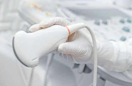
- transabdominal examination. With the help of a sensor located on the surface of the anterior wall of the abdomen, it studies the parameters of the gonads and gets the opportunity to identify gross pathology of the glands, determine the normal size of the uterus and healthy ovaries.
- A transvaginal examination allows you to establish the normal size of the ovaries in women or to identify pathology by inserting a probe into the vagina.
- The transrectal method allows for a full-fledged study of virgins who turn to a gynecologist with complaints of severe pain lower abdomen or menstrual irregularities.
Usually, best time for the study - 5-7 day of the cycle, but if necessary, the procedure is carried out several times. On 8-10, 14-17, 22-25 days of the menstrual cycle. Such studies in case of suspected dysfunction or the development of severe pathology should be carried out at least twice a year.
The remaining follicles degenerate and eventually disappear. The lead follicle will continue to grow until it is about 2 cm in diameter when it is ready to release an egg, a process called ovulation. As other follicles disappear, new ones will appear to replace them in preparation for the next cycle. This means that the ovary maintains a constant number of small follicles throughout the cycle.
Types of ultrasound diagnostics in gynecology
This occurs as a result of the failure to select the leading follicle, i.e. none of the follicles reach maturity. As a result, all follicles come out of degeneration and remain in the ovary. As new follicles continue to emerge with older ones retained, the total number of follicles in each ovary continues to increase. These follicles are hormonally active, producing mainly male hormone. This will release into the blood high levels androgens causing some manifestations such as extra hair on the body, acne, oily skin and baldness.
Maintaining a woman's health and reproductive function largely depends on timely access to a doctor and preventive examination. Such an examination will help in time to detect changes in the structure of the gonads, an increase or decrease in their size, indicating the development various diseases. Modern ways diagnostics allow for early stages detect pathology and take measures to prevent the development of diseases.
Discomfort, pain, incomprehensible discharge should always be a reason for a visit to a gynecologist who can deliver accurate diagnosis and prescribe treatment.
The best way to check internal organs is an ultrasound. Very often it is used in gynecology. After all, the result can be obtained quickly, safely and accurately.
What is an ultrasound for?
The reasons why a doctor prescribes an ultrasound of the uterus and ovaries are varied. So, for example, they could be:
- a woman may be disturbed by pain in the uterus and ovaries;
- permanent violations of the cycle;
- strong pain that appear during menstruation;
- upon occurrence suspicious secretions from the vagina, but not related to menstruation;
- in addition, such a study should be done by women in order to determine the presence of pregnancy and to exclude ectopic pregnancy.
Thanks to this method, any deviations from the norm and the onset of diseases can be detected.
Process
Many women are interested in how the ultrasound of the uterus and ovaries goes. Two methods are commonly used for this study.
- Transabdominal ultrasound. This method is based on the study of organs through the abdomen. To do this, the doctor applies lower part belly a little special gel that allows the sensor to glide better over the skin. In addition, if the doctor prescribes this type of ultrasound, it is necessary that the bladder is full. The fact is that ultrasound waves penetrate well through the aquatic environment, but through the air - on the contrary.
- Transvaginal ultrasound. In this case, a special device is inserted into the woman's vagina, with the help of which the examination is carried out. In order to avoid infections, a special condom is put on it. In this case, on the contrary, it is necessary that the bladder is empty. This method is considered more accurate than the previous one.
The process itself does not cause any pain at all, does not negative influence on the body and passes quickly enough.
Uterine parameters
During such an examination, the doctor evaluates certain parameters of the uterus in women.
- Position. The normal position is when the uterus is tilted to the side. Bladder or the rectum, i.e. forward. If the organ has a backward deviation, then this may well become a problem during pregnancy, because. this is not considered the norm.
- outer contours. The outer shell of the organ should be even and have a clear boundary. with myomas or neoplastic diseases the contours will, on the contrary, be uneven. If the boundaries are not clear, then this may indicate inflammation.
- Size. It is considered normal when the length of the uterus is in the range from 45 mm to 70 mm, depending on the age of the woman, the number of pregnancies. The width of the body is in the range from 45 mm to 60 mm and also depends on these indicators. Anterior-posterior size - from 34mm to 44mm. If the size of the uterus is less than normal, then this indicates its underdevelopment. If, on the contrary, the values are greater, then this may be a sign of pregnancy or tumor diseases.
- thickness of the endometrium. The doctor must examine this indicator. The fact is that the thickness of the endometrium is different depending on which day of the cycle the ultrasound is performed. Therefore, the doctor looks at the correspondence of this value to the day when the procedure takes place. Immediately after menstruation ends, the thickness of the endometrium is approximately 1-2 mm, but after ovulation occurs, its size varies from 10 to 15 mm.
- echogenicity. This indicator shows the density of the fabric. For the uterus, homogeneous echogenicity is considered normal. If any other indicators are present, then this may indicate the presence of fibroids or tumors.
- The structure of the uterine cavity. The cavity of this organ in healthy women is homogeneous, with clear contours. Its blurring indicates that endometrial diseases are present. In addition, ultrasound may show any neoplasms.
- Cervix. The normal size is from 35 to 40 mm. However, it must be homogeneous. The diameter of the cervical canal is approximately 2-3 mm. There must be liquid inside it. If the canal or the neck itself is expanded, then this indicates possible diseases.
- The presence of free fluid. After ovulation, women may have some fluid in the retrouterine space. However, on any other day of the cycle, the presence of such a liquid indicates possible diseases provoked by sexual infections.
Ovarian parameters

In addition to examining the uterus, doctors necessarily conduct an examination of the ovaries. These are paired organs, during the procedure the condition of one and the other is assessed. What parameters does the specialist consider, and what values \u200b\u200bare considered the norm?
- Location and shape. Both organs are located on the sides of the uterus. Moreover, this arrangement is most often asymmetrical. In healthy women, the ovaries are oval shape. The follicular apparatus is clearly defined, the follicles are clearly visible in it. If an ultrasound is done on the 8-9th day of the cycle, then the specialist will determine the dominant follicle, which at this time can be from 15 to 25 mm in size. If its size exceeds this value, then this indicates the possibility of a follicular cyst.
- Ovarian size. A normal indicator is when the width of the ovary is 25 mm, the length is about 30 mm, and the thickness is 15 mm. If these values differ upwards, then there may be inflammation or even serious illness these organs.
- External contours and echogenicity. The outer shell of the ovaries should be clear and bumpy (due to the growth of follicles). Echogenicity should normally be homogeneous. If the contours are blurred, then this indicates inflammatory processes.
- Structure. The ovaries are composed of follicles and a capsule. The number of the first may differ in the left and right organs.
The fallopian tubes
With ultrasound, the fallopian tubes, if they are in normal condition, should not be visible. If the specialist nevertheless detects them, then we can talk about the inflammatory processes present in them.
Diseases
Often a doctor, when comparing the data obtained with the results of the examination, can determine the presence of any disease. What is the diagnosis?
- Myoma. At the same time, the size of the uterus is larger than normal, its contours are blurred, and a node is determined in the myometrium.
- Endometriosis. This disease occurs as a result of the fact that endometrial cells begin to grow outside the uterus. On ultrasound, it can be seen as a number of bubbles that can be located in the uterus, and in its neck, and in fallopian tubes Oh.
- Incorrect development of the uterus. These may be her shortcomings in her development, for example, a bicornuate uterus or hypoplasia of this organ.
- Endometritis. In this case, the endometrium becomes thicker, its edema may occur. The size of the uterus also changes in the direction of increase.
- Uterine cancer. In this case, the ultrasound is determined large formations in the cavity of this organ.
- Cervical cancer. At the same time, the specialist sees that the size of the neck is much larger than the norm, and it is deformed due to the disease.
- Cyst. If a formation filled with fluid and exceeding 25 mm in diameter is found in the ovary, then most likely there is a disease such as an ovarian cyst.
- Polycystic. Both ovaries are larger than normal performance they get thicker. In addition, fibrosis is determined.
- Adnexitis. If this disease is present, then the ultrasound clearly shows that the fallopian tubes have rather thick walls, the ovaries become larger in size, their boundaries become fuzzy.
Pregnancy
Ultrasound of the uterus and ovaries during pregnancy is mandatory. Their sizes change. So, for example, the uterus is stretched to a length of about 40 cm. The ovaries also increase in size, but not much. And the reason for this is increased blood flow to the pelvic organs during pregnancy. Besides, ultrasound examination will help to identify pathologies of organs and the fetus if they ever show up. After childbirth, the uterus returns to its normal size, and the ovaries begin to function normally again.
Ultrasound of the uterus and ovaries - necessary procedure if you suspect any disease or pregnancy. This is not a terrible study at all, but it is it that gives the most complete and accurate answer to many questions.
