Echocardiography of the heart is normal. Echocardiography: indications for carrying out. Hypertrophy of the walls of the right sections
Leading place in the detection of heart disease in this moment occupies ultrasound - ultrasound examination. This diagnostic method is also called echocardiography. The first information about the use of ultrasound of the heart dates back to the middle of the last century. But the term "echocardiography" appeared a little later. It was proposed in 1965 by AIUM - the American Institute of Ultrasound in Medicine.
Ultrasound of the heart is an informative diagnostic method, characterized by harmlessness, reliability of results, and ease of implementation. It is prescribed to people only if there is evidence. These include:
- acquired and birth defects hearts;
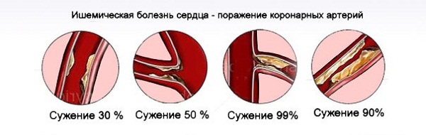
- IHD (ischemic heart disease);
- cardiomyopathy;
- endocarditis ( inflammatory processes that occur in the inner lining of the heart) caused by bacteria;
- heart tumors;
- hypertonic disease;
- thrombosis, thromboembolism;
- arterial hypertension;
- heart failure;
- diseases of the aorta;
- suspicion of other pathological conditions.
In some cases, Echo ultrasound of the heart, if indicated, is not performed. The reason for this lies in the existing contraindications. For example, transesophageal echocardiography is not performed if the patient being examined is very serious condition or has various diseases esophagus. For intravascular ultrasound diagnostic method, a contraindication is a person's being in an extremely serious condition.
Methods for conducting ultrasound of the heart
There are several different echocardiographic methods. The first to appear is one-dimensional scanning (M-method). It allows you to obtain graphic images of the structures of the heart, depending on the level of the cut and the direction of the ultrasound beam. One-dimensional scanning allows specialists to:
- find out the dimensions of the walls, chambers of the heart, valve openings;
- get an idea of the movement of organ structures.
The next method is B-mode. It allows you to get two-dimensional images. With this echocardiography, doctors:
It is impossible to evaluate the functioning of the heart without examining the blood flow in the blood vessels and heart chambers. This is done by specialists thanks to Doppler echocardiography.. This method study of intracardiac blood flow is based on the Doppler effect. The essence of Doppler echocardiography is to register a change in the frequency of the ultrasound signal directed by the sensor and reflected from moving elements, and to automatically determine the speed of blood flow and its direction.
Among the methods of ultrasound of the heart, it is worth highlighting transesophageal echocardiography.. The main advantage of this diagnostic method is the absence of interference in the path of the ultrasonic beam ( lung tissue, muscles, subcutaneous fat, etc.). With the help of transesophageal echocardiography, the functions of prosthetic heart valves are evaluated, aortic wall dissection is diagnosed, and infective endocarditis and complications of this disease.
Another method for doing ultrasound diagnostics- stress echocardiography. To implement this diagnostic procedure special ultrasonic systems having the necessary programs for processing and presenting the information received. Stress echocardiography records images obtained in standard positions at rest and during various stages of the exercise test in real time. This process is carried out in conjunction with synchronous ECG recording. 
3D echocardiography is becoming more and more popular and accessible.. This diagnostic method provides 3D images of the heart through the following options:
- when conducting a "live" three-dimensional echocardiography - in the volume of a truncated cone;
- when performing three-dimensional echocardiography in "full volume" - with a wider locating angle;
- when conducting three-dimensional color Doppler mapping.
How is an ultrasound of the heart performed?
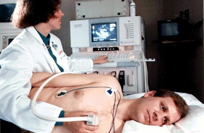
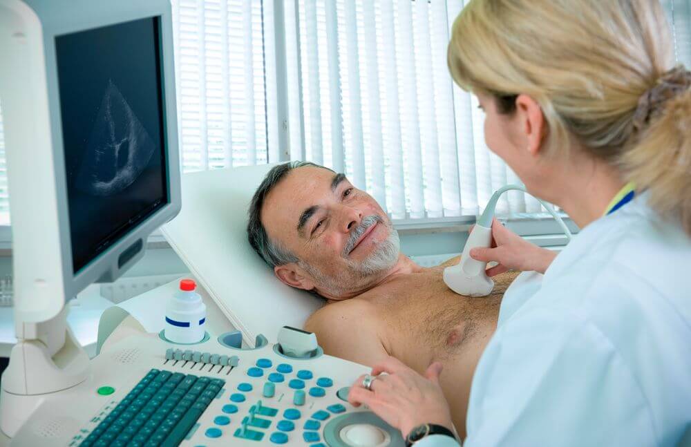
Scanning important body human body using ultrasound is performed in a specially equipped room. Before starting the diagnosis, the doctor conducts a patient survey, performs auscultation of the heart, reviews the necessary medical documents. Next, he puts on the hands and feet of the person being examined the electrodes necessary for synchronous ECG recording.
Specialists identify several main accesses for diagnostics:
- apical approach (examination of the heart is performed through a place located in the fifth intercostal space along the left mid-clavicular line);
- parasternal access (ultrasound is carried out through the area located in the third, fourth intercostal space along the left edge of the sternum);
- suprasternal access (examination is carried out through a part of the body located above the sternum in the region of the jugular "jugular" fossa);
- subcostal access (ultrasound is performed through a place located in the epigastric region).
During an ultrasound examination of the heart, performed from the first two accesses, the patient lies on his left side on a special functional bed. Its headboard rises 30 degrees. On examination internal organ of the last two accesses, the person lies on his back. The specialist is located either to the right or to the left of the patient. He can hold the sensor in his left or right hand.
The part of the body to which it is planned to attach the sensor is lubricated with a special gel. With it, the air gap between the body and the device is eliminated. The result is sharper images. The gel is safe for the skin. It can only cause slight discomfort due to the fact that it is cool.
Deciphering the echocardiogram of the heart
A patient who has undergone an ECG receives an echogram of the heart - the results of the examination with a conclusion. The document indicates by a specialist all the parameters measured during ultrasound internal organ. All values that have been defined in 1D and 2D modes are marked. Information obtained as a result of Doppler echocardiography is also indicated.
The lower part of the document contains the conclusion of the doctor. The specialist in it indicates what is normal, whether there are any pathological abnormalities. The conclusion also notes the exact or estimated diagnosis of the patient.
Normal values of indicators
For Echo KG performed in M-mode, the norms are typical, which are indicated in the table below.
Table 1. Some normal results echocardiography in one-dimensional mode
Below are normal values for echocardiogram indicators, B-mode.
Table 2. Some norms specific to B-mode
| Index | Mean |
| Parasternal position, long axis | |
| 1. Left ventricle: | |
| anteroposterior dimension during systole | 48 mm |
| anteroposterior dimension at the time of diastole | 31 mm |
| 2. Anteroposterior size of the right lower chamber of the heart during diastole | 28 mm |
| 3. End diastolic size of the left atrium | 36 mm |
| 4. Base of the aorta during the end-diastolic phase | 29 mm |
| Parasternal position, short axis, at the level of the base of the aorta | |
| 1. Aortic diameter during diastole | 30 mm |
| 2. Diameter pulmonary artery at the moment of diastole: at the level of the valve and in the middle part of the trunk | 23 mm and 28 mm |
| 3. Anteroposterior size of the left upper chamber of the heart at the time of systole | 36 mm |
| Parasternal position, short axis, at the level of chords | |
| 1. Anteroposterior size of the left ventricle: | |
| end-diastolic | 48 mm |
| End-systolic | 32 mm |
| 2. Anteroposterior size of the right ventricle, end-diastolic | 30 mm |
Doppler echocardiography is also characterized by certain norms. They are listed in the table below.
Table 3 Some Normal Doppler Echocardiography Parameters
In conclusion, it is worth noting that ultrasound of the heart, echocardiography, is considered highly informative. diagnostic method. And indeed it is. The survey allows specialists to obtain objective data about anatomical structure important organ of the human body and its functioning in real time.
At the appointment with a cardiologist, patients are faced with a variety of examination methods. One of these methods is echocardiography - instrumental method examination, which allows using ultrasound to see the structure of the heart and evaluate it contractile activity. Ultrasonic waves tend to be reflected from all tissues of the human body with different speed depending on the density of the fabric. After being reflected, the waves return in the form of an electrical impulse, which are processed by a computer and shown on the screen in the form of a specific pattern.
echocardiograph
Depending on the method of conducting, several types of echocardiography are distinguished:
- transthoracic - conducted through the chest with a sensor;
- transesophageal echo KG - the method is quite rare, it is used only if there are strict indications;
- stress echocardiography - evaluate the work of the heart during exercise.
Three types of CG echo are also distinguished:
- M-mode, or one-dimensional - the sensor produces a wave directed in one direction, resulting in a real-time image of the heart with a "top" view. This type helps to check all cardiac structures - atria, ventricles, aorta.
- 2D - ultrasonic wave with a high frequency of 30 m/s, passes at an angle of 90°, resulting in a high quality image, in which all structures are clearly visible.
- EchoCG with Doppler effect - is based on a change in the speed of reflection of a wave from a moving object in relation to a change in the frequency of the signal - in this case, the signal is displayed from red blood cells - erythrocytes. With this method, colored zones appear on the screen, displaying the blood flow in the ventricles and atria.
Cardiac ultrasound procedure
Cardiography using echo allows you to visually assess the work of the heart, its structural changes and structural features, look at the contractile activity of the myocardium, the work of the heart under stress, and using Doppler to determine hemodynamic parameters (for example, if there is a back reflux of blood).
When is an echocardiogram indicated?
During a scheduled examination:
- newborns - detection of malformations;
- children 12-14 years old - during the period active growth the development of the heart may be a little “delayed”, due to this, the load on the heart muscle increases;
- pregnant women - if available cardiovascular pathologies allows you to resolve the issue of self-delivery or caesarean section;
- people who are actively involved in sports.
For the diagnosis of diseases:
- endocardium and heart valves (thrombosis, malformations, endocarditis);
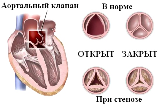
This is what it looks like aortic valve with stenosis
- myocardium (ischemic heart disease, cardiomyopathy, tumors, arrhythmias);
- pericardium;
- pathology large vessels(aneurysm), as well as the condition after surgical intervention on the heart, arterial hypertension, chronic heart failure).
Complaints that most often force an echocardiogram:
- violation of the rhythm in the heart;
- feeling interruptions in his work;
- dyspnea;

The appearance of shortness of breath with light exertion is a reason to see a doctor
- pain after stress and spontaneous;
- swelling of the limbs;
- fatigue and a feeling of lack of oxygen;
- chronic headaches.
How is the procedure?
Depending on which type of echocardiography is performed, the patient is required to perform different actions.
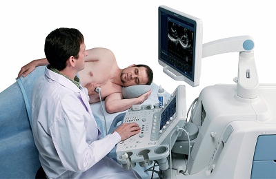
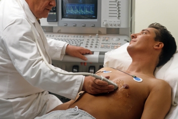
Transthoracic echocardiogram
The patient undresses to the waist and lies on the couch on his left side - this position of the body causes the best picture on the monitor. A special gel is applied to the body, and several electrodes are attached, which simultaneously record the ECG. The doctor guides the surface with an ultrasonic sensor chest, at the same time, a real-time image of cardiac structures appears on the monitor - a cardiogram.
Echocardiogram through the esophagus
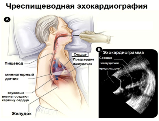
Scheme of transesophageal echocardiography
In the presence of acoustic obstructions, transesophageal echocardiography is performed. In this case, a probe, similar to a flexible tube, is inserted into the esophagus, which is in close proximity to the left atrium. Such a study helps to see the heart structures closer, but is very unpleasant for the patient and requires fasting before the procedure.
Stress echocardiography
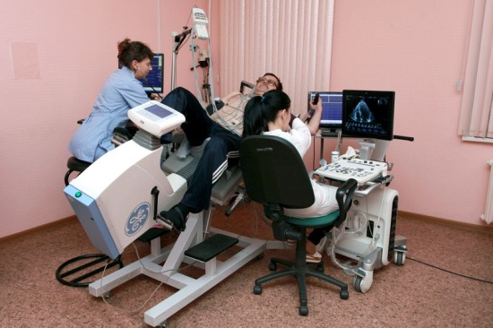
Recumbent ergometer for echocardiography
This method allows you to evaluate the work of the heart during the load, which is given using exercise or special medicines. Stress - cardiography reveals areas of ischemia. To prepare for a stress test, you should not eat a lot of food, drink plenty of fluids, clothing should be loose and comfortable, because a stress cardiogram takes about 45 minutes.
What is the difference between an echocardiogram and an ECG?

How is an echocardiogram different from an ECG?
- First of all, ECG allows you to evaluate the work of the conduction system of the heart, which generates electrical signals that make the heart beat, and echocardiography evaluates the work of the heart as a whole.
- An ECG is a static graph on paper, while an echocardiogram is a dynamic image on a monitor.
- ECG reveals cardiac arrhythmias and myocardial hypertrophy, while echocardiography reveals many structural and functional disorders.
Despite the clear advantages of an echocardiogram, ECG is a very affordable and convenient diagnostic method, and often an ECG is enough to correct setting diagnosis.
Interpretation of echocardiography indicators
Interpretation of the results of the cardiogram is carried out by a cardiologist. The main indicators are evaluated:
- The thickness of the heart walls.
- Measurements of the ventricles and atria are carried out.
- The presence of prolapse and the degree of prolapse.
- The presence of stenosis and the level of narrowing of the valves.
- The speed of blood flow and the presence of reverse injection - regurgitation.
- Diastolic function is the ability of the heart muscle to relax.
- The rate of blood flow in the aorta.
- Pressure in the pulmonary column.
- With a stress test, the presence of an ischemic site and blood flow velocity.
- Stroke volume is the volume ejected by the ventricle in one contraction.
- Mobility of valves, their volume and uniformity.
- The state of the pericardial sac.
Thickening of the walls of the myocardium on the cardiogram - feature arterial hypertension, a decrease in its contractility - a heart attack, changes in the valve leaflets may indicate malformations - stenosis or insufficiency. In any case, only a doctor can decipher the results of the cardiogram, and only make the correct diagnosis at an on-site examination, because even when using echocardiography, the need for other examination methods does not disappear. Self decryption results can cause irreparable harm to health, and only the doctor decides whether treatment is necessary and whether it will be inpatient or outpatient.
"url="http://dlyaserdca.ru/diagnostika/chto-takoe-exo-serdca.html">Both of these methods are accurate, but if cardiac pathologies are suspected, ECHO is usually used.
Echocardiography in a simpler sense is an ultrasound of the heart. By means of ECHO, the following features can be determined:
ECHO methods:
- Transthoracic (echocardiography is performed through the surface of the patient's body).
- Transesophageal.
- Stress-ECHO (the procedure is performed with a load on the heart muscle, which allows you to identify pathologies that are hidden).
Since such a study accurately characterizes cardiac activity, it is used very often. It can be performed even on newborns.
Is the reason for the ECHO:
ECHO should only be carried out in a medical facility, and should be carried out by a person who has the knowledge necessary to decipher the data.
Such a study has a number of advantages. This is the safety of ECHO (the same as when performing an ECG), the absence of discomfort for the patient and side effects, the accuracy of the results. There are no contraindications for cardiac echocardiography, only stress ECHO is performed with slight restrictions.

It contains 8 useful medicinal plants, which have extremely high efficiency in the treatment and prevention of arrhythmias, heart failure, atherosclerosis, ischemic heart disease, myocardial infarction, and many other diseases. In this case, only natural ingredients, no chemicals and hormones!
What diseases are diagnosed using this method?
ECHO can determine the condition of the heart valves. Also, such a study allows you to study the structural features of the organ. Thus, among the diseases that can be detected by this method, we can name the following:
- Heart failure.
- Stenosis.
- Prolapse.
- Heart attack.
- Aneurysms.
- Heart disease.
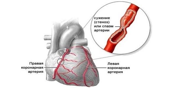
Vasospasm (angina pectoris)
Thanks to additional methods diagnostics, you can find out how the valve apparatus functions.
It is impossible to identify the causes of chest pain with the help of an ECHO of the heart. Also, this method does not report on the state of the vessels, does not detect arrhythmia and blockade.
Despite its safety and the absence of contraindications for performance, it cannot be considered that echocardiography alone is sufficient to be sure that there are no heart problems. Diagnostic methods should be chosen by the doctor, and only he should evaluate the results of the studies.
Execution features
Patients who are prescribed ECHO are interested in how this procedure is done. It is simple and does not require preparation. To get the maximum accurate information the patient is placed on the left side.
It is with this arrangement of a person that the heart is closest to the chest, and the picture becomes more accurate.
The data is captured by a sensor. The ultrasound beams from this transducer are able to study the chambers of the heart. When examining, it is important that the beam be correct form and went into the gap between the ribs. The ribs become an obstacle to the procedure and make it not effective enough.
 The examination begins with an examination of the aorta and the study of its condition to identify pathologies. After that, the ventricles and atria are studied, then the contractile properties of the heart muscle are assessed.
To carry out this study, special knowledge and experience are required, therefore only doctors do echocardiography. They also decipher the data obtained and, on the basis of this analysis, establish a diagnosis. Further treatment is prescribed.
The patient before this procedure, as well as before the ECG, does not need to take any action. There is no need to follow a diet, and there is no need to stop taking medications.
What influences the results?
Distortions in the results in this study of the heart may arise due to anatomical features patient. For a group of people, diagnosis in this way is very difficult.
These include people suffering from obesity, patients with an incorrect arrangement of organs inside the body or the structure of the chest.
Few people know that in addition to the standard electrocardiography procedure, there are other methods of examining the heart, for example, ultrasound, CT, MRI, which allow you to evaluate the work of the heart in a non-invasive way.
Ultrasound of the heart takes more time and is more expensive, but an ultrasound examination is more informative. This procedure is usually prescribed after the procedure, if its results are doubtful or unclear.
What is echocardiography and ultrasound of the heart? Advantages of the method
The ECG procedure is known to everyone. This examination allows you to detect malfunctions in the work of the heart, rhythm disturbances, etc. However, with the help of an ECG, it is not always possible to specify the diagnosis, therefore, further examination is prescribed.
Often patients are interested, and ultrasound of the heart is the same or not. Echocardiography is called cardiac ultrasound, as it is based on an ultrasound examination of the heart, but it also includes elements of electrocardiography. During the examination, an ultrasound and ECG procedure is performed simultaneously, which significantly increases the information content.The method is based on the ability of body tissues to reflect ultrasound and return it back to the transducer.
Because different tissues have different echogenicity, a 2D or 3D image is displayed.
Cardiac echocardiography has many advantages:
- Painless and non-invasive. The procedure is carried out as conventional ultrasound, but with several electrodes attached to the body. It's completely painless. No injections are given before the procedure. The only discomfort that the patient may experience is a slight cold from the gel and sensor. Any pain during echocardiography is not associated with exposure to ultrasound.
- Safety. The procedure is safe for patients of all ages. It has practically no contraindications. Echocardiography can be performed on the elderly, children, pregnant women. Ultrasound does not side effects and has no effect on the fetus.
- Availability of the procedure. The procedure can be carried out in any medical center, private or municipal. With a referral from a doctor, the procedure is free of charge. Equipment for echocardiography is available in almost every city.
- Small price. Compared to MRI, the cost of echocardiography is low. The price of an ECG is somewhat lower, but the procedure itself is less informative. The cost varies depending on medical institution.
- High information content. The EchoCG procedure is highly informative. By using ultrasound examination can assess not only heart rate and heartbeat, but also the size of the heart, the work of blood vessels, the presence of cysts and tumors.
Indications and contraindications

It is possible to carry out the EchoCG procedure both as prescribed by a doctor and for prevention. It is recommended to be screened for anyone who has symptoms of heart disease, suspected of having a cardiovascular disease, and for preventive examination already existing chronic diseases.
Appointment for examination:
- Disorders of the heart. If the patient often experiences attacks or angina pectoris, the heartbeat is very quick or slow, all this is accompanied by unpleasant sensations in the chest area, a heart examination is necessary. Usually, an ECG is prescribed first, and then an ultrasound.
- Dyspnea. AT healthy body shortness of breath may occur after physical activity and . But frequent or persistent shortness of breath even in calm state requires mandatory testing. Most often, it indicates a violation of the heart muscle.
- Edema. The occurrence of edema indicates fluid retention in the tissues of the body. This may be a sign of both kidney dysfunction and cardiovascular diseases. In this case, a comprehensive examination of the body is prescribed.
- . People with elevated blood pressure are at risk for heart disease. permanent high blood pressure significantly increases the load on the heart and blood vessels. For this reason, hypertensive patients are recommended an annual examination of the heart.
- Pain in the chest. Chest pain can only occur in a certain position, when walking or at rest. In any case, it is considered an alarming symptom in need of examination. EchoCG is also prescribed for frequent headaches.
- Very often athletes whose hobbies are associated with extreme sports undergo this procedure. Such sports increase the load on the heart, and they can be practiced only after a preventive examination.
The procedure has practically no contraindications. Both ECG and ultrasound are performed for everyone without exception. Only chest injuries, deformations, skin inflammations can interfere with the procedure. In this case, the examination will be difficult. In some cases, the procedure interferes excess weight. Then the EchoCG procedure is replaced by an MRI.
Preparation and procedure
It does not require any preparation procedure. For a standard EchoCG procedure, it is enough just to come to the office at the appointed time. In some cases, it is recommended not to overeat or eat for 3 hours before the examination. full stomach slightly raises the diaphragm, which can make examination difficult.
The EchoCG procedure is quite simple for the patient. It is carried out quickly and takes no more than 10-15 minutes.
- The patient enters the doctor's office, undresses to the waist and lies on the couch on his left side. The doctor lubricates the patient's skin in the chest area with a special gel and attaches the electrodes.
- The doctor leads the ultrasonic sensor to the chest. The image is displayed on the screen, the doctor fixes it and makes notes. The patient experiences only a slight cold and slight pressure. About any painful sensations you need to tell your doctor.
If conventional echocardiography is not possible, a transesophageal procedure is indicated. This procedure is similar to FGS. A sensor is inserted through the esophagus into the patient's mouth, through which information is transmitted to the monitor. Preparation in this case will be somewhat more complicated: before the procedure, you need to refrain from eating for at least 8-10 hours. During such an examination, a strong vomiting reflex. The patient is advised to breathe deeply through the nose and not strain the muscles of the larynx and esophagus.
There is another type of this procedure - stress echocardiography.
The examination is carried out during physical exertion, or the work of the heart is enhanced by special preparations. This procedure is considered more informative, because it allows you to identify hidden violations in the work of the heart.
The whole procedure takes about an hour. Loads are selected individually for each patient. The subject is asked to come in comfortable clothes so that nothing interferes with the procedure.
For more information about cardiac ultrasound, see the video:
Neither pregnancy nor breast-feeding are not contraindications for ultrasound. There is no need to interrupt breastfeeding. For greater convenience, the woman visits the doctor's office after feeding. Next feeding can be carried out in the same way as usual. Ultrasound has no effect on breast milk and does not harm the child.
Deciphering the results

All indicators are entered into a special protocol, which is then handed over to the patient. The indicators include the size of the myocardium, the size of the ventricles and the thickness of their walls, heart rate, stroke volume of the heart, the volume of ejected blood. With the help of Doppler ultrasound, you can evaluate the work of arteries and blood vessels, the movement of blood through them.
With the help of echocardiography, the following diseases can be detected:
- Aneurysm. With an aneurysm, part of the heart muscle bulges, forming a sac. Contractility heart in this case is reduced. Usually an aneurysm appears as a consequence of a heart attack. With this disease, weakness, shortness of breath,. A ruptured aneurysm is usually fatal.
- Heart failure. Heart failure can occur various reasons. It leads to disruption of the heart, which becomes unable to fully supply all organs and tissues with oxygen. The blood flow deteriorates significantly, which leads to various complications.
- Pericarditis. Inflammation of the pericardium usually occurs due to infection or past. At proper treatment pericarditis is cured completely and is not accompanied by complications.
- Heart defects. Defects can be congenital and acquired, but they all require regular examination. With heart defects, various defects of valves or partitions are observed, as a result of which normal work heart is broken. Some heart defects can be fatal.
- tumor processes. Echocardiography can detect various tumors and cysts on early stages. Malignant and benign neoplasms in the region of the heart are rare, but they timely diagnosis very important.
- Myocardial infarction. With a heart attack, the blood supply to part of the heart muscle is cut off, as a result of which necrotic processes begin. A heart attack can lead to both the death of the patient and various serious consequences.
It is worth remembering that the heart complex organ and its examination is also carried out taking into account many nuances. Only a cardiologist can decipher the result of the examination. It is impossible to interpret the results on your own, as this will lead to an erroneous diagnosis.
echocardiography is one of the most modern methods conducting a heart examination and contractile activity his muscles. It is an echocardiographic study that contributes to obtaining a visualization of a functioning organ and blood vessels.
It is based on the use of ultrasound, which is absolutely not perceived by the human ear. What kind of study is EchoCG, what does it show, and what is ECG, how to prepare for the procedure, how is ultrasound of the vessels of the heart done: find out the answers to all questions from this article.
ECG and echocardiography are among the most effective examinations of cardio-vascular system. They share common goals and objectives. But the methods and methods that are used in their implementation differ significantly from each other. What is the difference between EchoCG (ultrasound of the heart) and ECG and what does each of these studies give?
Method of carrying out. To take an ECG, you need to use a cardiograph and electrodes. At the same time, the electrostatic activity of the heart muscle is examined and recorded, and then the results are translated into a graphic drawing. It is clearly visible:
- whether the activity of the organ is characterized by a stable rhythm of pulsation;
- what are the numerical indicators of the beat;
- the presence or absence of arrhythmia.
For an echocardiogram of the heart, special electronic equipment, which is called a converter. It must be tightly attached to the chest, and then brought into working condition. This device is a generator of waves belonging to the ultrasonic spectrum. They are able to penetrate the body, fight off its tissues and come back.
Special equipment contributes to the processing of the received data and displaying them on the screen. At the same time, on his monitor you can see the big picture.
If the main purpose is to detect the electrostatic activity of heart tissues and study heart rhythms, then EchoCG is performed to study the ability of the cardiovascular system to pump blood.
With the help of the latter, doctors manage to establish and prevent the occurrence of organ failure, check valve activity, and determine the location of atrophied fractions of the heart muscle.
An echocardiogram is used to study the state of the heart of a patient who has had an attack identifying serious blood clots that are not moving. In addition, with the help of current echo transducers, it is possible to study the work of a vital organ in a 3D image.
In comparison with the ECG, the transducer is able to provide a more understandable picture of the examination, as it detects the presence of almost all diseases of the organ.
Varieties
An echocardiogram has several types, we will consider each separately.
transthoracic
The standard type of echocardiogram, which is characterized by painlessness and is somewhat similar to an x-ray, with the help of this procedure, an assessment of the state of health is carried out even before birth.
![]()
To conduct this type of echocardiography, a sensor that transmits high-frequency sound waves, applied to the chest. The heart muscles beat these waves. Thus, images and sounds are created, analyzing which the doctor determines the presence or absence of anomalies and diseases of the organ.
transesophageal
With transesophageal echocardiography, a transducer in the form of a swallowing tube connecting the stomach to the oral cavity inserted into the esophagus. Its close location to the heart helps to obtain a clear image of the structure of the organ.
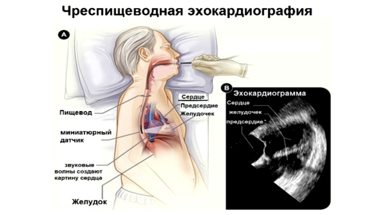
When conducting an echo ultrasound of the heart through the esophagus, there is no effect on the lungs or chest.
stress test
Echocardiogram, which is carried out during a stress test, using dobutamine or adenosine, refers to stress echocardiography. Only here it is not the physiological load on the organ that is applied, but the influence medical preparations that stimulate the work of the body.
With the help of this study, it is possible to assess the state of the organ in the case when it is not possible to use a path or a bicycle for these purposes, load tolerance, the possibility of coronary disease, the effectiveness of the therapy.
stressful
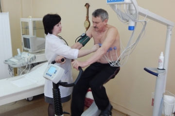 During the patient's sports activities using a jogging or cycling track perform stress echocardiography.
During this procedure, it is possible to visualize the movements of the heart walls and analyze its pumping function.
With the help of a stress echocardiogram, unlike other similar studies, it is possible to determine the lack of blood flow.
intravascular
For use intravascular ultrasound resorted to cardiac catheterization. At the same time, in the cavity blood vessels a special sensor is introduced. For this, a catheter is used.
In most cases, this procedure is performed in order to analyze the blockage inside the vessel.
Only experienced medical specialist will be able to determine which of the species suits you individually.
Types of echocardiography
Echocardiograms are of 3 types:
- 1D in M-Mode– the wave supplied by the device is placed along one axis. Therefore, the monitor shows a top view of the organ. By moving the ultrasound line, you can check the ventricle, aorta and atrium.
- 2D echocardiogram helps to examine the heart in two projections. Therefore, when it is carried out, it is possible to analyze the movement of cardiac structures.
- Doppler echocardiogram carried out in order to evaluate such parameters of the organ as the speed at which blood moves and its turbulence. As a result of the accepted results, it is possible to draw a conclusion about the presence of defects and the degree of filling of the ventricle.
Indications
An echocardiogram should be performed if the following symptoms are present:

- pain in the chest or heart;
- noises and disruptions of the rhythm during the activity of the organ;
- or ;
- symptoms that indicate the presence of heart failure;
- shortness of breath, rapid fatigue, lack of air, rapid blanching of the skin.
Be sure to carry out the EchoCG procedure for patients who have undergone surgery, with an injured chest. Also a referral for an ultrasound of the heart can be received by those who have:
- headaches of a chronic nature;
- artificial valve;
- hypertensive patients;
- is actively involved in sports.
Echocardiography is used to diagnose congenital or acquired defects, as well as in case of improper weight gain in newborn babies.
Preparation of the patient for the study and features of the conduct
Preparation for the procedure is not characterized by unusual complexity. The patient must take off your clothes to the waist and lie on your left side. This position provides the closest location of the chest to the top of the organ under study. This contributes to obtaining the clearest possible image.
 After that, the locations of the sensors are lubricated with a gel. Their various positions contribute to the most visual definition of the cardiac departments, as well as measuring and fixing the results of their activities.
Attaching these sensors not painful and does not cause discomfort. Actually, with their help, ultrasound is directed, which changes during its passage through the tissues, is reflected and returned back.
Then the sounds are converted into signals that enter the echocardiograph. Sound wave changes under the influence of modifications of the state of organs.
After signal processing a clear picture appears on the monitor, according to which the doctor makes appropriate conclusions about the patient's condition.
Find out how an ultrasound of the heart is done from the video:
Deciphering the results
A continuation of the echocardiographic study is. Precise and comprehensive only a cardiologist can analyze them.
And despite the fact that it seems simple and understandable to an ordinary person, the doctor as a result sees a complete picture of the state of the organ. In addition, the result of the study is influenced by the condition and age of the patient, as well as the purposes for which the study was carried out. this study.
In any conclusion of an ultrasound examination, there are unchanged, constant parameters that are characteristic of normal condition and functioning of the body. According to their meanings and the features of the functioning and structure of the chambers of the heart are determined. These include data characterizing the ventricles, the intergastric septum, valves, and the pericardium.
 When conducting echocardiography, set normal performance activity of the ventricles. Depending on the degree of deviation of real results from these indicators, the development or presence of the corresponding pathology is established.
More simple, in comparison with the parameters of the ventricles, is the decoding of the results of examinations of the heart valves. In case of deviation from the norm, you can say on the development of either insufficiency or stenosis. The reduced diameter of the lumen, at which pumping of blood is significantly difficult, indicates the presence of stenosis.
Formation of insufficiency provokes a slightly different process: leaky closing valves contribute to the return of blood back into the chamber, which significantly reduces the efficiency of the heart.
The most common pathology of the pericardium is pericarditis - between the pericardium and the myocardium, which significantly complicates the activity of the organ.
Find out more useful information about what and how the doctor evaluates during the study from the video:
The cost of echocardiography has a very wide range. Its performance is greatly influenced by the qualifications and reputation of the specialist who conducts this study, as well as the level and location of the medical institution. This is due to the fact that only a highly qualified specialist can fully and correctly decipher the information received.
In addition, only a specialist can establish the correct diagnosis based on the results obtained and prescribe the correct treatment. If you start to understand all this on your own, this will lead to incorrect conclusions and erroneous treatment.
And since the heart is practically the most important human organ, which ensures the vital activity of our entire body - then no need to risk it. Because very often it ends in death.
