How to understand the biparietal size of the fetus. How and why the biparietal head size (BSD) of the fetus is measured
Quick page navigation
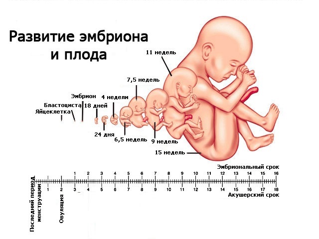
To monitor intrauterine growth and the development of the child during pregnancy, several mandatory ultrasound studies are carried out, which include fetometry - measuring the size of the fetus.
Starting from the 1st trimester (when the embryo is already visualized), one of the mandatory parameters that are determined during fetometry is the biparietal head size or BPS.
BPR - what is it in the ultrasound protocol?
BPR is the size of the head from one parietal bone to the other, measured in the transverse plane. To do this, the ultrasound specialist positions the sensor so that the fetal head is visible from above.
Usually the shape of the head is oval, closer to round. The size from the forehead to the back of the head is determined by a parameter called fronto-occipital size or LZR. The line that is perpendicular to the LZR will be the BPR of the fetus.
- We can say that in the transverse plane the BPR shows the width of the fetal head.
BPR - what is it (photo)
The measurement is carried out in two ways: externally - externally and internally - externally. Their difference lies in the location of the points between which the measurement is made: they can be located either on the outer and inner edges, or on both outer edges of the parietal bones.
This must be taken into account when analyzing the data obtained and comparing them with those tables of norms for which measurements were carried out in a similar way.
Why do you need BPR measurement:
- In the period from the 13th (but possibly earlier) to the 22nd week, this parameter determines the gestational age with an accuracy of 5 to 10 days. After the 28th week, the reliability of this definition becomes questionable due to the different individual growth rates of the fetuses and genetic characteristics, the appearance of “mobility” of the head shape;
- Along with parameters such as abdominal circumference and length femur, BDP indicates the expected weight of the fetus at the time of the study, which is especially important before upcoming birth, as it determines the method of childbirth: independently or with the help caesarean section;
- A dynamic change in this size indicates fetal growth and a normal increase in brain size;
- Is a marker of violations intrauterine development fetus and birth defects development.
Fetal BDP norm by week (table)
For an indicator such as fetal BPD, the norm is very variable and is never represented by one number. Nomograms (tables by which the obtained result is compared) are compiled using percentiles, that is, options that may occur when normal development.
The 90th percentile means that this indicator occurs in 90% or less of all subjects, the 50th percentile - in 50% or less, and the 10th percentile, respectively, in 10% or less.
Fluctuations within percentiles are not a pathology. Often, such nomograms are compiled for each region, even within the same country, which is associated with different ethnic and racial characteristics.
Therefore, the BPR measured in a fetus whose mother permanently lives in Moscow may differ significantly from the BPR of a fetus whose mother, for example, is from Makhachkala. To accurately determine what a parameter such as BPR provides, you need to use tables with regional standards or, if they do not exist, then based on the principle of territorial proximity.
Table - Biparietal size fetal head by week, mm(based on the internal-external measurement method, Moscow region)
| a week | percentile | ||
| 10 | 50 | 90 | |
| 13 | 21 | 24 | 28 |
| 14 | 24 | 27 | 31 |
| 15 | 29 | 31 | 34 |
| 16 | 30 | 34 | 37 |
| 17 | 35 | 38 | 42 |
| 18 | 38 | 42 | 47 |
| 19 | 40 | 45 | 49 |
| 20 | 44 | 48 | 53 |
| 21 | 47 | 51 | 56 |
| 22 | 49 | 54 | 60 |
| 23 | 53 | 58 | 64 |
| 24 | 56 | 61 | 67 |
| 25 | 59 | 64 | 70 |
| 26 | 62 | 67 | 73 |
| 27 | 65 | 70 | 76 |
| 28 | 68 | 73 | 79 |
| 29 | 71 | 76 | 82 |
| 30 | 72 | 78 | 85 |
| 31 | 74 | 80 | 87 |
| 32 | 76 | 82 | 89 |
| 33 | 78 | 84 | 91 |
| 34 | 80 | 86 | 93 |
| 35 | 82 | 88 | 95 |
| 36 | 84 | 90 | 97 |
| 37 | 86 | 92 | 98 |
| 38 | 87 | 94 | 100 |
| 39 | 89 | 95 | 102 |
| 40 | 90 | 96 | 103 |
What to do if the BPR deviates from the norm?
Deviation from the norm, which is detected once, special significance does not exist if it does not differ from the normal indicator within 2-3 lines in the table.
To suspect any developmental disorder in a child, it is necessary to record a change in the BDP several times in a row. For this purpose, ultrasound fetometry is carried out dynamically, usually at intervals of 1-2 weeks.
Increase in head size
Macrocephaly or large head
1. The reasons for a large head size may be hereditary factors. If big size head is detected in one of the parents, then no special measures are taken.
In this case, the attending physician must determine the possibility of independent vaginal birth or choose a cesarean section as a delivery method.
- If the large size of the fetal head is not associated with hereditary characteristics, then it is necessary to exclude developmental anomalies.
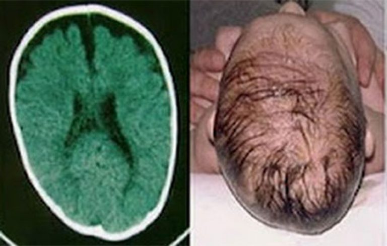
2. With hydrocephalus, dilation occurs internal cavities brain, which contain brain fluid - cerebrospinal fluid. The volume and pressure of the fluid increases - the size of the head increases.
The causes of hydrocephalus can be a violation of the development of the fetal brain, infection for a period of 13 to 27 weeks, tumors, injuries, hypoxia, spina bifida (non-fusion of the vertebrae, resulting in spinal cord does not have a full-fledged bone frame).
Detection of hydrocephalus before the period of fetal viability (3rd trimester) raises the question of termination of pregnancy. If the fetus is already at a viable age, then karyotyping, tests for viruses, a detailed ultrasound, and consultation with a neurosurgeon on the treatment of the child after birth are performed.
- If pressure in the ventricles increases, early delivery and further prompt elimination Problems.
3. Macrocephaly can be observed with disorders of the development of bone and cartilage tissue. This disorder is thanatoform dysplasia - congenital disorder skeletal development, which is associated with a lack of ossification.
An enlarged head, its shape in the form of a trefoil, can be detected on an ultrasound in the middle of the 2nd trimester. Another pathology is achondrogenesis, in which the production of cartilage tissue is impaired, also leading to the development of macrocephaly.
- This condition is a defect incompatible with life.

Hyperplastic development or large fruit
1. In this situation, heredity may also be the cause, and the increase in fetal size may be due to the family constitution. It is necessary to find out what body weight mom and dad were born with, as well as determine their body type.
- The large size of the fetus influences decisions on labor management tactics.
2. The second reason for a large fetal weight may be gestational diabetes, or diabetes pregnant women. It is necessary to conduct urine and blood tests for sugar. On an empty stomach, in the blood, the level of this indicator in pregnant women is lower than usual and amounts to 5.0-5.8 mmol/l.
There should be no sugar in the urine. If changes are detected in the tests, as well as for all pregnant women between 24 and 28 weeks, it is necessary to conduct an oral glucose tolerance test with 50 g. glucose. The test is carried out without preliminary preparation, Anytime. After taking the glucose solution orally, the amount of sugar in the blood is determined after 1 hour.
If this indicator is more than 7.8 mmol/l, further examination by an endocrinologist is necessary. Treatment for gestational diabetes consists of following special diet. If blood sugar continues to be elevated, insulin injections are prescribed.
- Any tablet preparations for lowering sugar during pregnancy are contraindicated.
Reducing head size
Microcephaly or small head
1. As with large head size, a small fetal head may be associated with small head sizes in the family. This feature does not require treatment.
2. Spina bifida is a developmental disorder of the spine, which can be indirectly suspected when the head shape changes to the “banana” or “lemon” type.
Refers to those defects in which termination of pregnancy is recommended if the fetus has not yet reached a viable age. In other cases, it is necessary to inform parents about possible manifestations this disorder after the birth of a child, such as impaired movement and sensitivity lower limbs, chronic pain, fecal and urinary incontinence, spinal curvature.
- If pregnancy continues, it is necessary to determine the date and method of delivery.
3. Intrauterine infection, which leads to damage to the embryo at a period of 3-12 weeks, can lead to the formation of malformations with a decrease in the size of the head. If the infection occurs at a period of 28 weeks or more, the infection is severe and spreads, that is, it becomes generalized - fetal development is delayed and an associated decrease in head size and BPD occurs.
If you suspect infection a number of studies are carried out on the mother aimed at identifying the pathogen: PCR, detection of antibodies in the blood, microscopy of smears, bacterial cultures to nutrient media.
Treatment depends on the type of pathogen and trimester of pregnancy. From 14-16 weeks in the background general treatment carry out prevention of placental insufficiency.
- If you become ill with rubella before the 16th week and if malformations are detected, it is recommended to terminate the pregnancy.
Hypoplastic development or small fetus
1. Hereditary low weight in children in the family, asthenic type of constitution in parents. Special measures not accepted at normal course pregnancy.
2. A decrease in body size may occur with genetic diseases, such as triploidy, trisomy 13 pairs of chromosomes - Patau syndrome, 18 - Edwards syndrome, or 21 - Down syndrome, which lead to early symmetrical growth retardation of the fetus: that is, both the head and the body of the fetus are mismatched with the gestational age.
In each case, the trip is individual. Children who are born alive have severe malformations of organs and their life prognosis is unfavorable - most die in the first year.
The exception is children with Down syndrome, who, when necessary treatment And social support able to have a fairly high quality of life.
Osteogenesis imperfecta - This genetic defect development, which leads to a group of diseases with impaired formation of bone and cartilage tissue. Has several types.
When performing an ultrasound, it is possible to identify only 2 types: shortened, curved bones and multiple fractures; deformation of the head occurs when the sensor presses on soft bones skulls Osteogenesis imperfecta type 2 is not compatible with life.
BDP is important when calculating the cranial index (CI, Y), which indicates the arrest of fetal development, its intrauterine death and approximate time finding a dead fetus in the uterus.
- Cranial index = BPR/LZRx100. If CHI is 83 or more, this is the norm.
After 4-5 days from the moment of death of the fetus, destruction occurs internal structures brain and a significant decrease in BDP: if CHI = 64-74, then fetal death occurred less than 3 weeks ago, if CHI = 64, then the fetus has not been viable for more than 3 weeks.
Change in head shape outside of pathology
Brachiocephaly or short head
The increase in the BPR in this case will not be due to a decrease in the volume of the head, but due to a decrease in the LZR, that is, a decrease in the distance between the forehead and the back of the head. The head has a more rounded or flattened shape.
- Brachiocephaly is often associated with individual characteristics. Often this head shape is observed among residents of the Caucasus and Asians.
Dolichocephaly or long head
With dolichocephaly, a decrease in the BPR is not a sign of a reduction in the head, but is caused by an increase in the LZR. The head has a more oval, elongated shape.
In the fetus common cause the formation of dolichocephaly is gluteal or breech presentation. Normally, this head structure is also genetically more characteristic of representatives of Nordic and Mediterranean peoples.
In these cases, to understand true size heads use the cephalic index: BPR/LZR, which normally ranges from 0.75 to 0.85. If extreme values of the cephalic index are detected, the gestational age is not determined using BDP.
Fetal developmental disability by week (table)
In this publication, we will try to understand the essence of the method for determining fetal BPD, what it is and how much important It has this indicator. This abbreviation stands for biparietal size (BPD). This is the size of the baby's head.
He is very important indicator when monitoring the development of pregnancy, and indicates the normal development of the fetal brain.
BDP values are constantly changing over the course of pregnancy, which is why a lot of attention is paid to this indicator.
What does BPR mean and what is it for?
During each mandatory and additional ultrasound, the fetal head is examined very carefully. After all, fetal brain development most important factor correct development of the child and the course of pregnancy. After all, here is located main body, which affects the entire body. With the correct size of the baby’s head, it is possible to accurately determine how the brain and the fetus are developing as a whole.
An ultrasound specialist measures the fetal BDP. A measurement is taken from temple to temple. If the doctor takes the wrong distance and does not take into account that the line connecting the contours of the temples should be clearly above the thalamus, then he may get results that are far from correct. Next, the fronto-occipital part is measured, which is measured from the forehead to the back of the child’s head.
But the biparietal size is considered more important, because it helps to more accurately determine the gestational age. It is for this purpose that fetal birth control is established. In addition, measurement values help resolve the issue of whether independent childbirth. For example, if the baby’s head is significantly more sizes birth canal, the child himself will not be able to pass them.
And then the need for a caesarean section arises. Although in most cases, the head is of a size that allows the baby to be born naturally.
Is there a norm for the biparietal size of the fetal head?
Having performed BPR measurements on ultrasound, the doctor compares them with normal sizes. For such comparisons, tables have been developed that record the average indicators of normal BDP for each week of pregnancy and acceptable deviations.
Any table is given from at least the twelfth week, because it is impossible to take measurements before this period. The fetus is too small for this BPD measurement.
And that’s why mistakes are common on ultrasound. Here is an example of such a table with normal indicators can be found on the Internet.
Normal indicators
BDP by week of pregnancy (table)
| BPR (BPD) |
Term on average (weeks) |
Range fluctuations (weeks) |
BPR (mm) |
Term on average (weeks) |
Range fluctuations (weeks) |
| 17 | 10,6 | 9,6-11,5 | 58 | 23,5 | 22,7-24,4 |
| 18 | 10,9 | 9,9-11,8 | 59 | 23,8 | 23,0-24,7 |
| 19 | 11,2 | 10,2-12,1 | 60 | 24,1 | 23,3-25,0 |
| 20 | 11,5 | 10,5-12,4 | 61 | 24,4 | 23,7-25,4 |
| 21 | 11,8 | 10,8-12,7 | 62 | 24,7 | 24,0-25,7 |
| 22 | 12,1 | 11,1-13,0 | 63 | 25,1 | 24,3-26,0 |
| 23 | 12,4 | 11,4-13,3 | 64 | 25,4 | 24,6-26,4 |
| 24 | 12,7 | 11,7-13,6 | 65 | 25,7 | 25,0-26,7 |
| 25 | 13,0 | 12,0-13,9 | 66 | 26,0 | 25,3-27,0 |
| 26 | 13,3 | 12,3-14,2 | 67 | 26,3 | 25,6-27,4 |
| 27 | 13,6 | 12,6-14,5 | 68 | 26,7 | 25,9-27,7 |
| 28 | 13,9 | 12,9-14,9 | 69 | 27,0 | 26,3-28,0 |
| 29 | 14,2 | 13,2-15,2 | 70 | 27,2 | 26,6-28,4 |
| 30 | 14,4 | 13,5-15,4 | 71 | 27,6 | 26,9-28,7 |
| 31 | 14,7 | 13,8-15,7 | 72 | 27,9 | 27,2-29,0 |
| 32 | 15,0 | 14,1-16,0 | 73 | 28,2 | 27,5-29,4 |
| 33 | 15,3 | 14,4-16,4 | 74 | 28,5 | 27,9-29,8 |
| 34 | 15,6 | 14,6-16,7 | 75 | 28,9 | 28,2-30,3 |
| 35 | 15,9 | 14,9-17,0 | 76 | 29,2 | 28,5-30,8 |
| 36 | 16,2 | 15,2-17,3 | 77 | 29,5 | 28,8-31,3 |
| 37 | 16,5 | 15,5-17,6 | 78 | 29,8 | 29,2-31,8 |
| 38 | 16,8 | 15,8-17,9 | 79 | 30,6 | 29,5-32,3 |
| 39 | 17,1 | 16,1-18,3 | 80 | 31,1 | 29,8-32,8 |
| 40 | 17,4 | 16,4-18,6 | 81 | 31,6 | 30,0-33,3 |
| 41 | 17,7 | 16,7-18,9 | 82 | 32,1 | 30,5-33,8 |
| 42 | 18,0 | 17,0-19,2 | 83 | 32,6 | 31,1-34,2 |
| 43 | 18,5 | 17,3-19,5 | 84 | 33,6 | 31,6-34,8 |
| 44 | 19,0 | 18,2-19,7 | 85 | 33,7 | 32,1-35,3 |
| 45 | 19,4 | 18,5-20,0 | 86 | 34,2 | 32,7-35,8 |
| 46 | 19,7 | 18,8-20,4 | 87 | 34,7 | 33,2-35,9 |
| 47 | 20,0 | 19,2-20,7 | 88 | 35,2 | 33,7-36,1 |
| 48 | 20,3 | 19,5-21,0 | 89 | 35,7 | 34,2-37,1 |
| 49 | 20,6 | 19,8-21,4 | 90 | 36,4 | 34,7-38,1 |
| 50 | 20,9 | 20,1-21,7 | 91 | 37,3 | 35,3-39,1 |
| 51 | 21,3 | 20,5-22,0 | 92 | 38,1 | 35,8-40,1 |
| 52 | 21,6 | 20,8-22,4 | 93 | 38,9 | 36,9-41,1 |
| 53 | 21,9 | 21,1-22,7 | 94 | 39,7 | 37,6-42,1 |
| 54 | 22,2 | 21,4-23,2 | 95 | 40,5 | 38,3-43,1 |
| 55 | 22,5 | 21,7-23,6 | 96 | 41,3 | 39,6-44,1 |
| 56 | 22,8 | 22,1-23,7 | 97 | 42,1 | 39,6-45,1 |
| 57 | 23,2 | 22,4-24,0 | 98 | 42,9 | 40,3-46,1 |
To install it yourself BPR norm, the same table that the ultrasound specialist uses will help. If you carefully study it, you can easily judge the dimensions that the doctor writes down in the conclusion. The table has three columns. The first gives the dimensions of the norm. In the second, the pregnancy dates are given, and in the third, they are discharged possible deviations from the norm. For example, at 22 weeks the size is 53 mm, deviations are possible from 21.1 m on the smaller side and up to 22.7 mm on the other side.
What to do if the BPR deviates from the norm?
BPD - Ultrasound image showing biparietal size
In case of detected deviations in the BPR, the ultrasound should be repeated a few days later with a different specialist and on a different device. If abnormalities remain, the doctor should evaluate all other fetal parameters. Maybe a very large baby will be born?!
In addition, it is necessary to take into account that the rapid growth of parameters at the beginning of pregnancy is replaced by a slowdown in recent months. And it is quite possible that the initially expected large fruit “will not happen.”
It happens that the baby develops in spurts. This may be another reason for deviations from the norm. As a rule, after some time the parameters become normal. If this does not happen, and the BDP on ultrasound still shows abnormalities, then you need to consult a doctor.
Parameters with a strong deviation from the normal BPD indicate such pathologies of the fetal brain as: tumor, hernia and others. Doctors may prescribe a course of therapy, or may suggest solving the issue of terminating a pathological pregnancy.
If, for example, the doctor establishes the BPD and all other parameters of the fetus are less than prescribed normal results and all other parameters of the baby, a diagnosis is made - intrauterine growth retardation. The reasons for this phenomenon may be various infections or chronic hypoxia, which appeared against the background of placental disorders.
If the diagnosis is confirmed, the woman is admitted to a hospital and prescribed an urgent course of therapy aimed at eliminating the causes of this phenomenon. At the same time, procedures are prescribed that improve blood flow in the placenta and improve the supply of oxygen and vitality. important substances to the child. Even small BPD parameters on ultrasound may indicate microcephaly.
Incorrect parameters indicate various pathologies fetal brain. Doctors may prescribe a course of therapy or may suggest terminating the pregnancy.
In conclusion, the importance of diagnosing BPD size
![]()
Expectant mothers are offered ultrasound examinations several times during pregnancy. After such an examination, the ultrasound doctor must give pregnant women a research protocol, which contains all the information about the baby, including the BPD.
If you undergo such an examination in time, you can get rid of many pathologies. Therefore, such examinations are strongly recommended for pregnant women. It will not cause harm, but there are many benefits from fetal ultrasound.
Today, obstetricians-gynecologists use different ways to measure the presence of various abnormalities in the fetus. In order to accurately measure the gestational age of the expectant mother down to the day, gynecologists use a special index called the biparietal size of the fetal head, which is the most accurate.
To make such a measurement, you need to conduct a special ultrasound examination, usually doctors carry it out from 12 weeks to 28. This article will help you correctly measure this index and describe in detail all the indicators, taking into account different terms maternal pregnancy and fetal development, and also tells the reader about all kinds of abnormalities.
The biparietal size of the fetal head is normal
To measure the BDP of the fetal head, you need to take the correct distance from one temple to another, but do not forget that the line that connects the contours of the parietal bones mandatory must pass strictly above the thalamus. If you arbitrarily change this long-established rule, the results of the study will be too distorted, and as a result, the gestational age will differ significantly from reality. At a certain stage of pregnancy, there are different values, which are the norm, so the longer the period, the more significant the BPR indicator changes, but do not forget that at the end of pregnancy, growth slows down significantly.
Fetal developmental disorder by week
- at week 12, is 21 mm,
- at 13 weeks approximately 24 mm,
- at 16 weeks – 34 mm
- at 24 weeks – 61 mm
- at 32 weeks - 82 mm
- at 38 weeks – 84 mm
- and at 40 weeks – 96 mm.
The doctor evaluates the BPR, also taking into account the fronto-occipital indicators; for this, all measurements must be taken at the level of the cerebral peduncles, as well as the visual thalamus. Because these data change strictly in proportion to the time of pregnancy of the expectant mother. It is worth remembering that if the fetus is at 38 weeks of development, then its head changes shape significantly, so if there is a dolichocephalic configuration of the baby’s head, then the index will be less than normal.
Ultrasound during pregnancy BPD of the fetal head, normal and pathological
Thanks to the use in today's medicine of the fetal head and other similar indicators, doctors are able to find and prevent various deviations in the baby's development in advance, for example, such as: very big child, hydrocephalus or intrauterine development. If these indicators are slightly different from the norm, then you should not despair, you just need to measure other indicators that will show the full picture. And if all parts of the body are evenly enlarged, then we can safely conclude that there will be a large baby. But if only the size of the head is increased, then you should start to worry, because the fetus is almost certainly diagnosed with hydrocephalus and caused it intrauterine infection.
When a gynecologist observes that the BPD is less than the norm and all other parameters of the baby are significantly less than those that correspond real time gestation, then a disappointing diagnosis is made - delayed fetal development in the womb. The reasons for such circumstances are different, for example, it could be an intrauterine infection, as well as chronic hypoxia, which arose due to fetoplacental insufficiency.
If such a diagnosis is fully confirmed, then the expectant mother is prescribed urgent treatment, which is entirely aimed at eradicating the causes harmful effects. Therefore, doctors prescribe a number of procedures that improve blood flow in the uterus and placenta, and also have a full effect on the fetus so that it receives oxygen and all the necessary nutrients, through the use of drugs such as Curantil for pregnant women, Actovegin and, of course, Pentoxifyline.
Doctors diagnose microcephaly if the fetal BPD and fronto-occipital size are significantly reduced.
During pregnancy, expectant mothers are required to undergo an ultrasound several times, after which the ultrasound specialist must give them a research protocol, where he describes in detail all the information about the development of the baby. Here in it he indicates all important information, including even the most important parameter - biparietal head size (BSD). We will now tell you why this is done.
BPR - transcript
When a doctor conducts ultrasound monitoring of a child, he first of all spends all his time on fully examining the baby’s head. The explanation for this is very simple: the brain is located in the head - the most important organ in the human body, it is it that fully influences the condition of the fetus as a whole. Therefore, if you correctly measure the size of the head, you can also assess the development of the brain, which is why the BDP exists. To put it another way, the biparietal size is the width of the fetal head, which is measured from one temple to another.
Of course, in modern medicine This is not the only parameter; there is also the fronto-occipital size (FOR), it will be measured from the forehead to the back of the head of the fetus. But still, the main one is the BDP, because it determines the development of the fetus. His doctor is trying to measure when future mom be between 12 and 28 weeks.
Doctors also operate on BPD when they consider the possibility of a woman in labor giving birth to a baby herself without surgical intervention. Otherwise, a caesarean section is prescribed.
Biparietal head size is normal
When I evaluate the BDP, I use specially designed tables that collect average data for estimating head size for a certain period using this method. In such a table, the data is indicated in percentiles, which are needed for special designation in medical statistics, that is, the average value is the 50th percentile.
When a doctor wants to use the data from this table, then, first of all, he finds its average value, and then completely determines extreme points proper development fetus Let's look at this table using an example when the fetus is 12 weeks old and its BPD for this period is 21 mm, while the norm ranges from 18 mm to 24 mm. Therefore, within the limits of such indicators, you should not worry, because the size of each baby is different and this is affected anatomical structure his body.
Fetal BPD in the table - deviations from the norm
Of course, these indicators may not always fall exactly within the boundaries. But this does not always indicate any pathology, and in order to fully convince of this, the doctor who is observing the pregnant woman must conduct further whole line various studies. If, when measuring, all elements of the body are proportional, then we can conclude that the fetus is simply large. Sometimes it is observed with such parameters that the baby grows in leaps and bounds.
But such deviations do not always mean good things; in most cases, they indicate problems with the child’s development. If the head is much larger established norm, then this may also indicate a tumor in the brain or hydrocephalus. In such circumstances to the expectant mother The doctor suggests terminating the pregnancy. If the brain is smaller than normal, then there is nothing comforting here either, because this indicates problems with the brain. In this case, the pregnancy is also terminated. If BPD is reduced in the third trimester, then this only indicates intrauterine growth retardation, which can be easily treated today.
During pregnancy, a woman has to undergo ultrasound examinations more than once to show how correctly the fetus is developing in the womb. To assess the condition of the baby, a number of indicators are used, one of which is the biparental size of the fetal head. In other words, the distance between the temples of the baby's head is measured, and the results are compared week by week.
BDP parameters change over the course of pregnancy; each week has its own norm of indicators. Active growth head is observed in the first two trimesters of pregnancy, the indicator is most informative in the period from 12 to 28 weeks and allows you to determine the approximate date of birth.
A discrepancy between the biparental size of the fetal head and the gestational age may indicate intrauterine growth retardation, hydrocephalus, microcephaly and other developmental anomalies of the child.
But deviations of the BDP from the norm are not always caused by pathological conditions. If the size of the head is comparable to the size of the arms/legs and abdominal circumference, then it is likely that the baby will simply be born large. Conclusions about the baby’s condition and deviations in its development can only be made by a gynecologist after assessing all fetal fetometry parameters.
The value of measuring the size of the fetal head on ultrasound
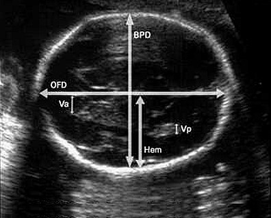
The main parameters measured during the study are the size of the skull, length humerus, the length of the femur and the volume of the child’s abdomen. When determining the parameters of the skull, two values are important: BPR (biparietal size of the fetal head) and LZR (fronto-occipital size). BPR means cross dimension head, the distance between the temporal bones. LZR is measured from the occipital to the frontal bone and means the longitudinal size of the skull.
Special tables have been developed for each parameter. They indicate the values of indicators corresponding to a certain stage of pregnancy. BPR, LZR and other quantities have three meanings: average rate, its top and lower limit. These indicators are developed for each week of pregnancy, starting from the eleventh. All values measured by ultrasound are indicated as a whole, since during normal fetal development they must be balanced among themselves and proportional to a certain week of gestation.
![]()
BPR - transcript
During ultrasound examination Special attention is devoted to the study of the baby's head. This is not surprising: the brain most important organ, the growth and development of which directly affect the condition of the fetus. BDP will help determine the size of the head, and therefore the level of brain development. Biparietal size is a kind of “width” of the head, measured along the minor axis, from temple to temple.
In addition to the BPR, the fronto-occipital size (FOR) is also determined - along the major axis, from the forehead to the back of the head. However, the main parameter remains the biparietal size: it is this that is used to determine the duration of pregnancy. With particular accuracy, this can be established in the period of 12-28 weeks.
The values of the biparietal size of the fetal head BPR are also important for determining the possibility physiological birth. If the size of the fetal head does not correspond to the size of the birth canal, a decision is made on a planned caesarean section.
In the BDP tables, fetal head size values are presented as percentiles. In order to use such a table and determine the norm of fetal BPD by week, it is necessary to find the value of the 50th percentile, the remaining values determine the boundaries of normal readings.

More than normal
When the biparietal size (BSD) of the fetal head is larger than normal, this may indicate the following phenomena:
- heredity. If someone in the family also has a large head volume, then in this case treatment is not required;
- large fruit;
- brachycephaly (short head) - the skull is smaller than usual;
- macrocephaly;
- hydrocephalus;
- disorders in the development of bone and cartilage tissue;
- tumor of the skull bones;
- brain tumor or brain hernia;
- diabetes mellitus in a pregnant woman.
Less than normal
The main reasons for which insufficient indicators of BPR of the fetal head may be recorded are:
- heredity. In this case, if at least one of the parents has a small head volume, then there is no reason for concern and no treatment is required;
- small embryo size;
- intrauterine growth retardation;
- genetic diseases;
- dolichocephaly (long-headed);
- disruption of the development of the spine, bone and cartilage tissues
- pathology of brain development or complete absence some of its parts;
- intrauterine infection.
Conclusion
Only a doctor can evaluate the full picture of an ultrasound. In turn, the doctor leading the pregnancy compares the data from the study of the fetal head with other studies, tests and complaints of the pregnant woman, which allows him to make the correct diagnosis and prescribe appropriate treatment.
