What is a fracture? Symptoms of bone fractures
Fracture
Fracture (fracture ossis) is a partial or complete violation bone integrity, which occurs when an external force or load exceeds the strength of the bone on which it acts. There are two mechanisms for bone fracture - trauma and decreased bone strength due to various diseases.
Causes
Bone fractures occur when exposed to excessive mechanical force that exceeds the strength of the injured bone. Due to the sharp increase in the power of modern technical devices, the strength of which is much greater than the strength of the human skeleton, complex fractures are becoming more common. For example, a hundred years ago, extension fractures cervical spine spine and bumper fractures of the shin bones were quite a rare occurrence due to the lack of mass use of cars and their low power. Now these are widespread types of fractures in car injuries.
Fractures can also be non-traumatic (pathological), that is, arising due to a decrease in bone strength. Among the diseases that can lead to pathological fractures are the following: acute and chronic osteomyelitis, bone cysts, osteogenesis imperfecta, benign and malignant neoplasms bones, or metastases in them, hyperparathyroid osteodystrophy, osteoporosis, etc. In such cases, fractures occur spontaneously without trauma or when exposed to an adequate load, which normally is not capable of causing a fracture. The second type of fractures is more common in older people.
Symptoms
General clinical manifestations for all fractures there is intense pain at the site of injury, which intensifies when attempting to move, swelling, redness (hyperemia) skin in the area of the fracture and bruising or bleeding (with an open fracture). It is rational to divide the symptoms of fractures into unconditional (reliable) and probable. Reliable ones include pathological mobility, shortening of the injured segment, as well as crepitus (crunching) of bone fragments. However, even reliable symptoms may be absent in metaepiphyseal fractures, short fractures tubular bones, incomplete fractures. TO probable signs may include local pain on palpation, deformation at the fracture site, characteristic position of the limb, increased pain with axial load, symptom of dysfunction, swelling in the fracture area, hematoma (may be pulsating with ongoing bleeding). With multiple fractures, the patient's general condition can be extremely severe.
Diagnostics
The standard method for diagnosing fractures of any location is radiography in two projections (direct and lateral). The image should show two joints adjacent to the damaged bone. At pathological fractures important role A carefully collected anamnesis plays a role in making a diagnosis. In some cases, magnetic resonance imaging may be used (usually for traumatic brain injuries).
Types of disease
There are many classifications of fractures. Based on etiology, fractures are divided into traumatic (caused by an external force) and pathological (caused by the presence of diseases such as tumor processes, tuberculosis, osteomyelitis, osteoporosis, etc.). According to the shape of the fracture, they distinguish the following types:
Longitudinal (parallel to the axis of the tubular bones);
transverse (perpendicular to the axis);
· wedge-shaped (more often with fractures of the spinal column);
· fragmentation (a single fracture line is not observed, the bones are crushed);
screw-shaped (with rotation of bone fragments);
·compression;
· impacted fractures.
Depending on the severity of the lesion, fractures are divided into complete (with or without displacement of fragments) and incomplete (fractures, cracks).
Based on the integrity of the skin, open and closed fractures are distinguished.
Based on the number of injured areas, the following types of fractures are distinguished:
single (single fracture of one segment);
multiple (fractures within several segments of the musculoskeletal system);
combined (fractures in different anatomical areas);
· combined (fractures in combination with skull fractures, injuries of internal organs, with polytrauma).
Patient Actions
If a bone is broken, you should call ambulance and before her arrival, if possible, immobilize the damaged area with a splint and, if necessary, try to stop the bleeding.
Treatment
The bone fragments are repositioned (closed or open), followed by fixation and immobilization for bone fusion. Can also be applied skeletal traction and surgical methods for fixation of bone fragments. After restoration of bone integrity, rehabilitation is carried out.
Complications
bleeding;
· damage to internal organs;
·infection of injury, osteomyelitis and sepsis as a consequence;
fat embolism.
Prevention
Prevention of fractures (non-traumatic) is the timely diagnosis and treatment of those diseases that can reduce the strength of bone structures.
A fracture is an injury in which a person's bones become deformed. Their anatomical integrity is compromised due to external influence. Bone tissues are damaged if their physical strength is lower than the force of the traumatic factor. Children and the elderly most often suffer from these injuries. The classification of fractures helps doctors correctly diagnose the type of injury.
Fractures and their symptoms
In some cases, damage can lead to serious complications: sepsis, bleeding, injuries to internal organs from bone fragments, traumatic shock, etc. Therefore, it is very important to provide assistance to the victim as soon as possible.
Dependence of injury on age
In infancy and childhood The bone tissue is not yet very strong and very elastic. Because of this, the child’s skeleton is more vulnerable to the influence of external factors than that of an adult. In addition, high injury rates in children are associated with their in a movable manner life and the fact that their instinct of self-preservation is still poorly developed. In children, two types of injuries most often occur: epiphysiolysis (in germinal zone bone fragments are separated) and a subperiosteal fracture.

Specific changes begin to occur in the body of older people. With age, calcium salts are gradually washed out of bone tissue, osteoporosis develops and bones lose their natural strength. IN old age the risk of falling increases as cerebral circulation is disturbed, and, therefore, dizziness may occur. Coordination of movements is also impaired.
Young people most often suffer from such injuries during the winter season and during excessive physical activity.
Exists international classification diseases, for which fractures are classified as class 19. These are poisonings, injuries and other damage that are consequences physical impact from outside.
Main symptoms
 It is not easy to immediately determine skeletal damage in a victim. But there are several by which they can be recognized:
- Unnatural mobility.
- Increase in the size and shape of the limb.
- Severe pain when moving.
- Bruising and swelling at the site of injury.
- Inability to commit certain types movements (in case of dysfunction of the limbs).
After injury, bone tissue is not completely destroyed. Trauma can lead to fractures, cracks, marginal and perforated fractures. In addition, an impacted fracture may occur, which is classified as a complete fracture. It is mainly observed in the locations of bone metaphyses. With this type of damage, one part of the bone fits tightly into another.
Classification
A correct diagnosis can be made by dividing the types of fractures into classes. Thanks to existing classification skeletal injuries, can be easily picked up optimal method carrying out therapy and making a further prognosis. Damage to bone tissue is divided according to the type of bone fragments, displacement of its fragments, the shape of bone tissue defects, the cause of the damage, etc.
Causes of injury
 First of all, doctors identify the etiology of the fracture, which can be pathological or traumatic. Pathological types:
- Bone thinning after surgery.
- The victim has osteoporosis, bone cysts and serious chronic diseases.
- Osteogenesis imperfecta.
- Malignant and benign tumors.
Traumatic injuries are divided into direct and indirect. Direct ones arise from strong blows, falls, violent actions, etc. They also include gunshot wounds (in this case, the fracture is classified as open). If the place of influence external factor does not coincide with the site of fracture formation, it is called indirect.
Communication of bone fragments
Depending on how bone fragments communicate with the environment, there are 2 types of fractures. If a wound forms at the site of the fracture, it is considered open. If there is no damage to external tissues, it is closed.
 a - closed fracture, b - open
 For open fractures soft fabrics and the skin are damaged, the victim develops a wound that communicates with external environment. This leads to bleeding, and there is a risk of pathogens getting onto the wound surface. There are primary and secondary.
With a primary defect bone tissue a wound is formed at the moment of injury. A secondary one may occur after some time if the victim is transported to medical institution was incorrect or, during unskilled repositioning of bone fragments, their sharp parts tore muscle tissue, blood vessels and epidermis.
May be:
- Combined. In addition to bone defects, the victim's visceral organs were damaged.
- Combined. The injury occurred under the influence of chemical, radiation and mechanical factors.
- Multiple. Several bones were broken at once.
- Single. One bone is broken.
- Full. The ends of the injured bone are completely separated, and it is displaced.
- Incomplete. The bone fragments remain in place. Such injuries include fractures, cracks, perforated and marginal fractures.
Often there are types of bone fractures with displacement of fragments - the most complex and dangerous injuries. The treatment and recovery process takes a very long time. When bone fragments are displaced, severe complications can develop: sensitivity decreases, paralysis occurs, bleeding occurs (closed and open), and the innervation of the limbs is disrupted. If large blood vessels and muscle tissue are damaged, hemorrhagic or pain shock, which can even lead to the death of the victim.
Location
Fractures, depending on their location, are as follows:
- Epiphysiolysis is a bone injury in children in growth zones.
- Epiphyseal - located in the cavities of the joints.
- Metaphyseal - in the joint area.
- Diaphyseal – injuries between the ends of long bones.
- Impression (impacted) – fractures of spongy skeletal elements.
- Separately, damage to tubular bones is distinguished.
An epiphyseal injury, in its essence, is not only a fracture, but also a dislocation. Because of this, it is much more difficult to treat the patient, and the recovery period lasts a very long time. Special attention it is necessary to pay attention to cases of epiphysiolysis, since with improper therapy the skeletal growth zones close prematurely. This is noticeably manifested in the fact that over time the damaged limb becomes significantly shorter than the healthy one.
Fracture line shape

Fractures are also divided according to the line of damage to bone tissue. The injury may be:
- Screw.
- Longitudinal.
- Transverse.
- Oblique.
For transverse fractures, the injury is considered stable. Displacement of bone fragments most often does not occur. In other cases, the bone moves after an injury because it is pulled by muscle tissue.
The comminuted type of fracture is characterized by the separation of one or more sharp fragments from the bone, which enter the soft tissue. With such damage, the patient will need surgical intervention and long lasting rehabilitation period. Such an injury can be large or small fragmented.
Help with fractures

Providing first aid for fractures is very important stage. It must be provided quickly and accurately. However, it is worth remembering that when different types Injuries require different manipulations. What types of fractures are there and how to behave correctly so as not to harm the victim?
Closed limb fracture
First of all, it is necessary to fix the injured limb using a splint, which you can make yourself. If there are no suitable materials at hand, then one lower limb you can tightly wrap it to another, and hang the top one using a scarf, scarf or scarf.
Thanks to these actions, the injured limb will be immobilized. This will avoid deterioration of the victim’s condition during transportation. In addition, it is advisable to apply ice cubes or another cold object to the site of injury to relieve swelling and give the patient an anesthetic.
Open limb fracture
 The open type of fracture is very dangerous. The victim's limb is severely deformed and often opens heavy bleeding. Wound surface It is necessary to treat as quickly as possible with an antiseptic and cover with a sterile bandage. Of course, as with a closed fracture, the limb must be fixed.
Under no circumstances should you try to straighten the damaged area yourself. This procedure can only be performed qualified specialist, after radiography. With such injuries, the patient may experience traumatic shock. To avoid this, the person must be given a medication that will help relieve pain and taken to the emergency room as quickly as possible.
Jaw bone fracture
The main one is deformation of the oval of the face. It also becomes difficult for a person to swallow, and his speech becomes slurred.
The victim must take a horizontal position. In this form, he needs to be taken to the hospital. During transportation, you can gently hold the broken jaw with your hands or tie it up in advance.
Spinal column fracture

The most dangerous are injuries to the spine. After this injury, a person may develop partial or complete paralysis. In some cases, the spinal cord may also be damaged, leading to the development severe complications. The first symptoms of a spinal fracture are the inability to perform certain movements and severe pain.
The victim must be immobilized as much as possible and placed on a hard surface in horizontal position. If there is no stretcher, you can use boards, doors, etc. Do not pull him by the arms or legs under any circumstances - this can lead to severe damage spinal cord. After this, the patient must be transported to a medical facility as quickly and carefully as possible.
Rib fractures
One of the most common types of fractures. If a person has, he will experience pain when deep breath, coughing, sneezing and sudden movements. If the victim produces blood and foam when breathing, attacks of suffocation and extreme thirst, which means he is injured internal organs. The most common injury is to the lungs.
After an injury, the victim must be brought to a lying or semi-sitting position and given an anesthetic. Then the patient must exhale, in this position they bandage him chest.
– destruction of the bone with subsequent separation of parts. May be caused by shock or various or inflammation.There are several complications that can occur after fractures:
once the bone is destroyed, its fragments can damage soft tissue, leading to additional injury and bleeding;
paralysis may occur due to damage nerve cells bone fragments or the bone itself;
with open fractures, the risk of infection with subsequent purulent inflammation increases;
a fracture can lead to life-threatening injuries important organs, such as the brain, if the skull is injured or broken, or the lungs, heart, etc., if.
Causes of fractures
Fractures can be divided into two large groups. The cause of fractures of the first group is the impact of various forces on the bone: a fall, a blow, etc. The cause of fractures of the second group is.
With the second type, the risk of fracture increases several times. It even gets to the point where a person can also break a leg while walking. Here the reason is that this is a pathology of the bone itself, and not an external influence on it. This is often affected by various diseases, such as various tissue tumors. If you have osteoporosis, then, as mentioned above, simply standing up may cause your bone to break. Hip fractures are very common in older people. As for open fractures, most often they occur in places of the lower leg, that is, legs, and also happens on the arms, where the layer of skin is thin. If you fall from a height, then most likely there will be a fracture of the spine or chest, that is, the ribs.
Types of fractures
There are two types of fractures: traumatic and pathological fractures:
Traumatic fractures appear due to the fact that a short but quite powerful force acts on the bone.
Pathological fractures- this is the effect of various diseases that affect the bone, destroying it. The fracture in this case occurs accidentally, you don’t even notice it.
There are also open and closed fractures:
Closed fractures are usually not visible, and skin deformation due to fragments does not occur.
As for open fractures, the opposite happens. As soon as the fracture occurred open type, then an infection immediately enters the wound, which can subsequently spread throughout the body. Gunshot-type fractures are very rare for the common people, but they also exist.
Fractures can also be divided by how many pieces the bone is broken into or whether it is displaced (displaced and non-displaced fracture)
Fractures can be divided according to the shape of the fracture itself, based on the direction of the fracture line into:
Transverse
V-shaped
Helical
Longitudinal
T-shaped
There may also be fractures depending on the type of bone:
After a fracture, damaged bones in most people heal according to the chondroblastic type. Chondroblasts are the youngest and most active cells cartilage tissue. They have a flattened shape, located inside the perichondrium and throughout the entire thickness of the cartilaginous tissue. At the stage of bone growth and fusion, the process of mitotic division and fermentation occurs in chondroblasts. In other words, a person owes the ability to grow the skeleton and restore it after injury to chondroblasts.
A cartilaginous bone callus forms at the fracture site. This process lasts several months and includes four main phases.
The first phase is catabolic (7-10 days):
Aseptic (that is, without the participation of microbes) inflammation develops in the soft tissues surrounding the fracture site;
Extensive hemorrhages occur;
Blood circulation in the tissues around the fracture is impaired as a result of blood stagnation;
Toxic products of aseptic inflammation are released into the bloodstream and spread throughout the body, which explains the general bad feeling patient (weakness, chills, nausea);
Enzymatic cellular activity increases around the fracture site;
Necrotic processes occur on the surface of the bone fracture (microscopic ulcerations and areas of death appear);
There are no signs of healing of the broken bones yet.
The second phase is differential (7-14 days):
The process of formation of fibrocartilaginous callus starts (new cells are actively produced at the fracture site: chondroblasts, fibroblasts, osteoblasts, osteoclasts and chondrocytes);
In these cells, the biosynthesis of glycosaminoglycans (polymer carbohydrate molecules) occurs, the main of which is chondroitin sulfate, which is contained in up to two-thirds of young cartilage tissue. Chondroitin sulfate is a substance whose carbohydrate chains are 90% identical to the monosaccharides galactosamine and glucosamine;
The basis for the future is gradually being formed callus– metrics. Collagen fibers are actively produced in the cells around the fracture site. At this stage, it is still fibrocartilaginous, that is, it lacks the beds of blood supplying vessels. It feeds on fluid from the extravascular space, which is almost ten times more than in the intravascular space. Due to this difference, the process of osmosis occurs - one-way diffusion of liquid through cell membranes towards greater concentration.
The third phase is primary accumulative (2-6 weeks):
From the surrounding tissues, small capillaries gradually grow into the fibrocartilaginous callus, which form vasculature future bone callus;
Chondroitin sulfate molecules, located in the mitochondria of cartilage cells, combine with phosphate and calcium ions;
The regulatory enzyme citrate synthetase and the main energy carrier in cells, adenosine triphosphate (ATP), help the active synthesis of calcium phosphate. Then the chondroitin sulfate molecules combine with calcium phosphate, enter the extracellular space and there they react with collagen;
During this period, the concentration of silicon and magnesium ions in the cartilage tissue also increases greatly. With the participation of these elements from calcium phosphate and collagen, a primary bone callus is formed at the fracture site. While it is still very weakly mineralized, it does not have an ordered crystal structure and therefore not strong enough.
The fourth phase is mineralization (2-4 months):
In the extracellular space of the primary callus, a molecular complex of chondroitin sulfate and calcium collagen pyrophosphate is formed;
These molecules react with phospholipids to produce crystalline hydroxyapatite;
Hydroxyapatite crystals, in turn, settle around collagen fibers in a special way - so that their axes are located at an angle of 41 degrees relative to each other;
From this tandem, the first nuclei of callus crystallization are obtained. Moreover, they can increase in size, feeding on inorganic ions from the fluid of the surrounding soft tissues. This process is called primary bone mineralization;
Then secondary mineralization occurs - intercrystalline bonds are formed around the nuclei. At the end of this stage, we can talk about the complete completion of healing of the fracture.
Features of the flow of phases
Above are averaged data on the course and duration of each phase of bone fusion. Calculations are made based on the fact that we have a relatively healthy patient, and the injury is not particularly complex.
But fractures are different, and the speed of recovery directly depends on many factors:
Type of fracture (open or closed, multiple or single, on one bone or on several);
The age of the patient (in older people, bone fusion can last more than six months, and in teenagers it can be completed in a month);
General health (level of bone mineralization, blood quality, muscle tone);
The presence or absence of aggravating factors (concomitant diseases and injuries) - the more damage to bones, organs and soft tissues the patient receives as a result of injury, the longer the rehabilitation process will last.
Treatment
With a closed fracture, the patient is sedated with some anesthetic that is injected into the fracture site. The broken area is strengthened, for example, with a splint, so that the bone and its broken bone are immobile. If the fracture is of an open type, then the pain is also relieved and the victim is brought to his senses, but only so that he is in adequate condition, then the bleeding should be stopped by squeezing. The bone is also secured in a splint and the victim is immediately taken to the hospital. If the bleeding does not stop, and this happens with arterial or venous damage, then a tourniquet is applied above the affected area.
Upon arrival at the hospital, the patient will have the bone set, but this will all happen only under complete anesthesia or, for example, general anesthesia. If the fracture is not visible enough, the skin is cut slightly. The bone is fixed with plaster.
On this moment In time, all treatment of fractures can be divided into two types:
Conservative - using the same plaster. This is how they treated it back in old times. Now only minor fractures or cracks in the bones are treated this way;
Operative - the bone can be reduced or pulled together using all kinds of knitting needles, tubes, and all kinds of chemical elements are also used.
Education: Diploma in General Medicine received in 2009 medical academy them. I. M. Sechenov. In 2012, she completed postgraduate studies in the specialty “Traumatology and Orthopedics” in the City clinical hospital them. Botkin at the Department of Traumatology, Orthopedics and Disaster Surgery.


Fractures are bone injuries accompanied by a violation of their integrity. In most cases, bone fractures are acquired due to injuries and diseases, rarely - congenital. In the latter case we're talking about O hereditary diseases skeleton, which lead to a decrease in the strength of the human bone apparatus.
There are many reasons for integrity failure musculoskeletal system, among them are mechanical influences, that is gunshot wound, fall, blow, and also pathological processes caused by tumors endocrine diseases, osteomyelitis and others. A strong impulse is not always needed to break bones. At pathological conditions even slight movement in a dream can provoke serious consequences.
Any damage to the bones also affects the surrounding tissues, muscles and nerves; in most cases, this condition is life-threatening, so urgent first aid is required, after which the patient must be taken to the hospital.
As already partially mentioned above, bone fractures can be traumatic and pathological. The former, in turn, have many varieties. The most well-known division among the majority of the population is:
- Closed bone fracture - which does not affect the skin, that is, the risk of tissue infection is reduced to almost zero;
- An open fracture is the presence of a wound and damage to the skin, and depending on the location of the bone and the depth of the lesion, injuries can be visually seen. In this case, there is a high risk of blood loss and infection entering the body.
Open fractures are:
- Primary open, as a result of the impact of traumatic force on a certain area, which leads to disruption of the integrity of the skin, soft tissues and bones. Often this condition is accompanied by a large wound and a comminuted fracture;
- Secondary open - a puncture with a sharp piece of damaged bone from the inside, which manifests itself as a wound on the skin and a smaller area of soft tissue damage.
Depending on the type of fragments formed, injuries are of the following types:
- Brokenness;
- Cracks;
- Transverse fractures;
- Marginal fractures;
- Oblique fractures;
- Helical fractures;
- Comminuted bone fractures.
You can also differentiate bone fractures by the location of the fragments:
- Displaced bone fractures - a change in the location of parts of bone tissue as a result of damage;
- No offset.
Due to a bone fracture, displacement can occur when adjacent muscles contract. A simple injury is said to occur in cases where the bone has split into two parts, but this does not mean that treatment is not difficult. In most cases, non-displaced bone fractures occur in children, while in adults, complete injuries are always associated with this problem.
Most often, fractures affect long tubular bones - the ulna, humerus, femur, radius and tibia.
Bone fractures in children can be of special types: apophysiolysis and epiphysiolysis. This means a displacement of the apophyses or epiphyses of the bones along the line of the fragile growth cartilage. A subtype of such disorders is osteoepiphysiolysis, accompanied by the passage of the fracture line also through cartilage with partial transition to the bone. Such a bone fracture in children is dangerous due to the possibility of damage to the cartilage and its premature closure. As a result, the child's limbs may become shortened or twisted.
Bone fractures in children are accompanied by severe swelling soft tissues.

Symptoms of bone fractures
Regardless of whether a person suffered from a closed or open bone fracture, the symptoms are always expressed by acute pain at the site of injury, edema and swelling, hemorrhage in the area of the fracture (in cases with closed bone fractures, hematomas can be seen), disruption of the contours of the joint or limb of the bone, significant limited mobility, unnatural mobility of the injured limb.
Treatment of bone fractures
Treatment of bone fractures is carried out only by a specialist in this field. But in any case, the victim needs first aid, on which not only his future health, but also his life may depend.
At open injury it is imperative to disinfect the wound with at least hydrogen peroxide, then apply sterile bandage to the damaged area. The task of those around him is to put the person in a motionless position so that the damaged bone is securely fixed. In case of a closed or open bone fracture, remove clothing from the affected area to make it easier for doctors to access this area. Often internal injuries, like open bleeding ones, also require surgical intervention.
The main goal in the treatment of bone fractures is the healing of broken parts and restoration of limb function. It is most difficult to achieve this with displaced bone fractures, since the result depends on the degree of damage, the timeliness of qualified assistance and the actions of the patient himself.
In the closed type, displaced bone fractures are restored using manual manipulation and the use of special devices for these purposes. If the patient has a large wound, that is, an open fracture, then an surgical incision is made and osteosynthesis of bone fragments is made.
In general, treatment can be divided into conservative and surgical, which, in turn, implies not only surgical measures, but also physical therapy, special therapeutic exercises, use of traction devices, taking medications (to eliminate infections) and fortified complexes (to restore bones). After providing the necessary medical care The patient is given a fixing plaster.
Rehabilitation after a broken bone
Rehabilitation for bone fractures means a period of recovery treatment, which is based on physical therapy. Along with special exercises massage, physiotherapy, mechanotherapy are indicated.
Therapeutic exercise is carried out in the form individual lessons and is appointed by a specialist. On initial stage rehabilitation after bone fractures is aimed at stimulating metabolic processes, prevention and elimination of the consequences of injury, muscle wasting, prevention of contractures, normalization psycho-emotional state person.
Also, for rehabilitation after bone fractures, physiobalneotherapy is prescribed to reduce swelling, relieve pain syndrome, improve blood circulation, improve bone fusion. For these purposes, ultrasound, UHF, inductothermy, electrophoresis, phonophoresis with drugs, electrical stimulation of muscles, UV irradiation, radon, pine, salt, and sodium chloride baths are used.
The adult skeleton contains more than two hundred bones (infants have more, but then some bones fuse together). And each of the bones can be broken. Can you imagine how many types of fractures and first aid methods students need to learn? medical universities? Of course, the scope of one article does not allow us to consider absolutely all varieties. bone damage. Here you will receive information about the main types of fractures, their signs and first aid to the victim before the doctor arrives.
Signs of open and closed fractures
Fracture- this is a complete or partial violation of the integrity of the bone as a result of mechanical damage.
Fractures can be open or closed, the differences between them are as follows:
- at closed damage there is no damage to the skin, bone fragments are located in the thickness of the soft tissues;
- at open damage bone fragments break through the surrounding soft tissue and skin.
At closed fractures A strong hemorrhage quickly appears at the site of injury. The intensity of pain can range from insignificant to unbearable; it intensifies not only from movement of the injured limb, but also from changes in body position. The limb itself either lengthens or shortens, and begins to bend in completely inappropriate places. When palpating the area, a crunching sound of bone fragments is detected. The affected limb completely loses its function.
In case of open fractures, the injury is accompanied by external bleeding, severe - in case of damage large vessels, minor - when small ones rupture; bone fragments are visible in the open wound.
Having identified the symptoms of a fracture, first aid should be provided immediately.
Clavicle fractures: types, symptoms and first aid
The collarbone may break in the middle or at the ends.
There are two types of clavicle fractures:
- straight - occur when falling on an outstretched arm;
- indirect - occur when the shoulder joints are compressed from the sides.
With a central fracture, the fragment moves upward and backward, and with a peripheral fracture, downward and anteriorly. The bone is deformed, moves in different directions. The site of injury swells, and when palpated, a crunching sound of bone fragments is detected. The arm, together with the shoulder joint, is turned inward, lowered down and shifted forward. Another symptom of a clavicle fracture is flattening of the supraclavicular fossa.
Before providing assistance for a fractured collarbone, the victim is given an anesthetic, for example 2 ml of ketorol. The effect of the drug can be enhanced by simultaneously administering suprastin, diphenhydramine or any other antihistamine. A good analgesic effect is obtained by injecting the fracture site with a 1-2% solution of novocaine (no more than 10-15 ml). Then, when providing first aid for a fractured collarbone, the injured limb is immobilized by hanging the arm on a scarf or bandaging it to the body.
The victim is transported to the emergency room in a sitting position.
Types and signs of shoulder fractures, first aid
 There are three types of shoulder fracture: upper third, middle third and the lower third of the bone.
If the upper third of the bone is fractured (about shoulder joint) the victim complains of severe pain in the shoulder joint, which intensifies with the slightest movement and touch. The joint itself is swollen. To reduce pain, a person bends his arm in a elbow joint and presses her to the body, supporting healthy hand. Additional feature In case of a fracture, there may be a crunching of the fragments, which is sometimes felt when palpating the joint area.
If the victim seeks help late (more than a day after the injury), a bruise appears in the area of the elbow joint or even on the forearm.
Fractures in the middle third may damage the nerve running along the shoulder. In this case, the victim, in addition to the classic signs of a fracture (such as shortening and deformation of the shoulder, pathological bone mobility, clearly noticeable crunching of fragments), has signs of nerve damage: the hand hangs passively, it can only be straightened passively, active movements are impossible. Another sign of a shoulder fracture in the middle third is the inability to abduct thumb hands.
In case of a fracture in the lower third of the bone, the protruding olecranon process is clearly contoured, above which the area of retraction is determined. The elbow joint swells and becomes sharply painful. At the site of injury, crunching of fragments is detected.
Before providing first aid for a fracture of the shoulder in the upper third, 2 ml of a 50% analgin solution is administered for pain relief (the effect of the drug can be enhanced if suprastin, diphenhydramine or any other antihistamine is simultaneously administered). The arm is suspended on a scarf; in case of severe pain, it is fixed to the body with a bandage.
If the middle part of the shoulder is fractured, the victim’s arm is fixed with a transport splint (instead, slats, straight sticks and tree branches, reinforcement, etc. can be used).
The splint is applied from the healthy shoulder blade to the base of the fingers. The arm is bent at a right angle at the elbow joint. For pain relief, 2 ml of a 50% analgin solution or 5 ml of baralgin solution are administered.
First aid for a fracture of the shoulder in the lower third consists of fixing the arm after anesthesia with a transport splint, bending the elbow joint at an angle of 90-100°.
Types of forearm fractures, symptoms and first aid
 There are many types of forearm fractures. At a fracture olecranon swelling and smoothness of the posterior surface of the joint occur; the person trying to keep his arm straight, pressing it with his healthy hand to the body; Active flexion at the elbow joint is possible, but extension is not.
When the coronoid process is fractured, swelling appears, maximum flexion is limited, and other movements are not impaired. A symptom of such a fracture of the forearm is the smoothness of the contours of the ulnar fossa.
Fracture upper sections radius occurs as a result of falling on an outstretched arm. The victim cannot fully straighten his arm, sharp pain appears when trying to turn the forearm outward.
At a fracture ulna active flexion and extension of the elbow joint is possible, other movements are carried out in a limited volume.
With a fracture of the radius, active movements are almost impossible due to severe pain.
A fracture of both bones is also possible. It usually occurs as a result of a direct blow to the forearm, the forearm and hand being pulled into moving parts of machines, or during a car accident. In this case, the forearm is noticeably deformed (while with a fracture of one bone, the deformation is not so pronounced), the bones are abnormally mobile, and when you feel the injured limb, a distinct crunching of the fragments is felt.
After administration of an anesthetic (2 ml of 50% analgin solution, 2 ml of ketorol, 5 ml of baralgin) apply to the injured limb transport tire from the lower third of the shoulder to the base of the hand, bending the arm at the elbow joint at an angle of 90-100°. To reduce swelling, when providing first aid for a forearm fracture, cold is used.
Fractures of the hand: types, signs and first aid
 There are three main types of hand fractures:
- fracture of the carpal bones;
- fracture of the metacarpal bones (I metacarpal and II-V metacarpal bones);
- fracture of the phalanges (main, middle and nail).
The main signs of hand fractures are acute pain, deformation (in case of displaced fractures) of the hand (or wrist), swelling, and crunching. For fractures nail phalanges hemorrhages appear under the nails.
First aid for a broken hand consists of anesthetizing and immobilizing the damaged area. The victim is given a small ball (a bottle, a roller made from scrap materials) into the sore hand, and the fingers are bandaged to it in a half-bent state. Then the hand is suspended on a scarf.
Types and symptoms of hip fractures, assistance
 Hip fractures can occur in several places. Fractures that pass through the attachment of the femur to the pelvis occur when falling on one side. This type of hip fracture is characterized by very severe pain in the zone hip joint, the limb is turned outward so that the outer edge of the foot becomes parallel to the surface of the bed. At the site of injury, swelling quickly develops and hemorrhage appears. At the slightest attempt to lift the leg, very severe pain occurs. Shortening of the leg is not typical for this type of fracture. An additional sign of a fracture is sharp pain in the hip joint when the heel is tapped.
With a neck fracture femur the pain is much less than in the previous case, but the victim cannot lift the leg on his own, while the doctor lifts it, causing virtually no pain. This symptom of a hip fracture is called a “stuck heel.” Tapping the heel causes slight pain.
A fracture of the femur closer to the knee joint is possible with a direct impact, with a fall on a bent knee, or with a fall on straight legs. The injury is characterized by severe pain in the knee joint. The joint itself is enlarged and deformed due to hemorrhage and displacement of bone fragments. Any attempt at active or passive movements is accompanied by increased pain and often causes crunching of the fragments.
After administering 2 ml of a 50% analgin solution to relieve pain, a splint is applied to the injured limb. To assist with a hip fracture, the ankles are wrapped in soft material (cotton wool), and two splints are placed on the leg - one with inside, the other - from the outside, capturing not only the leg, but also the torso; the splints are bandaged, from above - directly to the chest.
Fractures of the lower leg bones: types, signs, first aid
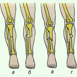 There are two types of tibia fractures: More often there is a fracture of both bones of the leg, less often - only one (tibia or fibula). The mechanism of injury is both direct (a strong blow to the shin, heavy objects falling on the leg) and indirect (sharp rotation of the shin with a fixed foot).
Fractures accompanied by displacement of bone fragments are quite easy to diagnose. With them, deformation and reduction in bone length are clearly visible. The site of injury is sharply painful, swollen, and hemorrhage quickly appears. When palpating the fracture, a crunching sound of the fragments is detected. The victim's attempts to lift his leg on his own end in failure.
Fractures of one bone are much more difficult to diagnose. In this case, the shape of the lower leg remains virtually unchanged, and the victim freely raises his leg. In this case, palpating the area of injury helps. At the site of the fracture, a sign of a tibia fracture is noted, such as local pain, which intensifies with passive and active movements of the foot, and tapping on the heel. In some cases, hemorrhage occurs here.
For pain relief, 2 ml of a 50% analgin solution is injected. Then, when providing first aid for a fractured leg, the victim is given a splint from the upper third of the thigh to the end of the toes.
Ankle and foot injuries, emergency care
 Sprain ankle joint characterized by a rapid increase in swelling in this area due to hemorrhage and severe pain when trying to move the joint. Sometimes the injury is complicated by a fracture of one of the bones of the dorsum of the foot. In this case, when palpating its base, acute pain occurs.
A fracture of the lateral malleolus also causes pain and swelling, but the area of greatest pain is located directly in the ankle area.
A fracture of both ankles with a subluxation of the foot is a very serious injury. The joint is very swollen, the foot is displaced to the side. Any movement, both passive and active, causes sharp pain, and in some cases, patients feel the crunching of fragments.
Fracture calcaneus characterized by an increase in the volume of the heel and its displacement outward. Often there is a flattening of the arch of the foot. Pain occurs when trying to lean on the sore leg, when feeling the heel, and also when moving the ankle joint.
Emergency care for injuries to the ankle and foot involves applying a splint from the knee joint to the toes. Pain relief is carried out using 2 ml of a 50% analgin solution intramuscularly.
Signs of a pelvic fracture and how to provide first aid
Similar injuries occur when the pelvis is compressed, when falling from a height and when there is a strong blow to this area (when hit by a car). A single, isolated fracture of one of the bones, uncomplicated, most often passes without any serious consequences for the body, which cannot be said about multiple fractures of the pelvic bones. This type of injury often leads to severe internal bleeding, the urinary tract is often damaged ( bladder, urethra). In most cases, this causes severe traumatic shock.
A sign of a fracture of the pelvic bones is severe pain at the site of injury, which intensifies even more when palpating the pubic area and ridges iliac bones. Over time, hemorrhage may appear in the area of the external genitalia. Characteristic sign fracture - a symptom of a “stuck heel” that appears on the side of the injury. In this case, the victim is unable to lift his heel off the bed.
Before assistance is provided for a fracture of the pelvic bones, the victim is placed on a rigid stretcher on his back. The knees are spread apart and a cushion is placed under them (the “frog” position). No splint required. If the stretcher is soft, then the victim is transported on his stomach. It is necessary to administer painkillers (analgin, baralgin).
Types of rib fractures, symptoms and first aid
 There are two types of hip fractures: isolated and multiple rib fractures. The causes of injury are most often a strong blow to the chest, a fall, etc. The older the person, the more likely fracture due to chest injury, since with age it becomes less and less elastic.
A sharp pain immediately occurs at the fracture site, which intensifies with breathing movements. Another symptom of a rib fracture is a decrease in the mobility of the chest on the side of the injury. When palpating the chest, it is possible to determine the place of greatest pain and feel the crunching of fragments. The victim breathes frequently and shallowly and tries to move as little as possible.
With multiple fractures of the ribs, when one or more bones are damaged in two places, during inhalation the area limited by the fractures sinks, and during exhalation, on the contrary, bulges, no matter how paradoxical it may sound. This condition very quickly leads to impaired respiratory function, and subsequently blood circulation.
Providing first aid for rib fractures begins with mandatory anesthesia with analgin solution (2 ml of a 50% solution). Then the victim’s chest is tightly bandaged (with a wide bandage, towel, sheet, etc.) and taken to the trauma center.
Spinal fracture: types, symptoms and how to provide first aid
 There are two main types of spinal fractures: injuries to the cervical vertebrae and injuries to the thoracic and lumbar regions.
Damage to the cervical vertebrae occurs when the neck is sharply flexed or hyperextended. They are observed when falling on the head, in divers, in car injuries, especially in cases where the seats in the car are not equipped with head restraints. Some victims are complicated by spinal cord damage of varying severity.
The injury manifests itself primarily as severe pain in the neck. To reduce it at least a little, a person supports his head with his hands and avoids turning and bending. If the integrity of the spinal cord is damaged, complete paralysis of the arms and legs occurs. The main symptom of this type of spinal fracture is the impossibility of active movements and loss of all types of sensitivity. In addition, it is developing acute delay urine.
Injuries to the thoracic and lumbar spine most often occur when falling on the back, less often from an impact, falling from a height, or excessive bending. A sign of a fracture is pain in the corresponding area, which intensifies when palpating the injury site. The protruding process of the damaged vertebra is often clearly visible.
When providing first aid, if there is even the slightest suspicion of a spinal fracture, under no circumstances should you turn over or carry a person in the usual way as it can cause vertebral misalignment and damage to the spinal cord. Before providing first aid for a spinal fracture, to avoid similar complication, the victim must be shifted so that the head and neck remain in the same plane with the body. First, he is turned over onto his back. To do this, you need at least three people: one holds the patient’s head and neck, the second holds the torso, and the third holds the legs. The rotation is performed synchronously. After this, it is necessary to secure the victim’s arms and legs. The arms are placed on the chest and tied at the wrists, the legs are extended, the knees and ankles are tied.
To transport a person with a spinal fracture, a solid stretcher or a wooden shield is needed (from improvised means, you can use boards, cabinet doors, sheets of plywood, tin, plastic, flat slate, etc.). When providing first aid for a spinal fracture, a cushion (made from clothes, towels, etc.) is placed in advance at the location of the lower back. The three of them also lift the victim, synchronously. At this moment, the fourth person moves the stretcher under the patient, after which he is lowered in the same synchronized manner. It is strictly forbidden to seat a sick person! Before transportation, you must also secure the head with a roller or rubber circle.
This article has been read 7,664 times.
