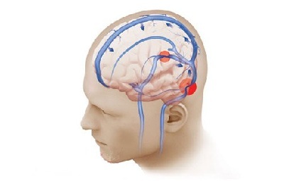
Single small focal changes in the brain substance. Focal changes in brain matter
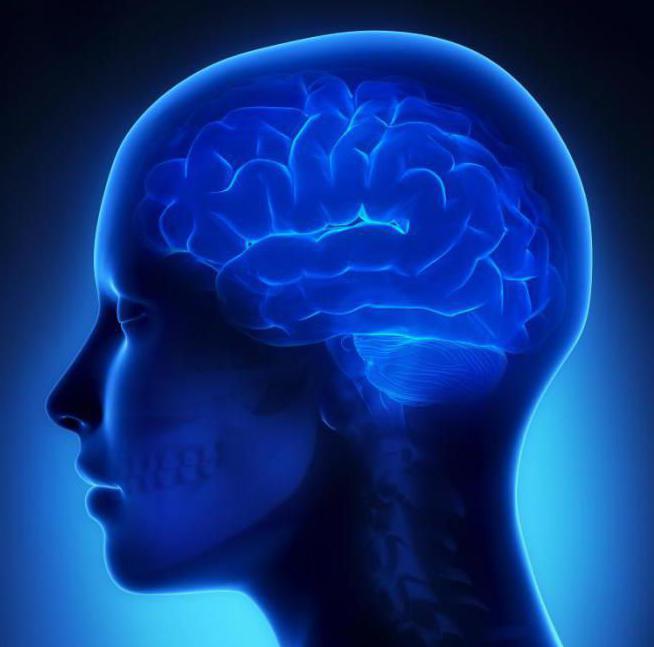
Every person sooner or later begins to grow old. The whole body ages along with it. Aging primarily affects the brain. A failure occurs in the heart and vascular system. The cause of such failures is insufficient blood circulation in the brain and spinal cord.
Cerebrovascular accidents are divided into:
- Focal,
- Diffuse.
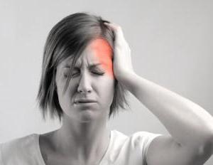
If a person is sick with ischemia, then in the brain there are local changes gray matter brain of the head, due to a lack of blood supply to the brain. This condition can be noticed after osteochondrosis of the neck of the spine or a stroke, when the main vessels through which blood flows to the brain are disrupted. Changes in the substance of the brain of the head can result from any injury or tumor.
Focal changes
Violation of the integrity of brain tissue in any one place is called a focal change in the substance of the brain of the head dystrophic nature. As a rule, these are those parts of the brain that receive virtually no nutrients. In this condition, tissue processes are reduced, and the affected part of the brain begins to malfunction.
Focal changes in the brain substance include:
- Small cysts
- Small foci of necrosis,
- Gliomesodermal scars,
- Completely minor changes in brain matter.
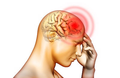
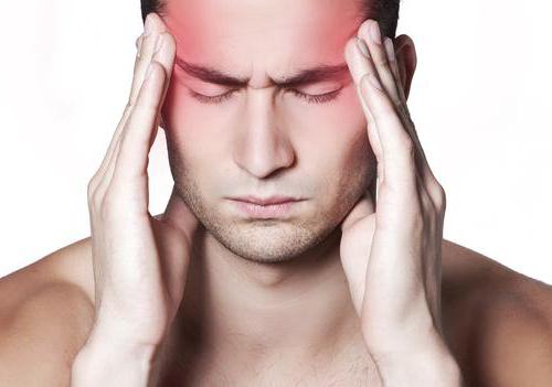
Single focal changes in the substance of the brain of the head of a dystrophic nature give the following symptoms that a person simply cannot help but notice:
- Frequent and painful
- Paresthesia,
- Dizziness,
- Hyperkinesis,
- Paralysis,
- Loss of coordination of movements,
- Decrease in intelligence,
- Memory loss
- Emotional disorders
- Sensitivity disorders
- Ataxia,
- Agraphia.
During the examination, the doctor will have to identify the cause of severe changes in the brain substance and accompanying diseases:
- Vasomotor dystonia,
- Atherosclerosis,
- Various somatic diseases,
- Arterial hypertension,
- Aneurysm in and spinal cord,
- Cardiocerebral syndrome.
When does the disease appear?
Local focal changes in the brain substance of a dystrophic nature occur after seventy years of age and are characterized by manifestations of senile dementia. With this disease, thinking disorder or dementia occurs. Dominant diseases include:
- Alzheimer's disease,
- Pick's disease
- Getington's disease.
By the way, single focal changes in the substance of the brain of a dystrophic nature can occur not only in old age, but also in young and middle-aged people. Any infection or mechanical trauma may compromise the integrity or patency of the blood vessels, which nourish the brain and spinal cord.
How to treat?
In treatment, the main thing is to recognize the disease in time, when the symptoms of focal changes in the brain substance are not yet so pronounced and the process of change can still be reversed. Many different therapeutic activities aimed at improving blood supply to the brain: normalization of rest and work regimes, selection proper diet, use of sedatives and analgesics. Medicines will be prescribed to improve blood flow to the brain. The patient may be offered sanatorium treatment.
Who is susceptible to the disease?
People who experience a single focal change in the substance of the brain of a dystrophic nature are:
- People suffering from diabetes mellitus
- Patients with atherosclerosis,
- Suffering from rheumatism. Such people need to first cure the main disease, follow special diet, monitor physical activity and, of course, regularly visit a doctor.
Local focal changes in the brain substance can be cured if this is approached skillfully and in a timely manner. Unfortunately, only senile changes in the brain matter are difficult to treat.
Vascular genesis of the brain in modern world occurs more and more every year. This depends on environmental degradation, the prevalence of stress factors and the consumption of low-quality food. Moreover, if one of the manifestations of diseases of vascular origin, such as stroke, used to occur in older people, now young people also suffer.
Pathological changes in the vessels of the brain are dangerous fatal outcome and cause brain damage that worsens future health. There are malfunctions internal organs, systems, paresis and paralysis occur.
Pathological processes
Vascular genesis is not equated to a separate diagnosis, so identifying exact reasons pathology is complex, but predisposing factors are identified that cause focal brain disorders. First of all, focal changes in the brain are divided into forms:
Vascular genesis is based on the process of brain damage by pathological foci, leading to the death of neurons. An island of cell tissue- gliosis. A dangerous gliosis lesion manifests itself in a state when, as a result of pathological processes, multiple foci of gliosis are formed, affecting the functioning of the brain itself.
 Physiologically, neuroglia are necessary to protect brain cells from pathological influences. For example, protection is activated during injuries and infections. Strong impact negative factors causes the death of neuroglia, and necrotic foci appear - a focus of gliosis.
At the site of gliosis, the brain stops functioning, and this is dangerous due to the appearance various symptoms– from headaches to disturbances in the functioning of internal organs. A focus of gliosis can form in any part of the brain, but the most characteristic is the appearance of a supratentorial focus in the upper part of the brain.
Supratentorial foci of gliosis initially cause headaches that worsen over time. With the progression and appearance of new foci of gliosis, disturbances in the functioning of the brain and internal organs appear. Lack of therapy and presence of injuries or inflammatory processes leads to the possibility of tumor formation.
Causes
The main predisposing factors that cause pathological changes in cerebral vessels, both focal and general, are identified. Arterial hypertension comes first.
High blood pressure causes unpleasant symptoms, forcing the body to work for wear.
That is why such a focus of pathology as hypertension most often leads to stroke, the formation of gliosis and other negative changes.
Feedback from our reader - Alina Mezentseva
I recently read an article that talks about the natural cream “Bee Spas Kashtan” for treating varicose veins and cleaning blood vessels from blood clots. Using this cream you can cure VARICOSIS FOREVER, eliminate pain, improve blood circulation, increase the tone of veins, quickly restore the walls of blood vessels, cleanse and restore varicose veins at home.
I’m not used to trusting any information, but I decided to check and ordered one package. I noticed changes within a week: the pain went away, my legs stopped “humming” and swelling, and after 2 weeks the venous lumps began to decrease. Try it too, and if anyone is interested, below is the link to the article.
Blood sugar levels affect nutrition and brain function. Coma, which occurs when there is a lack or excess amount of glucose in the blood, forms a focus of death in the head and forms gliosis in its place, therefore, treatment not provided on time threatens with dangerous consequences.
The focus of pathological changes occurs with alcohol abuse, smoking, consumption of fatty and junk food. Lifestyle largely influences the appearance of vascular genesis. The following factors are identified:
- regular overwork or excessive physical activity;
- weather dependence;
- stressful lifestyle:
- head injury;
- use of drugs and potent pharmacological drugs;
- sedentary lifestyle combined with obesity.
All these factors lead to conditions in which pathological processes occur in the white matter, a focus of inflammation is formed and blood supply is impaired. The following conditions predispose to the occurrence of diseases of vascular origin:
That is why, if there are risk factors, it is necessary to carry out timely diagnosis and treatment of pathologies of the heart, brain, metabolism and spine.
Symptoms
Symptoms that cause changes in the brain are closely related to the presence of increased intracranial pressure and degree of pathology. A person’s condition is aggravated by an extensive lesion in the white or gray matter of the brain, a focus of change or a tumor in its structure.

For the treatment of VARICOSE and cleaning blood vessels from THROMBUS, Elena Malysheva recommends a new method based on Cream of Varicose Veins. It contains 8 useful medicinal plants, which have extremely high efficiency in the treatment of VARICOSE. In this case, only natural ingredients, no chemicals or hormones!
Characteristic signs of constant or periodic increased blood pressure, which accelerate pathological process. Arrhythmia occurs.
The pulse may rise or fall sharply from 60 to 90 beats.
 Such a deterioration in the body’s condition with the indicated signs indicates the need to begin treatment so as not to cause damage to both the white and gray matter.
Over time, when focal changes progress, signs of weakness, fatigue, and headache appear. There is weakness in the arms and legs, impaired sensitivity. Failures in the functioning of organs and systems, as well as paralysis and paresis mean the threat of the appearance of tumors, which are divided into supratentorial (in upper parts brain) and subtentorial (in lower parts). If treatment is not carried out, then changes from oncological processes may become irreversible.
Diagnosis and treatment
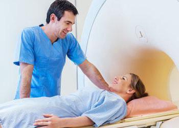
Among all head examination techniques, magnetic resonance imaging is considered the most relevant. By transmitting echo signals, human organs and tissues are displayed, so it is possible to identify a focus of gliosis, structural damage in the white or gray matter of the brain.
 You can effectively examine the arteries of the brain using ultrasound examination and neurological techniques. Difficult stage diagnostics involves making an accurate diagnosis, since symptoms of vascular origin relate to many pathologies. Therefore, the doctor collects anamnesis and prescribes various studies blood, urine, monitors the patient's dynamics.
IN complex diagnostics computed tomography techniques are used as prescribed by the doctor, duplex scanning, thermal imaging, positron tomography. If possible, it is recommended to study the results of magnetic resonance angiography and electroencephalography.
An important aspect in treatment vascular pathologies is to eliminate symptoms and restore health.
On early stages it is necessary to treat arrhythmia and hypertension. This will help prevent stroke and other heart diseases. vascular systems s.
A course of therapy is selected to combat obesity and impaired fat metabolism. Removed atherosclerotic plaques and replaced damaged vessels are included in the surgical intervention section of treatment. It is necessary to take all medications prescribed to restore cardiac function in a timely and regular manner. vascular function, brain work.
 The course of recovery is important. To do this, you need to follow a certain diet, undergo a course of physiotherapy, and therapeutic exercises.
Based on the above, we can conclude that the presence of symptoms frequent pain in the head, increased or low blood pressure may indicate disruptions in brain function. Even conditions such as gliosis lesions can be effectively treated if medical help is sought promptly.
DO YOU STILL THINK THAT IT IS IMPOSSIBLE TO GET RID OF VARICOSE VARICOSIS!?
Have you ever tried to get rid of VARICOSE? Judging by the fact that you are reading this article, victory was not on your side. And of course you know firsthand what it is:
- feeling of heaviness in the legs, tingling...
- swelling of the legs, worsening in the evening, swollen veins...
- lumps on the veins of the arms and legs...
Now answer the question: are you satisfied with this? Can ALL THESE SYMPTOMS be tolerated? How much effort, money and time have you already spent on ineffective treatment? After all, sooner or later the SITUATION WILL GET WORSE and the only way out there will only be surgical intervention!
That's right - it's time to start putting an end to this problem! Do you agree? That is why we decided to publish an exclusive interview with the head of the Institute of Phlebology of the Ministry of Health of the Russian Federation - V. M. Semenov, in which he revealed the secret of a cheap method of treating varicose veins and full recovery vessels. Read the interview...
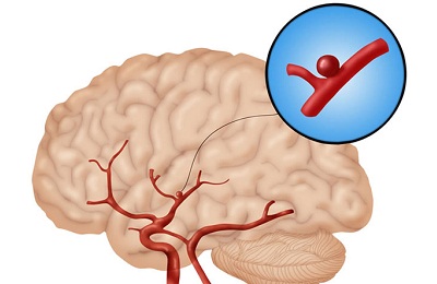
Vascular genesis refers to all possible diseases associated with blood vessels. What kind of illness is this? Vascular genesis means a violation of blood flow in, namely, its vascular and venous network. Now let's look at this pathology in more detail.
What is vascular genesis?
If a person experiences dizziness, noticeable memory deterioration, slow reactions and fatigue, then perhaps he has a constant deficiency of brain nutrition. Many people take such signs lightly. They attribute them to work pressure or lack of vitamins. In order for the brain to function normally, it needs energy. Blood provides it nutrients and oxygen. The body's functioning system is designed in such a way that the nutritional process of the brain is carried out through 4 arteries. Failure of its blood supply leads to various diseases. As a result, vascular genesis occurs.
That's why primary symptoms associated with headaches and fatigue should not be ignored. It is necessary to visit a doctor. He should be asked to conduct the necessary examination; perhaps the person has a vascular origin. Identifying disorders of the body at an early stage makes the treatment process more fruitful. It makes it possible to completely restore the body. Therefore, it is better to start treatment immediately if vascular genesis of the brain is detected. What this is, the doctor can explain, he will also prescribe necessary measures for the treatment of the disease.
Impaired blood supply to the brain
The main causes of malnutrition are hypertension and atherosclerosis. The first named disease is quite common. This disease affects both men and women. Often the origins of hypertension are unknown. But it can cause a person to be diagnosed with vascular genesis. The essence of hypertension is that the walls of blood vessels become denser, and the channel through which blood flows narrows. Sometimes complete narrowing occurs. In this case, the passage of blood becomes impossible. Next, vascular genesis of the brain occurs. What this is, we described above.

Atherosclerosis is associated with lipid metabolism disorders. Because of higher level cholesterol in the blood and other substances containing fat, deposits form in the vessels. They interfere with normal blood circulation. Its movement is hampered by the fact that plaques form in the vessels over time due to lipids. First, they clog blood vessels. Then they begin to fall apart. Their particles are spread with blood to other small vessels. As a result, they can cause blockages.
Also, a disease such as osteochondrosis can affect the blood supply to the brain. Since the movement of intervertebral discs can lead to pinched arteries. Thus, brain nutrition will deteriorate.
Symptoms of blood supply problems
With insufficient nutrition of the brain, neurons begin to die. Since the latter are associated with neurology, the patient may experience irritability, fatigue, insomnia or interrupted sleep. Depression is also common for this condition. contributing factor. If the disease progresses, the person may experience periods of severe excitability.
There is also a manifestation of egocentrism. At further development The disease results in indifference to anything and dementia. Insufficient nutrition of brain cells can cause other more serious illnesses. For example, a stroke. In our country, this disease occurs quite often. Not everyone can survive this disease. Moreover, it can lead to various severe consequences both for the person himself and for his close circle. There may also be epileptic seizures due to the fact that the brain does not receive enough nutrition.
Types of blood supply disorders
The types of brain nutritional disorders are classified as follows:
Stages
There are several stages that indicate the development of a disease associated with insufficient nutrition of the brain. The dynamics may be different, as it is influenced by some factors, such as heredity, lifestyle, ecological situation and so on.
At the first stage of the disease, people often have headaches, irritability, forgetfulness and sleep disturbances. In the second stage, memory deteriorates more severely; a person can sleep during the day, but at night sleep is disturbed. Also appear intrusive thoughts, the patient begins to think about the same problem. The gait becomes unsteady. Uncoordinated movements appear. Performance decreases. On last stage As the disease develops, dementia sets in, and the person stops recognizing relatives and finding his way around on the street.
Causes of the disease
As mentioned above, this disease has some cause. That is, not enough for normal functioning brain nutrition is associated with any disorders of the body. These include:
- Increased arterial pressure.
- Diseases of the cardiac system, such as arrhythmia, ischemic disease hearts and others.
- Diabetes.
- Excess weight.
- Sedentary lifestyle, lack of walks, sports, etc.
- The presence of high cholesterol levels in the body. This indicator is associated with poor nutrition, as well as the presence of fatty foods in the human diet.
- Heredity. If close relatives have suffered from diseases such as stroke and heart attack, then there is a possibility of of this disease.
- Availability bad habits such as alcohol and smoking.
- In men, in addition to the above reasons for the occurrence of insufficient nutrition of brain cells, there is such an indicator as emotional overstrain. This is primarily due to stress at work and at home. As a rule, a man feels responsible for the well-being of the family. Therefore, problems at work can affect his health.
Diagnostics
To identify that the brain receives little nutrition and to determine changes in vascular genesis on initial stage can be difficult, since in a given state of the body the electrocardiogram can be completely normal, without any abnormalities. But the ECG at physical activity can reflect changes that are present in the human body. It is also recommended to install a 24-hour monitor so that the doctor can see the work of the heart. But it is worth saying that these types of diagnostics may not show that some kind of malfunction is occurring in the body, for example, a single focus of vascular origin.

Other diagnostic methods
Fundus examination will help in making a diagnosis. This event will help to identify whether there are any changes in the brain or not. The patient's hearing may also deteriorate and swallowing reflex. Therefore, it makes sense to undergo an examination by an otolaryngologist. Also, if there is a suspicion of the presence of any abnormalities in the brain, the doctor should prescribe blood tests.
Computer diagnostics is good method identifying changes in a person. With its help, you can see supratentorial foci of vascular origin of the brain.
Conclusion
Now you know what vascular genesis is and how it manifests itself. We also looked at the features of diagnosing this disease, the causes of its occurrence and symptoms.
Carrying out MRI for focal brain lesions
Focal lesions Focal brain lesions can be caused by trauma, infectious disease, vascular atrophy and many other factors. Often degenerative changes are accompanied by problems associated with disruption of normal vital functions and coordination of human movement.
- Content:
- Signs of focal lesions
Signs of focal lesions
All disorders of brain activity are reflected in the natural daily functions of human life. The location of the lesion affects the functioning of internal organs and the muscular system.
Changes in vascular genesis can lead to mental disorders, cause high blood pressure, stroke and other unpleasant consequences. On the other hand, subcortical lesions may not have clinical manifestations and be asymptomatic.
One of obvious signs the presence of a focal lesion is:
- Hypertension - a lack of oxygen supply to the brain caused by vascular dystrophy leads to the fact that the brain accelerates and increases blood circulation.
- Epileptic seizures.
- Mental disorders - occur with pathology of the subarachnoid spaces, accompanied by hemorrhage. At the same time there can be observed congestion in the fundus. Characteristic feature pathology is fast education darkening, burst vessels and retinal rupture, which allows you to determine the likely location of the focal lesion.
- Strokes are clearly defined focal changes in the brain vascular nature MRI allows us to establish pre-stroke status and prescribe appropriate therapy.
- Pain syndrome - chronic headaches, migraines may indicate the need for general examination patient. Ignoring symptoms can lead to disability or death.
- Involuntary muscle contractions.
Signs of single focal changes in the brain substance of a dyscirculatory nature on magnetic resonance imaging mean that the patient has certain deviations in the functioning of the vascular system. Most often this is associated with hypertension. The diagnosis and explanation of the study results will be provided by the attending physician.
Diagnosing changes
The picture of focal changes in the substance of the brain of a dystrophic nature is observed, according to various sources, from 50 to 80% of all people as they age. Ischemia, as a result of which normal blood supply stops, causes provoking changes in soft tissues. Resonance tomography helps to identify the causes of violations and carry out differential analysis diseases.
Small focal changes that do not cause concern at first can eventually cause a stroke. In addition, the outbreaks increased echogenicity vascular origin may indicate oncological cause violations.
Timely identification of the problem helps to prescribe the maximum effective therapy. A focus of dyscirculatory origin, clearly visible on MRI, may indicate the following pathologies:
- In the hemispheres big brain- indicates the following possible reasons: blocking the blood flow of the right vertebral artery through congenital anomaly or atherosclerotic plaque. The condition may be accompanied by a hernia cervical region spine.
- In the white matter of the frontal lobe, changes can be caused by ordinary hypertension, especially after a crisis. Some anomalies and isolated small lesions in the substance are innate and pose a threat to normal life. Concerns arise about the tendency to increase the area of damage, as well as the accompanying changes in motor function disorders.
- Multiple focal changes in the brain substance indicate the presence of serious deviations in genesis. May be caused by either a pre-stroke condition or senile dementia, epilepsy and many other diseases, the development of which is accompanied by vascular atrophy.
If the MRI report indicates the diagnosis: “signs of multifocal brain damage of a vascular nature,” this is a reason for certain concerns. The attending physician will be required to establish the cause of the changes and determine methods of conservative and restorative therapy.
On the other hand, microfocal changes occur in almost every patient after 50 years. The lesions are visible in angiography mode, if the cause of occurrence is disturbances in genesis.
If a dystrophic focus is detected, the therapist will definitely prescribe a general medical history of the patient. With absence additional reasons for concern, it will be recommended to regularly monitor trends in the development of pathology. Substances may be prescribed to stimulate circulation.
Changes in the brain substance of a discirculatory-dystrophic nature indicate more serious problems. Pressure and lack of circulation can be caused by injury or other causes.
Signs of small focal brain damage with vascular etiology moderate expansion may cause the diagnosis of encephalopathy, congenital and acquired. Some medications can only make the problem worse. Therefore, the therapist will check the relationship between medication and ischemia.
Any pathological and degenerative changes should be well studied and tested. The cause of the focal lesion was determined and, based on the MRI results, prevention or treatment of the detected disease was prescribed.
Galina Mikhailovna asks:
The basal cisterns are moderately dilated.
The chiasmal area is without features; the pituitary tissue has a normal signal.
Subarachnoid convexital spaces and grooves are expanded, mainly in the frontal area parietal lobes and Sylvian fissures with moderately expressed atrophic changes in the brain substance.
The midline structures are not displaced.
The cerebellar tonsils are located normally.
In the white matter of the left frontal and parietal lobes, foci of demyelination (2) measuring up to 0.5 and 0.6 cm, respectively, are detected.
Conclusion: MR picture of the external replacement hydrocephalus. Focal changes brain substances of a discirculatory nature.
The patient is 62 years old and suffers from headaches in the crown area and noise in the right ear.
You can decipher the description, otherwise none of this is clear, is it worth seeing a doctor, is it serious. The doctor who made the description said that there was nothing wrong. Thank you very much in advance!
According to the examination, we can judge the changes in the brain typical for this age. However, if there is clinical symptoms, a personal examination by a neurologist is necessary.
Irina asks:
Hello!
I am 50 years old. I am very worried about headaches. I had an MRI of the brain. The picture shows moderate external replacement hydrocephalus. Multiple focal changes in the brain substance are probably of a discicular-dystrophic nature.
You can decipher the description, otherwise none of this is clear, is it worth seeing a doctor, is it serious. Thank you!
In this situation, you should definitely contact a neurologist for personal advice. Focal changes are most likely age-related. But signs of hydrocephalus indicate a violation of liquorodynamics, which is what causes headache attacks.
Elena asks:
I am 51 years old. I entered vascular department with a diagnosis of cerebral stroke, after treatment and discharge, she underwent an MRI examination of the brain, where it was determined: foci of demyelization were identified in the white matter of the frontal and parietal lobes, without signs of perifocal edema, most likely of dystrophic origin. The lateral ventricles of the brain were not dilated, with a moderately pronounced zone of gliosis on the periphery. The 3rd and 4th ventricles are unchanged, the basal cisterns are moderately dilated. The chiasmal area is without features, the pituitary tissue has the usual signal. The subarachnoid convexital spaces and grooves are unevenly expanded along the convexital surface of the brain and cerebellum, against a background of moderate cortical atrophy. The expansion of the perivascular liquor spaces of penetrating vessels is determined, mainly at the level basal ganglia on both sides. The middle structures are not displaced. The cerebellar tonsils are located at the level of the cerebral cortex. Conclusion: a picture of external replacement hydrocephalus. Focal changes in the brain substance of a dystrophic nature. Question: probable causes and forecast for the future.
Check your anthropological data, accompanying illnesses and current status. As well as the treatment received and the drugs currently used. Read more about
Elena comments:
Thanks for the answer! I’ll add: height 167, weight 80 kg. In childhood I suffered from rheumatism, pyelonephritis, until now I had vegetative-vascular dystonia hypotonic type 110/70, menopause since 2006, without any peculiarities. She was admitted to the hospital after several attacks of hypertensive crisis with all the symptoms ischemic stroke., after treatment with Actovegin IV, vitamin therapy, glycine, magnesia IM, was discharged under the supervision of a neurologist and further examination, continued treatment with indalamide, lisinopril, thromboasom, Sermion for a month, but dizziness, headaches, lack of coordination did not pass, I am currently undergoing treatment: IV Mexidol, vitamins and the same drugs in tablets, R-graphy of the cervical spine did not even detect age-related changes, my health has improved, but only slightly. Stress, physical activity (except for gymnastics to remove fat deposits), alcohol consumption were not the cause of my health condition. I would like to know other probable reasons for preventing the recurrence of attacks and the prognosis. Maybe you should not pay attention, because the most important thing is that your arms and legs are working and your speech is not impaired, but I really don’t want to wait for attacks with more serious consequences. I am very grateful in advance for your answer.
Elena, the most important thing is to set yourself up so as not to live in anticipation of the next attack. You need to achieve weight loss, constantly monitor your blood pressure, and take antihypertensive medications. Be sure to monitor your blood cholesterol levels. It is necessary to consult a neurologist about a possible replacement of the diuretic. The prognosis in your situation is favorable.
Elena asks:
Hello, I'm 23 years old. I did an MRI of the brain. We made the following conclusion - MR picture external hydrocephalus. Single focal changes in the brain substance in the right frontal and right parietal lobes (dyscirculatory nature? demyelinating process?). Tell me, do I need treatment and is this diagnosis dangerous?
You need complex treatment under the supervision of a neurologist. In the absence of adequate treatment, damage to the central nervous system will progress leading to irreversible consequences. First of all, it is necessary to prescribe drugs that would normalize intracranial pressure.
Elena comments:
Please tell me whether intracranial pressure can be treated and whether it could be the cause Full time job at the computer in sitting position?
Intracranial high blood pressure in some cases it is possible to stabilize with medication. In any case, to determine the cause of increased intracranial pressure and prescribe adequate treatment, a personal consultation with a neurologist is necessary. Long work sitting at the computer can be one of the factors that increases blood pressure.
Elena asks:
I'll repeat it just in case. In September of this year, she was admitted by emergency medical service to the vascular center with a diagnosis of ischemic stroke, the diagnosis was made according to clinical data, although CT data for stroke were not received, after a course of treatment she was discharged with a diagnosis of ischemic stroke in the vertebrobasilar system. Cerebral atherosclerosis. Hypertension 3 risks4. IHD: atherosclerotic cardiosclerosis., for further treatment and observation by a neurologist. In October, she was admitted to the hospital with a repeated TIA, after treatment she underwent an MRI of the brain, which also did not confirm the stroke (in conclusion: a picture of external replacement hydrocephalus. Focal changes in the brain substance of a dystrophic nature). Discharged with a doctor: consequences of an ischemic stroke in vertebrobasilar region with left pyramidal week, severe ataxia, dysphagia, elements of dysarthria. Discircular encephalopathy 2 with severe cognitive decline. Cerebrovascular disease. I was sent to a commission to establish disability, where it was refused, citing the fact that there was no stroke, and the rest did not correspond, although I needed constant medication and observation by a neurologist. Currently, my health is not satisfactory (constant headaches, unsteadiness, I can’t do the most light gymnastics, I only move around the house). Question: did I have a stroke, as the doctors claimed in vascular center? and is it worth applying again to establish disability, since you need to constantly purchase medications, and there are financial difficulties, but you really don’t want to experience the humiliation of proving your identity once again bad condition health.By work book I haven’t worked for 10 years (I worked part-time for a private owner, but now I can’t). Thank you in advance for your answer.
Unfortunately, if the diagnosis of stroke is not confirmed instrumental methods examination (MRI), he will not appear in the VKK documents. In the event that you want to submit documents for assignment of disability status again, you will need to carefully medical examination and consultation with a lawyer involved in similar problems, who can advise you on the legally justified possibilities for assigning this status in your specific case. You can read more about stroke, methods of diagnosis and treatment of this disease in our section: Stroke.
Ainura asks:
I want to know. What are you waiting for me? Thank you
Could you please clarify your question again? If you are sick, please check your full diagnosis to obtain adequate advice.
Marina asks:
Hello, please help! My father is 47 years old, his for a long time bothered by headaches, right ear Can't hear at all, numbness on the right side of the face. They sent me for an MRI, the MRI showed that in the right cerebellar corner a solid space-occupying formation was detected, with clear, even contours, irregular round shape measuring 27x 20x 17 mm with a heterogeneous hyperintense MR signal on T2WI, isointense MR signal on T1WI. Discicular lesions are detected in the white matter of the frontal and parietal lobes irregular shape up to 4 mm, without signs of perifocal reaction.
Tell me, how serious is this? what consequences? and what to do??? Thank you. Sincerely, Marina.
In this case, it is recommended to consult with an oncologist to conduct a personal examination, study the results obtained during the examination and decide on further treatment tactics and examination. In the event that this moment the above complaints are present, the situation is very serious, you cannot postpone a visit to the doctor, because any delay can only aggravate the situation and worsen general state. Read more about oncological examination by clicking on the link: Oncology.
Leah asks:
Hello! I have periodic severe headaches (several times a month). It starts with a headache, ends with vomiting, but there is no pressure. Recently I was vomiting all night with a temperature of 39. I donated blood, a swarm of 20, then 41. I did an MRI, changes only - mild expansion of the subarachnoid convexital spaces in the area of the frontal and parietal lobes. What kind of expansion is this? What to do? Where to look for the cause? Thank you!!!
Julia asks:
Hello, I was 30 years old and had an MRI examination because... I am bothered by frequent headaches. After the MRI, the conclusion “In the white matter of the frontal, parietal and left temporal lobes, small subcortical foci of increased signal intensity on T2 VI and FLAIR IP d up to 0.4 cm are detected. Please tell me what is this? And what could be the consequences?
An increase in signal intensity can occur for several reasons. It may be inflammatory or vascular origin, and can also be determined even in the absence of pathology. It is not possible to make any conclusion based on the data you provided. First of all, you need to see the images directly, which should be evaluated in conjunction with other studies and your complaints. Only in this case will it be possible to talk about possible violations. It is also not possible to make a conclusion about the consequences, since they are assessed only after an accurate diagnosis has been established and adequate therapy has been carried out. In your case, I recommend consulting with a neurologist. Read more about diseases of the nervous and vascular systems and the causes of headaches in the section: Headache
Julia comments:
Conclusion The MR picture shows focal changes in the brain substance, most likely of a dyscyclic nature. I would like to understand this seriously???? So what's this??? and can these foci, for example, develop into cancer or a stroke???
Discirculatory changes do not lead to the development of brain tumors, and strokes are caused exclusively by in rare cases. You need to be regularly monitored by a neurologist and receive appropriate treatment to restore normal microcirculation in the brain.
Tamara asks:
Tamara Leonidovna is 61 years old. Have been diagnosed diabetes, grade 3 hypertension, grade 3 angina, asthma, happened 5 months ago hypertensive crisis, on August 14, Bell's palsy, was sent for an MRI. MRI conclusion: a picture of moderate phenomena of external replacement hydrocephalus. areas of gliotic changes in the left frontoparietal region and pons of the brain, of a post-ischemic nature. Focal changes in the brain substance, discirculatory in nature. MR signs of intraosseous formation in the right parietal bone. At the moment the improvements are minor, headaches, noise phobia, trimer, weakness. The face has straightened out a little but is far from normal. Sugar is on average 10-14mmg.
Nellie asks:
Hello! My mother is 51 years old. She often suffers from headaches, her blood pressure is normal. My mother did an MRI and here is the diagnosis: “MRI picture of multiple finely focal changes in the brain substance of a discirculatory nature, not pronounced mixed replacement hydrocephalus.” Please tell me whether this is a serious diagnosis? And what will happen? further? How can this diagnosis be treated. THANK YOU in advance!
In this case, if there are changes in the brain associated with hemodynamic disturbances or complaints, it is recommended to consult a neurologist to conduct a personal examination and assess the current condition, as well as prescribe adequate treatment. Upon appointment timely treatment the condition may improve and the changes will not progress. Read more about the causes of headaches in a series of articles by clicking on the link: Headaches.
Natalya asks:
At my husband's high heart rate(120 - 140 beats) headaches bother me... There are attacks - very strange, the neurologist sent me for an MRI and EEG. This is what is written in the MRI report - a picture of external, internal hydrocephalus. Expansion of a large tank. Single focal changes of vascular origin in the white matter cerebral hemispheres. Cyst-like dilatation of the cistern magna. The pain is very disturbing, but the neurologist prescribed only pills for epilepsy... And my head hurts! and what to do?? what to drink for pain and how to remove this fluid from the brain?? I read that diuretics, but which ones are possible? I'm desperate.........
According to the survey data provided, there are pronounced violations: internal hydrocephalus, expansion of the ventricles of the brain, impaired hemocirculation, the presence of a cyst. In this case, you need to go through complex treatment, the prescription of antiepileptic drugs is justified, because all these changes can cause seizures. It is recommended to re-consult with a neurologist to decide whether hospitalization is necessary, to carry out complex treatment, or to prescribe adequate therapy for outpatient setting. It is also recommended to consult with a cardiologist to prescribe adequate treatment, because pulse is much higher than normal. Read more about headaches in the section of the same name by following the link: Headache.
Ekaterina asks:
She did an MRI of the brain, angiography of the cerebral arteries. Conclusion: MR signs of expansion of the subarachnoid convexital and perivascular spaces. Single focal-dystrophic changes in the brain substance measuring 0.2-0.3 cm. Based on the MR picture, no data for pathological changes in the cerebral arteries were identified .Please tell me what this is? And is it dangerous?
If such changes occur, the blood supply to the brain may be impaired. You need a personal consultation with a neurologist to evaluate the result obtained in combination with clinical picture, complaints and anamnestic data. IN currently There are no threatening or dangerous changes, but the selection of corrective treatment is required, which the treating neurologist can do for you. Read more about this study You can find out from the thematic section of our website: MRI
Lyudmila asks:
MR picture of focal changes in the brain substance of a dyscirculatory nature, moderately pronounced diffuse bihemispheric atrophy
According to the results of the examination, there are signs of atrophy of brain tissue; perhaps the atrophy is associated with cerebral circulatory insufficiency. To clarify the situation, a personal consultation with a neurologist is necessary. You can read more about deciphering MRI results in our section dedicated to this method diagnostics: MRI. You can read more about examining a neurologist and what questions you should ask this specialist in the section: Neurologist.
Varvara asks:
MRI showed focal changes, white matter brain, we see it is of a vascular nature. How dangerous is this and how can it be treated?
Nadya asks:
MRI picture of uneven expansion of the subarachnoid convexidal space. A single focus of gliosis of a discirculatory nature
Unfortunately, based on the examination results you provided, it is impossible to give a conclusion about the severity of the brain damage. You need a personal consultation with a neurologist to evaluate the results of the examination. You can read more about the examination of a neurologist and why it is needed in the section: Neurologist.
Nina asks:
MR picture of external replacement hydrocephalus, slightly pronounced. Single foci of demyelination in the brain substance of a dystrophic nature. what does it mean? Do I need to see a doctor? I am 45 years old.
In this case, you must be examined by a neurologist, since if you have replacement hydrocephalus, the doctor will be able to prescribe treatment for you depending on the data general examination, neurological status and existing complaints. You can learn more about this from the section: Hydrocephalus
Andrey asks:
MRI picture of post-traumatic, postoperative zones of cystic-gliotic changes in the right hemisphere of the brain and in the right hemisphere of the cerebellum. Internal non-occlusive and external hydrocephalus.
Based on MRI, hydrocephalus (internal and external), as well as post-traumatic and postoperative changes. In this situation, it is necessary to consult a neurologist or neurosurgeon, a detailed study of the medical history and an assessment of the current neurological condition, which will allow you to select adequate treatment (drugs that reduce brain swelling, improve microcirculation). I recommend that you personally consult with your treating neurologist. You can learn more about hydrocephalus from the thematic section of our website: Hydrocephalus
Marina asks:
Please explain the diagnosis:
On a series of MR tomograms, weighted by T1 and T2 in three projections, sub- and supratentorial structures are visualized. Lateral ventricles of the brain regular sizes and configurations. The subarachnoid convexital space is locally unevenly expanded, mainly in the area of the frontal and parietal lobes. The midline structures are not displaced. In the white matter, in the area of the basal ganglia and semioval centers, expansion of the perivascular spaces of Virchow-Robin is determined. The cerebellar tonsils are located at the level of the foramen magnum. In the white matter of the frontal and parietal lobes, subcortically, single small foci of increased T2 and FLAIR signal are detected, without signs of a perifocal reaction, probably of a dystrophic nature. Conclusion: MR picture of a single expansion of the arachnoid spaces in the area of the frontal and parietal lobes. Single focal changes in the brain substance of a dystrophic nature. Thank you in advance.
Denis asks:
Volumetric and focal formations in the brain are not identified. The ventricles of the brain are not expanded, the lateral ventricles are symmetrical. The dimensions of the lateral ventricles (at the level of the foramen of Monroe): right 8 left 8 The middle structures are not displaced. Moderate uneven expansion of the external liquor spaces subconvexitally in the fronto-parietal regions, lateral fissures. The occipital cistern is reduced in volume, the cerebellar tonsils prolapse in the BZ up to 5 mm. The sellar, pineal region, cerebellopontine angles, craniospinal junction are usually visualized. The course and caliber of the great vessels - b\o Good afternoon, I am 30 years old Lately bothered by burning and tingling in the head and in different places, then in the frontal area, then in the back of the head, then in the temples! Help me, tell me what’s wrong with me and how to treat it please!
Unfortunately, only based on the provided research results in the conditions online consultations It is not possible to prescribe treatment for you. I recommend that you personally visit a neurologist who can compare the research protocols with your complaints and data clinical examination. Only after this will it be possible to establish a diagnosis and begin treatment. You can obtain more information on this issue in the corresponding thematic section of our website by clicking on the link: Computed tomography (CT)
Elena asks:
I am 36 years old. For 10 days now I have had a very bad headache in the lower right back, but I have never suffered from headaches before. I did an MRI, this is what they wrote - focal changes in the white matter frontal lobes, genesis is questionable (foci of beliosis of a vascular nature? hemeelinating process?) moderate external hypotrophic hydrocephalus... it’s kind of scary. Please tell me what to do and what awaits me?
Elena comments:
Good afternoon, thanks for your participation! Passed additional examination, everything is fine with the eyes, the vessels of the neck are also normal, but the X-ray did not please me - osteochondrosis and uncovertebral arthrosis, instability. They prescribed Milgamma and physiotherapy, but the headache does not go away. Can osteochondrosis be the cause of focal changes?
Natalia asks:
Hello. I am 35 years old. It is not yet possible to contact a neurologist, so if possible, I would like to hear here an explanation and transcript of my conclusion, as well as possible consequences, preventive/treatment measures. MRI conclusion: “There is an expansion of the subarachnoid spaces around the penetrating vessels of the brain in the basal nuclear zones. In the area of the anterior horn of the left lateral ventricle single lesions of a dystrophic nature 1-3 mm in diameter are visualized. initial dyscirculatory changes in the GM." Everything else according to the scripture has not been changed. Thank you in advance!!!
These changes are age-related, moderate character. In the presence of clinical complaints A personal examination by a neurologist is required. Treatment can be prescribed only after examination by a doctor, depending on the indications. You can get more information on this issue in the thematic section of our website: MRI
Natalia comments:
Thanks for the answer! Forgive me, I understood correctly that the presence of such complaints as poor memory, absent-mindedness, inattention, mental lability and instability, a tendency to depressive states, are not indications for contacting a neurologist? and can the above complaints affect the size and number of lesions in the future? and also, such foci of a dystrophic nature and dyscirculatory changes as mine are not yet indications for the use of some drugs, for example, those that improve blood supply to the brain?
In any case, if you have any complaints, you should personally visit a neurologist who can prescribe you adequate treatment. You can get more information on this issue in the thematic section of our website by clicking on the link: Neurologist and neuropathologist
FATINHA asks:
Hello! I am 22 years old. They did an MRI of the brain. Conclusion: Single change dystrophic brain. Tell me how this is treated? Is it dangerous at all? Could it be in the background cervical osteochondrosis, there was an exacerbation recently.
A single change in the brain of a dystrophic nature, as a rule, is a consequence of impaired blood circulation and vascular patency. As therapeutic measures drugs are prescribed to improve cerebral circulation, microcirculation, and strengthen blood vessels. This condition not threatening, but requires correction, so I recommend that you personally visit your attending neurologist, who can prescribe you the appropriate treatment. You can get more information about this study in the thematic section of our website:
Lily asks:
Hello! I am 54 years old. I have frequent headaches on the right side of my head and face, and sometimes I feel numbness on the right side of my head. Nausea, dizziness and weakness. I did an MRI. Conclusion: Based on the MR image, no evidence of a space-occupying lesion in the brain was obtained. A single lesion in the left temporal lobe, probably of a dyscirculatory nature. Cystic thickening of the mucous membrane of the right maxillary sinus. Option for the development of the Circle of Willis. A marked decrease in the signal from the blood flow along the intracranial segment of the right VA (hypoplasia?). Areas of stenosis in the A2 segment of the left ACA and in the P1 segment of the right PCA cannot be excluded.
Please tell me how serious this is? Which specialist should I contact?
Thank you.
In this situation, your complaints are most likely related to a violation cerebral circulation. With adequate treatment, existing symptoms can be eliminated. I recommend that you personally visit a neurologist who will prescribe you appropriate drug treatment. Read more about this study in the section: MRI
Anna asks:
Hello!
My husband is 37 years old; he has been suffering from constant headaches for about 10 years, but his blood pressure is normal. I had a concussion in my youth. Examinations (several years ago) by a neurologist did not reveal anything; painkillers were prescribed. Lately my head has been hurting really bad. I did an MRI of the brain, according to the results: “Based on the MRI picture, there are data for focal and diffuse changes not received. Slight expansion of the main and quadrigeminal cisterns. Swelling of the mucous membrane of the left upper quadrant, cavity of the mastoid process on the left." What may this conclusion indicate? What other necessary examinations what needs to be done to make a diagnosis? Thank you in advance!
These changes are possible in the presence of increased intracranial pressure, which may be a consequence of previous injuries. I recommend that you personally visit a neurologist who, based on the available results, as well as taking into account your medical history and clinical symptoms, will be able to prescribe adequate treatment for your spouse. You can get more information on this Question in the corresponding section of our website by clicking on the link: Magnetic resonance imaging (MRI). Read about the causes of headaches and their diagnosis in the information section of our website: Headache
Oksana asks:
Hello! I am 43 years old, I had an MRI of the brain, the conclusion was a picture of moderate dystopia of the cerebellar tonsils. Single focal changes of vascular origin in the white matter of the cerebral hemispheres. What does “focal changes in white matter” mean? I am periodically bothered by dizziness (when changing the position of the head, when bending down), pain in the back of the head.
Focal changes may indicate that the blood supply is impaired in certain areas, which requires drug treatment. Dizziness may be associated with dystopia of the cerebellar tonsils, since it is this organ that is responsible for the coordination of movement. I recommend that you personally visit a neurologist to prescribe adequate treatment. Read more about this study in the section of our website: MRI
Alena asks:
help me decipher what it is -Focal lesion left parietal lobe, Deterotopia of gray matter? Are they recruited into the army with this diagnosis?
Marina asks:
Hello! My son is 18 years old, a psychiatrist diagnosed him with depersonalization, direalization, MRI conclusion: A single focus of dyscirculation in the left frontal lobe. Pineal gland cyst 11x8x6 mm. Intermediate velum cyst. Moderate open internal hydrocephalus of a replacement nature. Could these changes cause psychiatric illness?
Unfortunately, these changes can cause the development of psycho-neurological disorders. You need to personally consult with a neurosurgeon regarding further treatment tactics, and I also recommend that you visit a psychologist who can provide real help in the correction of such manifestations. Read more about this in the section: Psychologist
Victoria asks:
Based on the MRI results, I received the following conclusion:
MR picture of uneven expansion of subarachnoid spaces. A single focus of demyelination in the right parietal lobe (probably dystrophic in nature). Tell me, is this something scary???? What to do?
Expansion of the suarachnoid space is often observed due to traumatic brain injury, increased intracranial pressure and past infections central nervous system. Foci of demyelination are often found in diseases such as multiple sclerosis. In this case, it is necessary to personally study the obtained images, so I recommend that you visit a neurologist, who, after studying the study protocols, will be able to make a conclusion and establish correct diagnosis and prescribe appropriate treatment for you. You can get more information on this issue in the section of our website: MRI
Galina asks:
I had an ischemic stroke twice (July 2008 and November 2011 - there are MRI study protocols). Today in July my legs gave out again and I felt weak again. I made an MRI report in November: an MRI picture of focal changes in the brain of a dystrophic and post-ischemic nature (previous lacunar infarctions). Mixed replacement hydrocephalus. Doctors send to a commission (VTEC). Is it worth it or not? (I have already been refused 2 times (after the first and now on October 1). Age: 60 years old, weight: 58 kg, height: 164.
In your situation there are all indications for obtaining a disability group, the issue in this case is resolved medical commission. I recommend that you prepare all the documents and visit the VTEK commission. You can get more information about your disease, its course and treatment in the thematic section of our website by clicking on the link:
Svetlana asks:
Hello. My 27-year-old husband began to suffer from severe headaches. We did an MRI: on a series of T1- and T2-weighted MRIs, sub- and supratentorial structures were visualized in three projections.
The lateral ventricles of the brain are of normal size and configuration. 3rd and 4th
the ventricles and basal cisterns are not changed. The chiasmal area is without features, the pituitary tissue has a normal signal.
The perivascular Virchow-Robin spaces are expanded, mainly in the area of the basal structures.
Roots 8 pairs CMN in the area of the cerebellopontine angle are traced on both sides, symmetrical.
The subarachnoid spaces are locally expanded along the convexital surface of the brain and in the area of the lateral fissures. The median structures are not displaced. The cerebellar tonsils are located normally.
In the white matter of the right parietal lobe, a rounded focus of gliosis is detected subcortically, measuring 0.5 x 0.4 cm, without a perifocal reaction.
The mucous membrane of the nasal turbinates is thickened, the nasal passages are narrowed, and patency is preserved. Deviation of the nasal septum to the right by 0.5 cm is determined.
Conclusion: MR picture of external replacement hydrocephalus. Focal changes in the brain substance of a residual nature. Curvature of the nasal septum.
Consultation with a neurologist, otolaryngologist.
The ENT specialist said that everything is fine. We live in the region and will not see a neurologist soon. I am very worried about what this is, how serious it is and whether it is curable.
According to this conclusion, there are signs of replacement hydrocephalus, which happens in such cases: increased intracranial pressure, changes in blood vessels and metabolism, encephalopathy, etc. Treatment in each case is prescribed by a neurologist based on a study of the medical history, research protocols, personal examination and patient complaints. There is no need to worry ahead of time, but try to get to a neurologist in a timely manner, who can prescribe adequate treatment. For more information on the issue you are interested in, you can find information in the thematic section of our website: Replacement hydrocephalus
Andrey asks:
MRI of the brain reveals residual focal changes in the left cerebral hemisphere.
MRI of cerebral vessels reveals a disruption of the intracranial section of the right vertebral artery.
explain in simple words What is this? and is it treatable?
Residual changes is a term meaning residual effects encephalopathy, that is, those changes that could have formed as a result of injuries, hypoxia, intoxication, etc. If you have any complaints, you must personally visit a neurologist for an examination and prescribe adequate treatment. Find out more detailed information on this issue you can in the thematic section of our website by clicking on the following link: Magnetic resonance imaging
Vera asks:
In the white matter of the frontal and parietal lobes, numerous small foci of gliosis with fuzzy outlines, without signs of perifocal edema. What does this simply mean that there was a micro stroke before?
Foci of gliosis can be figuratively compared to scars that develop in the tissues of the central nervous system as a result past diseases, in particular: encephalitis, tuberous and multiple sclerosis, hypoxia, chronic hypertensive encephalopathy, epilepsy, long-term hypertension, disorders fat metabolism etc. This change does not indicate a micro-stroke. I recommend that you personally visit a neurologist to prescribe adequate treatment. You can get more detailed information on the issue you are interested in in the thematic section of our website by clicking on the following link: Neurologist and neuropathologist
Galina asks:
Hello..I am 46 years old. I recently had an MRI of the brain...because last month I had two hypertensive crises. I have hypertension and I take medications. Biprol, indopamid and lisinopril.. Until recently there were no crises and I felt not bad.. After the last attacks my blood pressure dropped very much to 95 and frequent headaches. MRI findings are as follows: MRI picture of arachnoid changes of a liquorocystic nature. Focal changes in the white matter of the brain, dystrophic in nature. Periventricular zones of gliotic changes.. Tell me what this means and whether I need to see a doctor.. Thank you..
Polina asks:
Hello! I am 20 years old.
The history of my illness is as follows: at the age of 4 I was diagnosed with epi syndrome, there were 3 attacks, after 5 years the diagnosis was removed. At the same age there were 2 traumatic brain injuries - strong blows on the back of the head.
At the age of 10, migraines began, and every year they became more frequent and severe. No painkiller pills help anymore.
Hypotension.
With a migraine, the right side of the head hurts, spasms radiate from the temple, the eye, cheekbone and jaw turn out. It makes me very sick. It hurts to walk and talk.
Sometimes there are very strong sharp and dull pain in the back of the head and to the left of the crown: a few blows and everything goes away.
Two months ago pain began in the arms and legs: as if they were pressing on the elbows and knees. pain points, such sudden attacks pain, and then weakness.
I had an MRI a few days ago and duplex study vessels of the neck. The doctor diagnosed: VSD and moderate angioencephalopathy.
I had doubts because some symptoms, such as seizures, are not explained by these diagnoses.
Here is what is written in the MRI: in the subcortical parts of the frontal lobes of both hemispheres, single foci of gliosis of a dystrophic nature are detected. There is an expansion of the perivascular spaces along the perforating vessels of the brain at the level of the basal ganglia at the supraventricular level.
In conclusion on the duplex: signs of slight extravasal influence of the V3 segment of the VA on the right with a slight difference.
Tell me, is it possible that the doctor made a mistake and I have something else or something besides this?
Unfortunately, it is possible that previous convulsions were a manifestation convulsive syndrome, which could be a consequence of injuries. I also recommend that you do an EEG, which will allow you to judge the presence or absence of a tendency to develop a convulsive syndrome. You can find out more detailed information on this issue in the corresponding section of our website by clicking on the following link: EEG
Polina comments:
This is clear, thank you. What about gliosis foci? I read that a brain tumor - glioma - consists of gliosis. Could this develop from isolated outbreaks into something more serious?
Foci of gliosis and glioma are various concepts. Foci of gliosis represent the replacement of nervous tissue with neuroglial cells. Foci of gliosis appear as a result of hypoxia, encephalopathy, encephalitis, prolonged arterial hypertension, multiple sclerosis and many other diseases. In this case, treatment is carried out for the underlying disease. You can get more detailed information on the issue you are interested in in the corresponding section of our website by clicking on the following link: Computed tomography - the latest diagnostic method
Lyudmila asks:
Hello! I had an MRI done on me, 52 years old. In the white matter and subcortical parts of the frontal, temporal and parietal lobes, foci of gliosis measuring up to 0.4 cm are detected, without a perifocal reaction: please explain what this means? And the conclusion is MRI signs of focal changes in the brain substance, more likely, dystrophic in nature. MR signs of moderately expressed mixed replacement hydrocephalus! How to understand this, please explain, and is it worth raising a panic about this!!! or is it not scary!!!
These changes do not cause panic - they are age-related and could arise as a result of hypertension, atherosclerosis, traumatic brain injury, hypoxia, etc. In this situation, you need to routinely visit your attending neurologist, who, after an examination, a thorough examination of your medical history and evaluation of research results, will be able to prescribe you adequate treatment. You can obtain more detailed information on issues that interest you in the thematic sections of our website by clicking on the following links: Magnetic resonance imaging (MRI)
Marina asks:
Hello, I'm 20 years old. Here are the MRI results.
T2 weighted and FLAIR tomograms of the brain were obtained in the axial projection, FLAIR tomograms - in the frontal projection, T1 weighted - in the sagittal projection. focal formations, pathological change no MR signal intensity was detected in the cerebral hemispheres, brainstem, and cerebellum. The midline structures are not displaced. The ventricular system is not deformed, normal sizes. The lateral ventricles are slightly asymmetrical (S>D). The cisternal spaces of the brain are usually expressed and symmetrical. Convexital subarachnoid grooves are expressed unevenly, the pattern of grooves in the cerebral hemispheres is enhanced. The subarachnoid space of the parietal lobes, left occipital lobe and right lateral fissure is slightly expanded. The pituitary gland is differentiated and not enlarged. Cerebellar tonsils on Chamberlain's line. Cerebellopontine angles without additional ones volumetric formations, internal ear canals not expanded.
Tell me, is this serious and who should I contact for treatment?
These changes are not threatening and can be observed with intracranial hypertension, with replacement hydrocephalus and other pathologies, therefore I recommend that you personally visit a neurologist to prescribe treatment. You can get more detailed information on the issue you are interested in in the corresponding section of our website by clicking on the following link: Computed tomography
Nikolai asks:
Conclusion: MRI picture of ischemic stroke in the basin of the terminal branches of the left cerebral, middle cerebral and partially anterior cerebral artery (acute-subacute stage) against the background of small-focal changes in the white matter of the occipital, parietal lobes of vascular origin from both sides.
Woman, 54 years old, 110/65 sugar and normal cholesterol. Prospects for recovery. Thank you.
The existing changes are quite serious and therefore require monitoring over time. You need to receive comprehensive treatment under the supervision of a neurologist, as well as continue monitoring, which will determine the prospects for recovery. You can get more detailed information on the issue you are interested in in the thematic section of our website by clicking on the following link: . You can get additional information about magnetic resonance imaging in the corresponding section of our website: Magnetic resonance imaging (MRI)
Lyudmila asks:
Hello, my son is 13 years old, in the last month he started having seizures, he had never had them before. We were sent for an MRI.
An MRI of the brain revealed no volumetric lesions. Supra, paraventricularly at the posterior horns, small single foci are detected, up to 2 mm in diameter, with a hyperintense MR signal in the T2w image. The subarachnoid spaces are moderately locally expanded over the surface of the brain. Sylvian fissures are not widened. The differentiation of gray and white matter of the brain is not impaired.
Basal cisterns of the brain (peri-sellar, interpeduncular, big vein brain, pontine) not expanded.
The lateral ventricles are not dilated, symmetrical, at the level of the bodies on the right 8 mm, on the left 8 mm.
The third and fourth ventricles are not dilated.
The midline of the brain is not displaced.
The basal ganglia are unchanged.
Stem sections, area h.ch.ya. without features. The cerebellopontine angles are unchanged.
Sella turcica - location, shape, contours, dimensions are usually visualized. The pituitary gland, its infundibulum and epiphysis are located normally, the shape and dimensions are not changed.
The craniospinal junction is unchanged.
Eyeballs, retroorbital tissues and optic nerves without features.
Paranasal sinuses - local swelling of the mucous membrane of the ethmoidal labyrinth on the right, mastoid processes, middle and inner ear usually visualized.
No bone destructive changes were detected.
At the level C1-C4 in spinal canal- without pathological formations.
Please tell me how serious and dangerous this is for my son?
According to this conclusion, signs of intracranial hypertension cannot be excluded. To find out the nature of the seizures, I recommend that you do an EEG and personally consult with a neurologist. You can get more detailed information on the issue you are interested in in the thematic section of our website by clicking on the following link: EEG
Tatyana asks:
I am 39 years old and had an MRI without contrast.
Conclusion: MRI signs of calcification of the falx, single vascular focus in the parietal lobe on the left, expansion of the cerebrospinal fluid spaces.
I didn't understand a single word. What is this? The unknown is scary.
These changes in themselves do not constitute a diagnosis; they reflect visible changes that were identified as a result of a computed tomography scan. In this situation, intracranial hypertension and vascular changes cannot be ruled out, so you need to personally visit a neurologist for an examination, study of research protocols, and comparison of existing results with clinical symptoms etc., after which the attending physician will be able to establish the correct diagnosis and prescribe adequate treatment. You can find out more detailed information on this issue in the thematic section of our website by clicking on the following link: Computed tomography (CT)
MARINA asks:
Hello! A 10-year-old son was diagnosed with focal heterotopia of the gray matter in the central part of the left lateral ventricle. Please tell me what is this?
Heterotopia of gray matter is not a diagnosis; this change characterizes a shift in the localization of gray matter in a certain area, which is a malformation of the brain. Clinical changes however, they may be absent. To determine further management tactics, a set of therapeutic and diagnostic measures, you need to personally consult with your attending neurologist, who will conduct a personal examination and evaluate changes in dynamics. You can get more detailed information on the issue you are interested in in the thematic section of our website by clicking on the following link: Computed tomography
Natalya asks:
Good afternoon Age 36, frequent headaches. According to the MR picture, there are single foci of gliosis of a vascular nature in the white matter of the frontal lobes. Mild external hypotrophic hydrocephalus. According to the X-ray data of the SHOP - osteochondrosis, period 2-4. Tell me, what is this all together? Thank you.
Foci of gliosis represent damage to the tissues of the central nervous system of various nature. Proliferating glial cells are supporting cells of nervous tissue that protect and help repair nerve tissue. Considering the vascular nature of gliosis, it probable cause are disorders of a vascular nature that could arise against the background of vascular disorders - arterial hypertension, encephalopathy, impaired microcirculation of the brain, as a result of injuries, etc.
I recommend that you personally visit a neurologist for an examination, careful study of research protocols and the appointment of adequate treatment. You can get more detailed information on the issue you are interested in in the thematic section of our website by clicking on the following link: Magnetic resonance imaging (MRI)
Natalia comments:
Thank you very much for your answer. Is it possible to clarify how serious this diagnosis is?
This conclusion in itself is not a diagnosis, but only reflects the changes that have developed against the background of the disease. Install accurate diagnosis your attending physician, a neurologist, can do so after studying the study protocols, reviewing your medical history, complaints, and conducting a personal examination. You can get more detailed information on the issue you are interested in in the thematic section of our website by clicking on the following link: Neurologist and neuropathologist
Natalya asks:
Please tell me what the conclusion means
MRI picture of vasogenic foci of the brain, I am 49 years old, thanks in advance
what to do next
This conclusion indicates vascular changes, which may be age-related, associated with cerebrovascular disease, encephalopathy, hypertension, etc. In this situation, I recommend that you personally visit a neurologist to prescribe adequate treatment. You can get more detailed information on the issue you are interested in in the thematic section of our website by clicking on the following link: MRI
Elena asks:
Small focal process in the white matter of the frontal and parietal lobes, the picture is nonspecific, possible as a result of perinatal damage, with stage 1 angioencephalopathy. Suspicion of pituitary microadenoma, prima clinic. Left-sided sinusitis. Decipher it please. 7 years ago I had an operation on the thyroid gland, hemithyroidectomy with removal of paratracheal tissue on the left, I take L-teroxin, TTg 1.9
Considering the existing suspicion of pituitary microadenoma, a personal study of the study protocols and dynamic observation is required, which will make it possible to establish the correct diagnosis and prescribe adequate treatment, therefore I recommend that you personally visit a neurologist.
You can obtain more detailed information on the issue you are interested in in the corresponding section of our website by clicking on the following link: Computed tomography (CT) Neurologist and neuropathologist.
To adjust the dose of L-Thyroxine, it is necessary to evaluate the function thyroid gland, therefore, you need to take a detailed test for thyroid hormones, including indicators: TSH, T3, T4, AT-TPO, and also do an ultrasound of the thyroid gland, and then personally consult with your attending physician, an endocrinologist. You can obtain additional information on this issue in the sections: Thyroid gland - hypothyroidism, hyperthyroidism, as well as in the section: Endocrinologist
Svetlana asks:
Good afternoon! On tomography, a 2-year-old child has a white matter cyst on the left and an enlargement of the cerebrospinal fluid spaces. Is this dangerous? When should a repeat tomography be done?
If such changes are present, dynamic monitoring is recommended, including repeat tomography after 6-12 months. You can get more detailed information on the issue you are interested in in the corresponding section of our website by clicking on the following link: Computed tomography. Additional information You can also get it in the following section of our website: Neurologist and neuropathologist
Julia asks:
Good afternoon. They did the child 2 years 3 months MRI in conclusion they write signs focal education cystic in nature in the white matter of the temporal lobe of the left hemisphere. brain - local expansion of the Virchow-Robin perivascular space, or a small cerebrospinal fluid cyst. Please tell me. what does this mean and what does it threaten? The child during sleep apnea. Could this be the reason for what is shown on the MRI to be apnea?
Unfortunately, it is not possible to draw a conclusion without personally studying the study protocols. However, a symptom such as apnea can be caused by a cerebrospinal fluid cyst, so I recommend that you personally consult with a pediatric neurologist or neurosurgeon, and also continue monitoring over time - MRI should be repeated at least once a year. You can find out more detailed information on this issue in the thematic section of our website by clicking on the following link: MRI. You can also get additional information in the following section of our website: Neurologist and neuropathologist
Elena asks:
conclusion: MRI signs Arnold-Chiari I anomalies. Small focal changes in the frontal lobes of both hemispheres of discirculatory origin
Arnold-Chiari malformation is congenital pathology rhombencephalon, often combined with hydrocephalus. If the only symptom of this disease is pain symptom, then it is assigned conservative treatment, including muscle relaxants and non-steroidal anti-inflammatory drugs. If the effectiveness of such treatment is not observed and signs of neurological deficit appear (numbness and weakness in the limbs), it is recommended surgery.
I recommend that you personally visit a neurosurgeon, who, after conducting an examination and studying the research protocols, will prescribe you adequate treatment. You can get more detailed information on the issue you are interested in in the thematic section of our website by clicking on the following link: Computed tomography (CT). You can also get additional information in the following section of our website: Neurologist and neuropathologist
Valentina asks:
Hello, please explain the MRI diagnosis - a picture of external internal hydrocephalus. Dystopia of the cerebellar tonsils. Focal changes of vascular origin in the white matter of the cerebral hemispheres
The changes discovered as a result of the study are mainly related to vascular disorders, that is, they could arise due to arterial or intracranial hypertension, atherosclerosis, etc. I recommend that you personally consult with your attending neurologist, who, after carefully studying the study protocols, conducting an examination and studying your medical history, will prescribe you adequate treatment.
Magnetic resonance imaging (MRI). You can also get additional information in the following section of our website: Neurologist and neuropathologist
Natalia asks:
Good afternoon Please decipher the MRI. My dad is 55 years old and has frequent headaches; lately he often gets chills, his whole body shakes for 10-15 minutes, then it goes away. MRI reveals multiple foci of hyperintense MR signal on T2WI and FLAIR ranging in size from 3 to 9.4 mm without perifocal edema.
The third ventricle, up to 4.4 mm wide, is located centrally. On MR angiography, the main artery is elongated and wide. There is asymmetry of the vertebral arteries, the right one is narrower than the left one. Conclusion: focal changes in the brain of a vascular nature, elongated basilar artery.
Is it dangerous? retrocerebellar cyst
Thanks in advance for your answer!
A retrocerebellar cyst is a formation in the form of a cavity or bubble filled with fluid. Such a formation can occur in any part of the brain. In modern practical medicine such formations are not considered pathological and are considered as one of the options for the structure of the brain. Most often they are discovered by chance during an MRI, since they are not accompanied by any complaints.
The expansion of the perivascular spaces in the area of the basal structures, as well as the expansion of the subarachnoid convexital spaces are signs of external replacement hydrocephalus - accumulation of cerebrospinal fluid in the ventricular system of the brain due to the fact that its movement to the absorption sites in the brain is difficult. circulatory system. I recommend that you personally consult with your attending neurologist to conduct an examination and prescribe adequate treatment.
You can get more detailed information on the issue you are interested in in the thematic section of our website by clicking on the following link: Magnetic resonance imaging (MRI). You can also get additional information in the following section of our website: Neurologist and neuropathologist
Nadezhda asks:
I was suffering from headaches, so I consulted a neurologist and was sent for an MRI. The MRI gave the conclusion: an MR picture of a single focus of demyelination in the brain substance. It is also written that in the right frontal lobe, a focus of increased intensity of the MR signal up to 0.5 cm in size with unclear contours is identified subependimally - a focus of residual demyelination. I would like to know the decoding of the diagnosis, because my neurologist cannot decipher and how to treat this disease, I still suffer from headaches, the right side of my face becomes numb, my neck hurts.
The appearance of foci of demyelination is not a diagnosis, it is a symptom that occurs in a group neurological diseases autoimmune nature and in some cases multiple sclerosis. Also, foci of demyelination can occur after encephalitis, meningitis, influenza, borreliosis, yersiniosis, and brain injuries. In this situation it is recommended detailed study neurological status, therefore I recommend that you visit your attending neurologist, and a study is also required immune status, in connection with which you need to do an immunogram and personally consult with an immunologist.
Neurologist and neuropathologist. You can also get additional information in the following section of our website: Immunity is the basis of good health
Alice asks:
Hello!
I am 21 years old. From 14 years of age, VSD and hypotension, anemia. They treated me inpatiently, gave me injections, prescribed massage, exercise therapy, physiotherapy, and drank mainly Piracetam, Cavinton, Glycine, and vitamins. In June there was an exacerbation: headaches, deterioration of memory, vision, bad dream, emotional outbursts, depression, increased fatigue, nosebleeds appeared due to heat and weather changes fainting. previous x-ray diagnoses: signs of hypertension. I contacted a neurologist and was sent for an MRI.
in the white matter of the left frontal lobe, a linear section of a hyperintense T2-WI T2-TIRM mri signal up to 0.4 by 0.2 cm is determined paraventricularly. In the area of the zone of changes, the lumen of the vessel is visualized.
everything else is within normal limits.
The MRI concluded:
MRI picture of a single focal change in the left frontal lobe.
Please decipher the MRI diagnosis and give recommendations if you can.
Such changes may be of vascular origin, but, unfortunately, assessment requires personal examination of study protocols. I recommend that you personally visit your attending neurologist, who will conduct an examination, study the study protocols and then be able to give further recommendations.
You can get more detailed information on the issue you are interested in in the thematic section of our website by clicking on the following link: Magnetic resonance imaging (MRI). You can also get additional information in the following section of our website: Neurologist and neuropathologist
Ella asks:
Hello! Help me understand my problem. I am 36 years old. Six months ago, my head began to hurt sharply. There was a strong jump in pressure 170/110. At the moment, I constantly have a headache (temple, occipital part - the pain varies), at night, including stuffy ears, periodically the pressure rises to 150/110, sometimes my hands go numb, or there is a tingling sensation in my fingers, I feel that vision decreases. The neck and spine in the upper part also hurt. I went through some examinations. Here are the results. SCT SHOP-Determines a decrease in the heights of the intervertebral spaces in segments C2-7; compaction of the endplates of bodies C2-7; marginal bone growths along the anterior contour of apophyses C4-6. Manifestations of uncoarthrosis C4-5 and C5-6 are noted. In the atlanto-axial joint, uneven narrowing of the joint space and subchondral sclerosis are determined articular surfaces, marginal osteophytes. Conclusion: CT signs of widespread osteochondrosis of the cervical spine. Artorosis of the atlanto-axial joint, uncoarthrosis. MRI of the head - subarachnoid convexital spaces are slightly expanded in the parasagittal sections of the frontoparietal areas, single grooves are slightly deepened. MRI picture of slight external open hydrocephalus. MRI signs of moderately pronounced inflammatory changes in the mucous membrane of the maxillary sinuses. ECHO results - no displacement of the median structures was detected. Hydrocephalic signs poorly expressed. The M-echo signal is split. Ultrasound of the heart - conclusion: moderate hypertrophy of the left ventricle, impaired diastolic function. Ultrasound of the brachiocephalic vessels - conclusion: the tortuous course of the vertebral arteries on the right and left is hypodynamically significant in segments 1 and 2. Speed indicators in vertebral arteries on the right and left are increased (more on the right) - impaired blood flow. Venous dysfunction along the VAV on the left. Ultrasound of the kidneys - no pathologies. Examination by an ophthalmologist - angiopathy of the retinal vessels (signs of moderate cranial hypertension). Blood test for the lipid spectrum - everything is normal, except for lipoprotein 18.89 (normal 0.00-11, 00), lipoprotein high density 1.13 (norm >1.15), atherogenic index 3.04 - risk of developing atherosclerosis and coronary artery disease (norm 0.00-3.00). Are these test results the cause of my headaches, and what treatment is needed? Thank you in advance!
Considering the data provided, it is possible that your condition is associated with many reasons, including hypertension, coronary artery disease, common osteochondrosis and intracranial hypertension syndrome. I recommend that you personally consult with your attending cardiologist and neurologist, who, after an examination, will prescribe you comprehensive, adequate treatment.
You can get more detailed information on the issue you are interested in in the thematic section of our website by clicking on the following link: High blood pressure. Arterial hypertension and in a series of articles: Osteochondrosis
Svetlana asks:
in the cerebral hemispheres there are foci hyperintense on Flair (residual changes). What does it mean?
Please indicate the patient's age, after which we will be able to interpret the changes. You can find out more detailed information on this issue in the thematic section of our website by clicking on the following link: EEG (Electroencephalogram). You can also get additional information in the following section of our website: Neurologist and neuropathologist
Popova asks:
Age 22 years. Violations have been observed throughout the year. menstrual cycle, concrete-daub in the middle of the cycle. My period always comes on time. I was examined, no infections were found, and the harmonica also seemed to be normal. They sent me to do an MRI of the pituitary gland. Results: the gtpophysis is located usually, has dimensions: sagittal - 1.2 cm; vertical - 0.7 cm; frontal -1.6 cm.
The contours of the pituitary gland are clear, even, its structure is uniform due to a small cyst of the intermediate zone with a diameter of up to 0.2 cm. The neurohypophysis is clearly differentiated on T1 VI. The funnel is located sagittally, the optic chiasm is without features, the distance from the superior pituitary cone to the chiasm is 0.3 cm. The siphons of both ICAs are without features. Mediobasal sections temporal lobes th are not changed, the distance between them is 2.9 cm. The sagittal size of the entrance to the sella turcica is 0.7 cm. Conclusion: MR picture of a small cyst of the intermediate zone of the pituitary gland.
Tell me what does this all mean? And is this related to menstrual irregularities? What to do with a cyst?
The presence of a pituitary intermediate zone cyst can lead to menstrual irregularities, provided that this feature accompanied by a violation hormonal levels. To get an objective picture, we recommend that you take a blood test for sex hormones and personally visit an endocrinologist. You can obtain more detailed information on this issue in the thematic section of our website by clicking on the following link: Hormonal tests - types, principles of implementation, diagnosed diseases.
You can also obtain additional information in the following section of our website: Hormonal disorders in men and women - causes, symptoms, treatment methods and in a series of articles: Vascular changes detected as a result of MRI may be associated with dyscirculatory encephalopathy, intracranial hypertension, and previous head injuries etc. The presence of a maxillary sinus cyst requires a more detailed examination and careful study of research protocols, so we recommend that you personally visit both a neurologist and an oral surgeon, who will examine you and prescribe you adequate treatment (conservative or surgical, depending on the exact location of the cyst, its size , growth dynamics, etc.).
You can get more detailed information on the issue you are interested in in the thematic section of our website by clicking on the following link: Magnetic resonance imaging (MRI). You can also get additional information in the following section of our website: Neurologist and neuropathologist
Lusine asks:
Hello! Thank you in advance. I am 29 years old, due to frequent headaches, I had an MRI of the brain and MRA. Conclusion: MR picture of single small foci of altered signal in the white matter of the frontal lobes, most likely of a vascular (dystrophic) nature. Single small cysts in the right maxillary sinus. Option for the development of the Circle of Willis.
These changes are not pronounced or threatening: the development option for the Willis Circle means anatomical feature and is not a pathology; single small lesions in the white matter can form with long-term vegetative-vascular dystonia, arterial hypertension, intracranial hypertension, etc. I recommend that you personally visit your attending neurologist for an examination and prescription of adequate treatment.
You can get more detailed information on the issue you are interested in in the thematic section of our website by clicking on the following link: Magnetic resonance imaging (MRI). You can also get additional information in the following section of our website: Neurologist and neuropathologist
Sergey asks:
MRI picture of the white matter of the brain, more likely of discircular origin, expansion of the subarachnadal convex space. This is the last MRI and this is the EEG.
according to the EEG data it can be assumed general changes electrical activity of the brain of an irritative nature against the background of dysfunction of stem structures with parosysmal activity from the fronto-central region and local parosysmal activity from the right fronto-centre-temporal region. the threshold of convulsive readiness of the mediobasal formations of the temporal lobes of both hemispheres is reduced. indirect signs of intracranial hypertension .
doctor what is this and what should I do?
According to the report provided, you are experiencing moderate changes bioelectrical activity of the brain, signs of moderate intracranial hypertension are observed, as well as a decrease in the threshold of convulsive readiness, that is, the possibility of developing a convulsive syndrome cannot be excluded. We recommend that you personally visit a neurologist for an examination and prescription of adequate treatment.
