What examinations of cerebral vessels. Various methods for studying the vessels of the head
Acute violations cerebral circulation is currently one of the leading causes of death in the population. The earlier diseases of the vessels of the brain and neck are detected, the more likely good forecast for the health and life of the patient.
 The brain is a very thin structure that controls all organs and systems. human body. For its activity, the brain requires a large number nutrients and oxygen. Their lack instantly impairs the performance of the brain. Any disturbances in the state of the brain affect the functioning of a particular organ or the whole organism.
Feedback from our reader Victoria Mirnova
I was not used to trusting any information, but I decided to check and ordered a package. I noticed changes within a week. constant pain in the heart, heaviness, pressure surges that tormented me before - receded, and after 2 weeks they disappeared completely. Try it and you, and if anyone is interested, then below is a link to the article.
For timely detection cerebral vascular diseases it is very important to regularly conduct not only examination of the vessels of the brain, but also the vessels of the neck.
Examination of cerebral vessels is indicated for many diseases of the brain, neck, heart and other organs and systems of the body:

Study vascular system head and neck can be carried out by various diagnostic methods, depending on the objectives of the study, indications and contraindications. There are diagnostic methods that allow:
Research methods
How to check the vessels of the brain? To the most common diagnostic methods cerebrovascular diseases include:

Dopplerography (duplex scanning)
This is the name of the variety ultrasound vascular bed of the head and neck using a special sensor. This sensor senses impulses after they are reflected from blood cells(Doppler effect).
 Through this research, you can:
- establish the degree of narrowing and severity of atherosclerotic lesions of the vessels of the head and neck;
- detect cerebral vascular aneurysms;
- assess the state of blood flow in the vascular bed.
The advantage is absolute painlessness, the absence of contraindications and radiation exposure to the patient.
The disadvantage of the method can be called its insufficient information content in comparison with contrast arteriography, CT or MR arteriography.
Contrast angiography (arteriography, venography)
This is the method X-ray examination with preliminary intravenous administration radiopaque substance. Using this diagnostic method, you can examine the arteries (arteriography), veins and venous sinuses brain (venography).
 The essence of the method lies in the fact that using a catheter, contrast is injected as close as possible to the site of spasm, stenosis or occlusion of an artery or vein. After its introduction, a series is made x-rays, on which against a colorless background medulla and translucent bones of the skull, contrasted vessels are clearly visible.
The advantage is high information content, visualization and the ability to perform therapeutic manipulations during the procedure. The disadvantage of the method is its invasiveness and high radiation exposure.
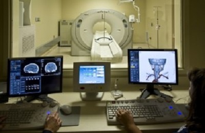
CT scan
This is another method radiodiagnosis. Sometimes, radiopaque agents are used during the procedure.
For cleaning VESSELS, preventing blood clots and getting rid of CHOLESTEROL - our readers use a new natural preparation recommended by Elena Malysheva. The composition of the drug includes blueberry juice, clover flowers, native garlic concentrate, stone oil, and wild garlic juice.
 Research can provide information about:
- able vascular walls(thickness, narrowing, expansion, rupture);
- pathologies of the form of vessels (malformations, aneurysms, hypoplasias);
- the presence of neoplasms in the brain.
The advantage is high information content and the possibility of conducting it in people with pacemakers and metal products in the body.
The disadvantage is the inability to assess the characteristics of blood flow in motion and high radiation exposure.
Magnetic resonance angiography
Currently, this method is the most informative and modern diagnostic study of the brain and its vascular bed, especially if performed with contrast (MR-arteriography, MR-venography).
 It allows you to get the most objective pathoanatomical picture of vascular diseases of the brain and assess the degree of violation of its blood supply. As a result of the study, a three-dimensional image of the scanned organ is formed.
The advantage is high information content, the ability to study functional pathologies in real time and the absence of radiation exposure. The disadvantage is the impossibility of its implementation in persons with pacemakers and non-removable metal products.
Many of our readers for CLEANING VESSELS and lowering the level of CHOLESTEROL in the body actively use the well-known method based on Amaranth seeds and juice, discovered by Elena Malysheva. We strongly recommend that you familiarize yourself with this method.
Radiation (radionuclide) scintigraphy
it radioisotope method obtaining two-dimensional images of the brain. It is carried out using radiopreparations, which include the so-called vector and marker. The vector tends to accumulate selectively in the tissues of the body, and the marker emits gamma rays, which are captured by a special sensor of the device.
Allows you to evaluate the blood supply to various parts of the brain and identify even the slightest violation.

The disadvantage of the method is the high cost of the study and less information content in comparison with CT or MR angiography.
Rheoencephalography
The method is based on the registration of the electrical resistance of the vessel walls during the passage of a weak high-frequency current through them. The study allows you to evaluate the filling of the vessels of the head and neck, the speed of blood flow, the elasticity and reactivity of the vascular walls, the tone of the arteries and veins.
Advantage – ability to assess blood supply different departments brain. The disadvantage is the inability to visualize the pathology.
Electroencephalography
Using this method, the biopotentials of the brain are recorded. The study allows you to detect violations cerebral blood flow and decline nerve conduction brain matter. Among the advantages are non-invasiveness, no radiation exposure.
The disadvantages include low information content, requires additional diagnostic manipulations.
Ophthalmoscopy
This is a study of the state of the vessels of the fundus, conducted through the pupil. Diseases of the vessels of the brain and neck often lead to impaired blood supply to the eyes. These disorders can be detected by conducting a non-invasive study - ophthalmoscopy.
 The advantage is the simplicity, accessibility and painlessness of the procedure, the absence of radiation exposure. The disadvantage is the impossibility of carrying out in patients with photophobia, a decrease in the transparency of the media of the eye (the lens, the vitreous body).
Only qualified specialist can suggest how to check the vessels of the brain as efficiently as possible. The choice of method for diagnosing diseases of the vessels of the head and neck depends on the decision of the doctor prescribing it, the type of suspected pathology, the advantages and disadvantages of the methods, and their availability to the patient.
Do you still think that it is completely impossible to RESTORE blood vessels and ORGANISM!?
Have you ever tried to restore the functioning of the heart, brain or other organs after suffering pathologies and injuries? Judging by the fact that you are reading this article, you know firsthand what is:
- often occur discomfort in the head area (pain, dizziness)?
- You may suddenly feel weak and tired...
- constantly felt high blood pressure…
- about shortness of breath after the slightest physical tension and nothing to say...
Did you know that all these symptoms indicate an INCREASED level of CHOLESTEROL in your body? And all that is needed is to bring cholesterol back to normal. Now answer the question: does it suit you? Can ALL THESE SYMPTOMS be tolerated? And how much time have you already "leaked" on ineffective treatment? After all, sooner or later the SITUATION WILL AGAIN.
That's right - it's time to start ending this problem! Do you agree? That is why we decided to publish an exclusive interview with the head of the Institute of Cardiology of the Ministry of Health of Russia - Akchurin Renat Suleimanovich, in which he revealed the secret of TREATMENT of high cholesterol.
The brain, like any structure of the human body, is subject to serious illnesses. Sad statistics show that mortality from strokes is 12-15% of total mortality, ranking 3rd after heart disease and malignant tumors. According to WHO, for every 100 million people in the world, there are 500,000 strokes and cerebral vascular crises per year. To protect yourself from brain diseases and their severe consequences should be examined in a timely manner and diagnose problems.
Methods for studying the human brain: from ultrasound to MRI
The most common diseases of the brain include: stroke, Alzheimer's and Parkinson's diseases, epilepsy, brain oncology. In Russia, up to 400 thousand cases of stroke are registered annually, and 35% of them end lethal outcome from the disease itself or its consequences. Unfortunately, in this moment there is no tendency to improve the situation. In addition, diseases associated with cerebral vessels (subarachnoid hemorrhage, cerebral hemorrhage, thrombosis, embolism, malignant tumors of the brain, atherosclerosis, hypertension, and others) today affect not only older people, but also very young people. Scientists attribute this to unfavorable ecology, aggressive information environment, unsatisfactory biochemical indicators and personal risk factors: smoking, alcoholism, unbalanced poor-quality nutrition, in general - with in an unhealthy way life.
The risk group includes people over 50 years of age and those who are regularly subjected to psycho-emotional and physical overload - they are shown a study of the brain. It can also be assigned:
- with diabetes mellitus, which causes destructive changes in the work of many organs and provokes the development of atherosclerosis;
- with atherosclerosis, which is dangerous because over time it causes complete or partial blockage of cerebral vessels;
- with inadequate blood supply to the brain. Disturbances in coordination of movements, persistent severe headaches, vomiting, general weakness and bad feeling- symptoms of this ailment;
- upon detection of a neoplasm in the brain;
- in a pre-stroke state;
- with vertebrobasilar insufficiency;
- with craniocerebral injuries and bruises of the head and spine;
- before elective cardiac surgery.
The doctor determines the diagnostic method depending on the purpose of the study, but in any case, the specialist needs to know:
- whether there are blockages or narrowing of the vessels of the brain;
- how the patient's disease affects blood flow;
- whether the tone of the walls of blood vessels is in order;
- whether there are aneurysms, deformities, angiospasm and congenital abnormalities in the structure of cerebral vessels.
It is interesting
prototypes modern diagnostics brains have existed since ancient times. So, in France, during archaeological excavations, a skull was discovered with a competently, even by modern standards, carried out trepanation of the skull. The age of the find was determined at 7000 years. What the primitive surgeon was trying to find out - either to diagnose the brain of his fellow tribesman in this way, or to get rid of unbearable headaches - remained a mystery. Now there is no need to make a hole in the skull unless absolutely necessary. Modern methods of diagnosing the brain and blood vessels open up opportunities for doctors that even a few decades ago no one imagined.
Study of the vessels of the brain
There are a lot of modern diagnostic methods with which you can check the state of the vessels of the brain. Let's consider them in more detail.
Doppler ultrasound (USDG)
The examination is based on a combination of ultrasound with Doppler ultrasound. Due to its information content, safety, and effectiveness, the method has gained recognition in the medical environment. It can be used to determine the speed of blood flow, narrowing in the lumen of blood vessels and atherosclerotic formations, occlusion of blood vessels, the presence of a change in the direction of blood flow, provoked by osteochondrosis or tissue deformation, as well as identify brain aneurysms. The only drawback of ultrasound is its inaccessibility. Only in modernly equipped clinics can you undergo this examination. With all the informativeness of the method, there are practically no contraindications to its implementation. Only serious condition patient and inability to lie down may interfere with the procedure. Ultrasound does not include special training.
Rheoencephalography (REG)
It is similar in principle to the method of electroencephalography (which will be discussed below). According to her testimony, the doctor evaluates blood circulation in the brain, vascular tone and the state of blood filling. When conducting REG, special preparation is not required, the method is harmless, contraindications are not noted.
Magnetic resonance angiography (MRA)
The method is most informative when studying small brain structures. Extremely accurately determines the state of the nerve trunks of blood vessels, the medulla. Much depends on the apparatus used, the power in such a study is high (0.3 T). The doctor directs for this examination for such disorders in the work of the vessels of the neck and brain, such as microstrokes and thrombosis. MRA has the same contraindications as for MRI, which will be discussed below.
If the attending physician needs a comprehensive "picture" of hemodynamics, blood flow rate, functionality and blood vessel filling, he suggests that the patient undergo dopplerography. Transcranial dopplerography uses a digital study, while the depth of passage of ultrasound beams increases to 9 cm. Scanning takes place in “slices”, which gives a complete and detailed visualization of the state of the arteries and veins of the head.
At duplex scanning vessels of the head apply the principle of spectral analysis and Doppler digital coding. The procedure helps to display the color "picture" of the lumen of the vessels, the tone and structure of the walls of the vessels, the branching and deformation of the vessels, the presence of blood clots, atherosclerotic plaques and their sizes.
Dopplerography, like duplex brain scanning, is so harmless that this study can also be performed on small patients.
Diagnosis of diseases of brain structures
Echoencephalography (EchoEg)- Ultrasound examination of the brain. A special apparatus is used - an oscilloscope, which captures the state of the brain with the help of ultrasound and reproduces the result in the form of a diagram. The doctor receives information about the state of the vessels of the head, the performance of all parts of the brain, and brain activity.
Neurosonography (NSG) also called the children's method, because it is applicable to newborns and children early age. It is absolutely harmless. NSG allows you to determine the state of the medulla, soft tissues, brain vessels, the presence of aneurysms, tumors, and various pathologies.
Electroencephalography (EEG) is to register the electrical impulses of the brain. The method is well studied and tested by generations of doctors. Explores brain activity and degree of functionality comprehensive examination brain, circulatory system of the brain and network nerve fibers. Before the procedure, it is necessary to stop taking antispasmodics and anticonvulsants. The method is harmless to everyone age categories. Indications, in addition to diagnosing the state of the vascular system, may be sleep disorders, mental disorders, brain injuries.
craniography- This is an X-ray diagnostic method that will “tell” the attending physician everything about the patient’s skull: its structure, changes in injuries and diseases of the brain. Applicable in the diagnosis of Paget's disease, the detection of myelomas, neoplasms, indirect signs intracranial hypertension. Since craniography is often performed using contrast agents injected into the CSF receptacles (cerebral ventricles), this procedure is poorly tolerated by the patient. Today this method research doctors prefer to replace CT or MRI.
Electroneuromyography (ENMG)- This is a research method that evaluates the patency of impulses along the nerves. Determine the area where the nerve impulses insufficient or non-existent.
Positron emission tomography (PET)- the most modern diagnostic method, which is based on the use of radiopharmaceuticals. Creates a three-dimensional "reconstruction" of the processes occurring in the brain. Diagnoses, unlike all other methods, functional activity brain. Another, everyday, name for PET is “ functional tomography". PET is adequately evaluated by oncologists. Tumors larger than 1 cm and without obvious clinical manifestations can be diagnosed and differentiated into benign and malignant. In most cases, glucose is used as the radiopharmaceutical. It has been observed that neoplasm cells consume glucose more intensively than normal tissues. Glucose is unevenly distributed throughout the body, and this allows the doctor to make the right conclusions. PET, in addition to diagnosing oncology, is also used in the determination of Alzheimer's disease, epilepsy, ischemic disorders, and the consequences of concussions. A categorical contraindication for PET is pregnancy or breastfeeding.
Indications for referral for research computed tomography(CT) covers many conditions, because CT is able to detect almost all pathologies:
- inflammatory processes in the substances of the brain and membrane;
- elevated intracranial pressure;
- brain cysts, neoplasms, anomalies in the development of the organ;
- multiple sclerosis and others.
CT “shows” the state of the necessary brain structures in layers. The tomogram allows the doctor with a very high degree of probability to put final diagnosis and start therapy. The state of white and gray matter brain, pituitary gland, hippocampus, meninges, ventricular system, cranial nerves, vessels - the study will objectively show. The method is safe, radiation exposure is low. CT scans are also allowed for children.
Magnetic resonance method is as informative as a CT scan, and it also takes layer-by-layer images of the brain. The essence of the method is different than with CT. The object is not affected X-rays, and radio waves. The object of study is placed in a created magnetic field. Thus, resonant vibrations are created in molecular nuclei, which are fixed by the program. The result is a series black and white tomograms with high contrast, each of them is a "slice" of the brain. Pictures are taken in different planes, the device allows you to see the brain in three-dimensional format. Thus, the specialist comprehensive information about the structure of the brain.
Indications for MRI:
- uncertainty of the result when conducting other research methods;
- complaints of severe headaches, convulsions and other "general cerebral" symptoms;
- increased intracranial pressure and head trauma;
- neoplasms and inflammatory diseases brain, anomalies in the structure of the brain and blood vessels;
- examination before surgery.
Special preparation for the study is not required. It is possible for children. The method has contraindications. For example, its implementation in the presence of metal prostheses, implants, pacemakers in the human body is impossible.
"Food for brain"
The scientific journal "Psycholoqy Today" published the results of studies on which foods improve brain function. As it turned out, the fish, which is traditionally considered the best "food for the mind", is not the leader of the rating. First on the list is the cranberry. Next come blueberries, beets, cabbage. Spinach is in fourth position. Fatty fish closes the top five. Anchovies or sprats will not help you become smarter. Only salmon, tuna, sardines and other fish containing fatty acid capable of breaking down harmful enzymes. These same foods contain phosphorus, which nourishes the brain.
Which brain imaging method is right for you
Statistics show that the most popular methods for studying the brain are MRI, CT, ultrasound, PET, while CT is more suitable for research. bone structures and MRI for soft tissues. The appointment of a doctor when choosing a research method remains a decisive factor, but the information content and safety of the method and the amount you plan to spend on treatment are equally important.
Where Can I Get a Brain Test?
CT, MRI, ultrasound, PET and other types of diagnostics can be done in many medical institutions countries. The main thing is to correctly prioritize when choosing, health is worth it. Low price should not be at the top of the list of priorities. And the quality of equipment and the qualifications of doctors should be at the top positions.
We recommend that you pay attention to the network of diagnostic laboratories - it has a positive reputation in the market medical services. Laboratories "INVITRO" characterize the highest standards of diagnostics and service, the clinics of this network occupy one of the dominant positions in the domestic market for the provision of quality medical services, including the study of blood vessels and brain structures.
License of the Ministry of Health of the Moscow Region No. LO-50-01-006731 dated June 17, 2015

Modern diagnostic methods make it possible to identify wide range pathologies of the brain early stages.

X-ray of bones and soft structures skull is one of the leading and available methods research.
The quality of the functioning of the vessels of the brain and neck determines the state of the brain itself. nervous tissue constantly in need of oxygen and nutrition. If the state of the vessels does not allow for a full blood supply to the brain, its functions are impaired, and this affects the health and quality of life of a person. Many people wonder how to check the vessels of the brain, but thanks to modern methods diagnosis, it can be done quickly and easily. The decision to send the patient for examination is made by the doctor.
- signs of impaired blood supply to the brain: headaches, decreased ability to work, problems with memory, vision, hearing;
- the presence of risk factors (burdened heredity, hypertension, obesity, atherosclerosis, diabetes addiction to alcohol and smoking);
- preparation for;
- brain tumors;
- increased intracranial pressure;
- migraine;
- hypotension;
- traumatic brain injury;
- heart defects;
- age over 50 years.
Examination of cerebral vessels allows not only to see violations in them, but also to detect the development of ischemia.
Ultrasonic research methods
- Echoencephalography (one-dimensional and two-dimensional) is a non-invasive and simple method of ultrasound examination of all vessels and organs (brain, heart, liver, kidneys, etc.). The procedure does not require special preparation. Thanks to the high-quality equipment on which the study is carried out, echoencephalography allows you to evaluate not only inner part structures of the brain, but also the state of the periosseous space of the skull. Also, using this method, it is possible to track the strength and nature of the median pulsations, and this allows you to determine the amount of intracranial pressure. Echoencephalography is prescribed for suspected brain abscess, intracranial hematomas, concussion, tumors, and circulatory disorders. The procedure is performed in the supine position, while the patient must remain motionless. A gel is applied to the scalp and neck, after which echoencephalograph sensors are attached. The procedure takes up to 20 minutes.
- Doppler ultrasound allows you to assess the speed of blood flow in the vessels of the head and neck, as well as the course and diameter of not only cerebral, but also carotid and vertebral arteries. Such diagnostics also makes it possible to detect the presence of atherosclerotic plaques in the vessels. Before the procedure, you should stop taking vasodilators. For the study, the patient must lie on the couch. The doctor will move the probe of the ultrasound machine over the areas of the head and neck to be examined. During the procedure, you can not move and talk. During the diagnosis, the doctor can press a finger on the neck area for a few seconds. The procedure takes no more than 20 minutes.
- Duplex scanning is a study of blood vessels based on the principles of dopplerography. Modern equipment allows you to build a color scheme of blood flow in the brain, which makes the study more informative. Duplex scanning reveals initial stages development of vascular pathologies such as aneurysms, atherosclerosis, stenosis and occlusions. The principles of diagnostics resemble those that are characteristic of the above ultrasound methods.
- Neurosonography is ultrasound diagnostics vessels, applicable to patients younger than 1 year. The study is carried out through an open fontanel. Neurosonography makes it possible to assess the state of blood flow and the functioning of the cerebrospinal fluid pathways, as well as the presence of congenital pathologies organ. Applicable to infants born with brain hypoxia or birth trauma.
Radiographic methods
When answering the question of how to check the vessels of the brain, it is important to take into account other types diagnostic procedures. Method X-ray diagnostics veins and arteries is called angiography. It allows you to determine areas of vascular stenosis, aneurysms, the presence of internal bleeding and dissemination tumor process. To conduct a study, it is necessary to introduce into the patient's vessel radiopaque agent containing iodine. Angiography is used to study the heart and blood vessels, including those providing cerebral circulation. The most popular types of angiography are:
- Computed angiography: invasive method studies requiring the installation of a catheter and the introduction of a contrast agent into the vessel. Such a check of the brain vessels is carried out in preparation for a neurosurgical operation, as well as in case of suspicion of cysts, tumors, aneurysms and vascular thrombosis. Computed angiography is not performed for heart (liver, kidney) failure, mental disorders, allergies to iodine, problems with blood clotting, as well as during pregnancy and lactation. The procedure is performed on an empty stomach. Before puncturing the vessel into which the contrast will be injected, the patient is given local anesthetic. After that, the installation of the catheter and the introduction of radiopaque begins. This process is followed by angiography. Then on the device are made x-rays. The procedure takes no more than 40 minutes. After the catheter is removed, a tight bandage is applied to the puncture site. On the first day after angiography, a person should arrange for himself plentiful drink in order to remove the contrast agent from the body as soon as possible.
- Magnetic resonance angiography: checking blood vessels is carried out when vegetative dystonia, vasculitis, heart defects, stenosis, aneurysms, strokes, increased intracranial pressure and atherosclerosis of the vessels of the brain and neck. Magnetic resonance angiography allows you to build a three-dimensional layered image of the network of cerebral vessels and examine its condition. The study is carried out with or without radiocontrast. contrast agent administered when a patient is suspected of having a brain tumor. There are such types of magnetic resonance angiography:
- sinusography: the study of the veins of the brain and their collectors, which is performed with suspected thrombosis;
- study main arteries brain;
- Diffusion-weighted magnetic resonance imaging is performed for suspected ischemic disease brain.
Magnetic resonance imaging does not require special preparation. During the procedure, the person must remain still and listen to the doctor's instructions. The study is not performed on patients who have metal implants, a pacemaker or artificial joints. Weight more than 150 kilograms is also included in the list of contraindications.
Other Methods
High-frequency studies applicable for the diagnosis of cerebral vessels include rheoencephalography. The method allows you to assess the blood circulation in the body. Thanks to rheoencephalography, it is possible to determine pulse fluctuations, blood circulation speed and vascular patency, as well as the elasticity of their walls.
An extract with the results of the study is not information about a person's diagnosis. The data obtained during the diagnosis must be deciphered by the doctor. If necessary, the patient can be referred to diagnose the state of the vessels not only of the brain, but of the whole body. The diagnosis and further prescription of therapy is carried out exclusively by the doctor.
The treatment of any disease is most effectively started at an earlier stage, and this requires timely identification of the problem. With damage to the vessels supplying the brain, some clinical symptoms may occur - pain, dizziness, decreased performance, and memory impairment. But all these symptoms can be signs of diseases that are completely different in nature. Therefore, the diagnosis of cerebral vessels is required, which gives an objective picture. There are several effective research methods.
Diagnosis of cerebral vessels
There are quite a few indications for this kind of examination: suspicion of acute or chronic insufficiency blood circulation, migraine and tumors, increased intracranial pressure, etc. In any case, the decision on the appropriateness of the procedure is made by the doctor.
Ultrasound of cerebral vessels
Doppler ultrasound - USDG of vessels brain. This procedure is also called duplex scanning brachiocephalic vessels. This survey is carried out to assess blood flow in large arteries and veins, not only of the head, but also of the neck. Of the major trunk lines, the state of vertebrates, carotid and subclavian arteries, and from large vessels brain - posterior, middle and anterior arteries. Also, the specialist gives a conclusion about the condition venous outflow from the skull.

This ultrasonic method The study allows real-time assessment of the condition of the walls and indicators of the blood flow of arteries and veins. In this case, the assessment is issued in graphical, quantitative, as well as audio form. The examination allows to draw a conclusion about the quality of blood flow in cerebral arteries, its uniformity. If there are any narrowings, blockages, the presence of blood clots or plaques in them, then the specialist will see changes in blood flow.
During this study, the anatomical structure arteries - their tortuosity, the presence of aneurysms and deformities. If there is vasospasm, then the specialist will make a conclusion about its severity, as well as what exactly is squeezing the artery - a vertebra or spasmodic muscles.
As for the venous outflow from the cranial cavity, with the help of Doppler sonography it is possible to assess the patency and viability of the venous valves.
The uniqueness of the ultrasound scan method lies in the ability to detect disorders and pathologies even before the first symptoms appear.
The study using Doppler ultrasound has no contraindications and is usually performed on an outpatient basis. A day before the appointed date, the patient is advised to stop drinking alcohol, drinks with high content caffeine and smoking.
MRA of cerebral vessels
Magnetic resonance angiography - MRA. This study involves the use of the principle of spectral analysis and at the same time Doppler digital coding. Scan at this study passes through the sections, resulting in a color image of the lumen of the vessels, their branching, the presence of deformations, atherosclerotic deposits, blood clots.
MRA technology allows you to create a three-dimensional reconstruction of the network of veins and arteries, as well as obtain thin sections, which increases the efficiency of diagnostics.
This method (along with Doppler ultrasound) is considered the most informative.
Magnetic resonance angiography has a number of contraindications: pregnancy, kidney failure, the presence of metal implants in the body, mental disorders.
REG of cerebral vessels
Rheoencephalography -. This research method is similar to encephalography. But in this case, the state of blood circulation in the study area, the degree of filling of the vessels, their tone are assessed. Among the parameters to be determined, one can note the speed of blood flow, blood viscosity, the speed of propagation of the impulse wave, the degree of severity of the reaction from the side of the vessels.

Many modern experts consider this method already outdated, but in some cases it is advisable to use it to clarify the diagnosis. It is traditionally used in the study of hemodynamics (circulation) of the fetus during childbirth.
Other Methods
Common survey methods are:
- echoencephalography (EchoEg), which is carried out using an oscilloscope that captures the reflected ultrasonic signals;
- magnetic resonance imaging (MRI) - effective method, allowing to identify, among other things, manifestations multiple sclerosis and inflammatory demyelination;
- electroneuromyography (ENMG), the principle of which is the registration of muscle biocurrents);
- computed tomography (CT), performed using a special scanning device;
- neurosonography (NSG) is a method by which small children are examined, with an open fontanel.
The choice of the method of examination is carried out only by the attending physician.
