What examinations of cerebral vessels. Dopplerographic (uzdg) examination of the vessels of the brain and neck
Ultrasound is by far one of the safest and most promising directions diagnostics of internal and surgical diseases. Its diagnostic importance as a method lies not only in its penetrating ability, but also in the ability to view tissues in cross section, similar to tomographic methods. A relatively new type of ultrasound is Doppler scanning of blood vessels, which greatly expands the possibilities of modern non-invasive diagnostics.
Doppler study is to determine the frequency of the returning sound. Waves of a certain frequency are sent from the piezocrystal to the projection area of the cerebral vessels, the blood in which reflects this sound.
In addition, the reflected sound, due to the presence of a certain speed in the blood, is reflected from it with a changed frequency, which is fixed. This change makes it possible to judge the velocity of blood flow in a given section of the vessel. So, passing along the length of the vessel, you can find a narrowing or vice versa expansion.
In addition, special load tests are used to determine the performance of anastomoses, the importance of which lies in compensating for the blood flow of a certain area when the lumen of the artery that brings blood here, or the vein that drains blood is blocked. Such functional tests have no analogues. Doppler study is very revealing and greatly facilitates the diagnosis of diseases, including topical.
How is a Doppler vascular study performed?
What is being examined?
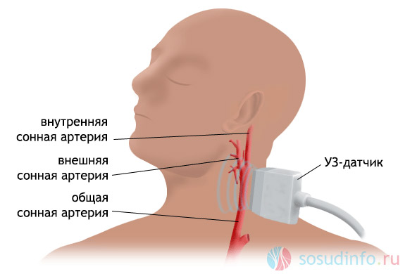
During dopplerography, a specialist uses a sensor to examine the vessels of the brain or neck over their projection. Carotid and vertebral arteries are examined in the neck area, jugular veins(internal external and anterior) and venous vertebral plexus, subclavian arteries and veins.
Of the vessels of the brain, the external and internal carotid arteries, the basilar, posterior, middle and anterior cerebral arteries, the cerebral arteries, the communicating arteries (Velisius arterial circle), veins.
The vessels of the brain take part in the formation of the so-called Velizian circle, which is an anastomosis connecting the blood supply of the right and left halves big brain. Smaller arteries can also be used to study blood flow in the brain: supratrochlear, ophthalmic.
The procedure itself
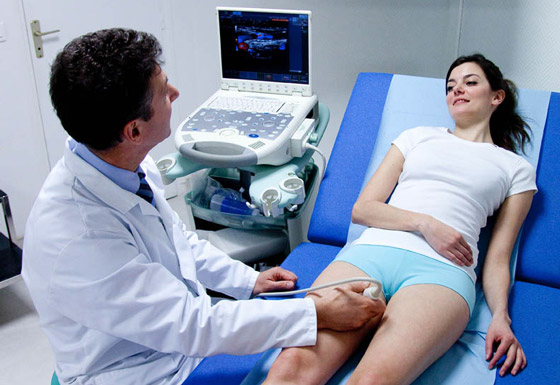
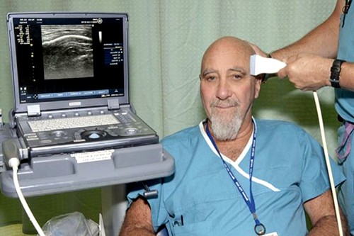
Often, a neurologist directs such an examination. During the examination, the patient sits with his head tilted to the side, or lies with a cushion under his neck.
Special load tests are used to investigate possible functional disorders in hemodynamics. The patient is asked to turn his head to the side, cough, hold his breath, squeeze one of the common carotid arteries.
When coughing, blood pressure rises chest cavity, therefore, blood from the veins of the neck and head does not pass to the right atrium, which can give neurological symptoms, such as headaches. With the help of a Doppler examination, the degree of slowing of blood flow is assessed. The same happens during breath holding.
When clamped carotid artery on one side, the region of the velius circle (the base of the brain) is examined with a sensor. The direction of blood flow in the anterior communicating artery(anastomosis between the anterior cerebral arteries different parties large brain), which indicates the viability of the roundabout blood circulation. The procedures are carried out for a rather short time so as not to disturb the blood circulation of the brain. Also, a Doppler study of the vessels of the head and neck can be performed after taking nitroglycerin, which dilates the vessels.
For transcranial examination of vessels, points are used where bone presented in smaller quantities. Temple area, suboccipital (access to cerebral arteries and veins) and ophthalmic (the study is carried out through the supratrochlear and ophthalmic arteries to the internal carotid artery).
Why is this study being done?
Doppler study of cerebral vessels is one of the best ways to monitor the state of blood supply to a given area. This non-invasive and completely painless method allows you to solve two problems at once, to understand the cause of the headache that the patient complains about and at the same time save the attending physician from the headache.
Ultrasound of cerebral vessels - duplex scanning - allows not only to examine the vessel (its structure, wall, surface, lumen, the presence of deposits on the intima), but also using the scale to trace the dynamics of blood supply and blood flow through these vessels. This not only allows you to detect a pathology, but also to make a prediction about a future disease, to assess how big the risk is. ischemic stroke with this blood flow.
Results display duplex scanning
The first thing the doctor pays attention to when prescribing this method of examination is the patient's complaints:
- headache
- dizziness
- ringing or noise in the ears,
- violation of sensory and motor innervation, respectively, of the skin and muscles of the head
- complaints about "flickering flies"
- visual impairment, including partial and transient
Doppler scanning allows you to see the anatomy of the vessels of the head and neck, the state of their lumen, wall thickness, visualize organic disorders, to assess the circulatory disorders created by this pathology, allows you to identify the disease itself (stenosis, occlusion of the lumen, atherosclerotic plaques, compression, and others), without using methods with penetration into human body directly.
Goals of duplex examination of cerebral vessels
- early diagnosis of vascular diseases
- detection of narrowing or compression of cerebral vessels
- detection of defects in the development of blood vessels, which are congenital and may not manifest themselves in any way before the examination
- functional assessment of the adaptation of the brain to a given type of vascular development or the present state
- detection of obstructions for the outflow of blood from the intracranial cavity through the veins
- detection of local changes in the arteries of the brain and neck associated with any systemic disease(atherosclerosis, etc.)
- dynamic assessment of the state of blood vessels and the brain against the background of ongoing treatment or in connection with the disease
Indications for Doppler vascular studies
Indications for the procedure for examining the vessels of the brain and neck are various violations activities of the central nervous system, possible cause which is vascular pathology, namely:
- relevant complaints (pain in the head, visual impairment, hearing impairment, persistent memory problems, and others)
- diabetes
- hypercholesterolemia (including the shift of the lipoprotein index towards low density lipoproteins)
- some transferred acute diseases (ischemic disease heart (myocardial infarction, postinfarction cardiosclerosis, arrhythmias), stroke)
- endocarditis
- thrombophilia
- stenosis or compression of the vessels of the neck
- vegetovascular dystonia
Ultrasound signs of major pathologies
Why are these indications for this procedure singled out? 
Atherosclerosis
Conditions such as arterial hypertension, hypercholesterolemia are the risk of developing atherosclerosis, including the vessels that feed the brain. atherosclerotic plaques, firstly, they themselves block the lumen of the artery, and secondly, they can rupture and serve as a site for the formation of blood clots.
The first will be the reason chronic ischemia the brain and as a result - memory impairment, mental activity, etc.
The second one is dangerous. acute condition, such as thromboembolism or simply complete blockage of the cerebral vessel in place, which will cause the development of a cerebral infarction - a stroke. 
The state after a stroke is an indication for monitoring the condition of the vessel wall that caused the disease.
Thrombosis and thromboembolism
The condition after myocardial infarction (post-infarction cardiosclerosis, heart aneurysm), cardiomyopathy, endocarditis, arrhythmias can serve to form blood clots. Blood clots in the heart are the cause of thromboembolism for any organ. A good blood supply to the brain can lead to the blockage of its vessels. Therefore, vascular Doppler in people who have had such diseases is quite important. 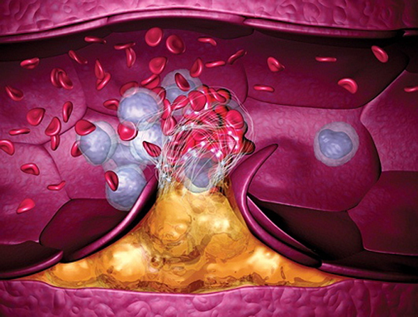
Thrombophilia is a condition of increased blood clotting. In such cases, again, many small blood clots appear in the vascular bed, which can serve as emboli and block the vessels of the brain. DIC syndrome(disseminated intravascular coagulation), is also manifested by increased thrombus formation, which occurs when severe course infectious processes, complications of pregnancy and others. With the appearance or increase of neurological symptoms, people suffering from such a pathology are recommended to undergo a Doppler study of the vessels.
Violation of tissue trophism
Diabetes mellitus is very dangerous because its main target is small vessels, including the brain. Sugar (glucose) begins to accumulate in the wall of the vessel and it thickens. As a result, the transfer of oxygen and nutrients from the blood to the tissues is disrupted, and nervous tissue very sensitive to starvation, more than others.
Compression of blood vessels
Compression of the vessels can be the cause of neurological symptoms, which the patient will complain about. Compression of the arteries can lead to cerebral ischemia, dizziness, headaches, loss of consciousness. Violation of the outflow of blood from the brain through the vessels of the neck will lead to headaches due to increased pressure on the dura mater. 
Conclusion
Doppler of the vessels of the brain and neck is now widely introduced in medical practice and is used in many indications. It allows differentiating pathological conditions, including acute ones, in a non-invasive way.
It is very important that the Doppler examination makes it possible to assess the state of blood supply in dynamics and observe functional deviations in the work of the cardiovascular system, which is so important for human life.
Ultrasound diagnostics (ultrasound) of blood vessels: how and when they do it, advantages, particular situations
Vascular diseases have become a real plague of our time. Scientists believe that the vessels have changed little in the course of evolution, but the way of life of people has changed dramatically: physical inactivity, eating refined foods, polluted environment, information overload. All this negatively affects the state of human organs, including blood vessels. However, medicine is also not dormant - it is developing. Her arsenal contains a rather impressive collection of methods for treating vascular pathologies. The most effective and safe are Doppler studies.
Today, to study the state of blood vessels and blood flow, it is most often used doppler ultrasound (USDG) And duplex scanning of vessels (DS).
What are the advantages of these methods?
- They are highly informative.
- Allows you to identify the earliest preclinical signs vascular diseases, evaluate vascular lesions and blood flow disorders.
- They are non-invasive and absolutely safe.
Doppler techniques are so named because they are based on the same Doppler effect (by the name of an Austrian physicist). The essence of the phenomenon is the reflection of an ultrasonic beam from moving objects, for example, blood cells. This changes the frequency of the signal. These changes help determine the speed of blood flow, the direction of blood flow, atherosclerotic stenosis, volume minute blood flow, occlusion of the vessel, evaluate collateral circulation, the presence of pulsation. All these indicators are determined using ultrasound.
Doppler ultrasound as an isolated study is used less and less today. DS is mainly used - duplex scanning, which combines ultrasound and traditional ultrasound. Conventional ultrasound (B-mode) informs the clinician about vascular anatomy through 2D images in black and white. The use of this mode supplements ultrasound dopplerography with a graphic image or areas, an assessment of the characteristics of changes in vascular patency, a study of the size and density of a thrombus, and a study of the state and diameter of the vessel wall. If there are fistulas, a study of their size is carried out. Possibly more precise definition localization of the damaged vessel.
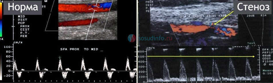
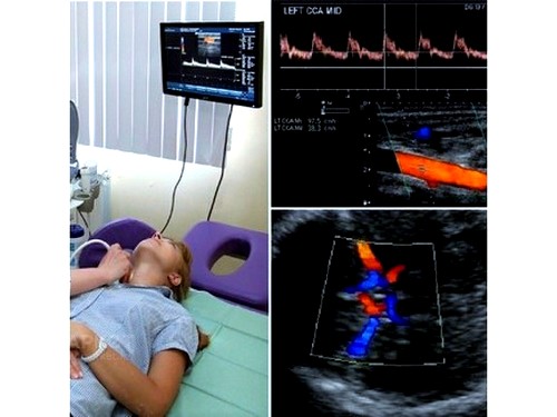
A healthy (left) and stenotic (right) artery in a duplex ultrasound image.
In the first case, the blood flow has a normal course, speed (about 60 cm / s) and color
In the second - the blood flow is disturbed, due to the narrowing of the artery, its speed in the affected area reaches 507 cm / s
Other variants of Dopplerography do not fundamentally differ from those described above. Eg, color mapping translates blood flow information into more comfortable shape: deoxygenated blood on the monitor is colored in Blue colour, and arterial - in red.
Triplex scanning is a technological mode of DS. It is not an independent ultrasound technique. During this study, the ultrasound machine operates in three modes: B-mode, ultrasound and color Doppler mode.
Important! The determining factor in choosing a research option should not be the price, but the doctor's recommendation. Depending on the localization of the vessels and the nature of the lesion, one or another research method is prescribed.
Video: the doctor explains the principles of conducting ultrasound diagnostics of blood vessels
Doppler studies of the vessels of the lower extremities
For vascular research lower extremities use two options: actually USDG of vessels lower limbs and duplex scanning. These procedures reveal such ailments as arterial thrombosis, and others.
The main objectives of the study of the vessels of the legs:
- Diagnostics ;
- Identification of an aneurysm;
- Assessment of the state of peripheral vessels;
- Diagnosis of pulsating formations;
- Monitoring dynamics;
- Definition ;
- Diagnosis;
- Preoperative marking of veins.
We can safely say that ultrasound techniques are the most advanced methods for examining the vessels of the legs. For example, duplex scanning of the veins of the lower extremities can be used to examine the vessels in detail, determine the degree of change in them, and correct diagnosis. It is important that these methods reveal vascular disorders on the very early stage which is not always possible in other ways.
When to contact the doctor?
Signs of pathology arteries:
- Feeling is often noted;
- There are complaints of weakness in the limbs;
- Occur for no apparent reason;
- There may be pain when running, walking;
- The legs become cold to the touch and pale, as there is a decrease in temperature in them;
- A person limps, although there are no inflammatory processes in the joints.
For problems with veins indicate:
- , intensifying in the evening;
- Brownish skin of the legs;
- Emergence;
- Long-term non-healing ulcers may appear;
- They become noticeable.
It should be noted that numbness, weakness, cramps and other signs may appear in completely healthy people. Therefore, you do not need to immediately diagnose yourself and, even worse, prescribe treatment. Consult a doctor, tell about your suspicions, if necessary, you will be prescribed an examination and only then treatment.
Benefits of ultrasound examination of the vessels of the legs:
- Absolute security;
- Painlessness;
- informative;
- Non-invasive (no skin damage);
- The speed of manipulation (the patient receives the result immediately);
- Can be done at any age
- Low price.
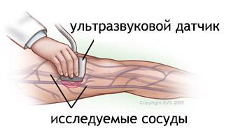
Ultrasound of the vessels of the legs is performed on an outpatient basis. After applying a special gel, the doctor examines the vessels using a sensor. Pathological changes are clearly visible on the device monitor. With color mapping, it is easy to distinguish veins from arteries. The whole manipulation lasts less than an hour. There is also no need to prepare for the examination.
Important! The conclusion of Dopplerography of the vessels of the lower extremities is not yet a diagnosis. The degree of pathology is determined only by the attending physician.
Video: doctor about ultrasound of the veins of the legs
Ultrasound examination of blood vessels during pregnancy
This is the most safe way receive information about the child's condition. Dopplerography of the fetus helps to trace the dynamics prenatal development. Already at 21 weeks pregnant future mom and her attending physician receive extensive information about the blood flow in the uterus, placenta and fetus, about the pressure in the vessels, their elasticity. Thanks to this study, you can find out about the lack of nutrition of the fetus, about possible pathology in development.
Degrees of change in fetal blood flow:
- Change in the blood flow of the uterus and placenta.
- Violation in the blood flow of the fetus and placenta.
- Non-critical change in the blood flow of the fetus, uterus and placenta.
- A critical change in the blood flow of the fetus, uterus and placenta.
If mild degrees of changes in blood flow are detected, the pregnant woman should be examined weekly. If the condition worsens, it is necessary to be examined every day. With this pathology, childbirth at term in a natural way is possible.
If the second degree of pathology is diagnosed, then it is recommended to be examined every other day. If necessary, after 32 weeks, a caesarean section is performed.
If the third degree of pathology is detected, then control should be carried out daily.
Doppler examination of pregnant women is carried out unscheduled, in the case of:
- Multiple pregnancy;
- fluctuations in blood pressure;
- Abnormal sized fetus
- Delays in the development of the child;
- Violations in the state of the placenta;
- Maternal diabetes.
Video: Ultrasound during pregnancy
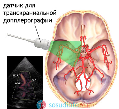
Ultrasound of the vessels of the brain
This examination is also known as transcranial dopplerography
. In essence, this is a duplex scan of the vessels of the brain. The term "transcranial" means that the study is carried out inside the skull. Usually carried out in combination with duplex scanning main vessels neck and head (BCA).
The worst thing that can happen to the vessels of the brain is their blockage. As a result, necrosis occurs, and as a result - necrosis. Such a state is called. Some people recover safely after a stroke, others - alas. It all depends on the location of the damaged vessel and the degree of brain damage.
Happen, of course, acute disorders blood flow. But more often the first "bells" appear long before the sad event. They are manifested by certain symptoms (which are discussed below), but there are also asymptomatic. It is important to notice these "calls". Indeed, many people do not consider insomnia or memory impairment as a symptom. Very rare, but there are congenital. However, more often they make themselves felt at a young age.
To assess the state cerebral blood flow, it is enough to do the procedure of transcranial dopplerography.
Transcranial dopplerography informs the doctor and the patient about:
- The state of the inner choroid;
- damage vascular wall;
- Elasticity of blood vessels;
- The presence of formations inside the vessels;
- Changes in the anatomy of the vessels.
What can be revealed?
- (inflammation of the vascular walls and their change);
- Anomalies in the location or course of blood vessels;
- Traumatic lesions;
- Angiopathy (toxic, diabetic, hypertensive);
By performing a Doppler study of the vessels of the head, you can understand:
- Degree pathological process resulting in damage arterial vessels;
- Causes of strokes,;
- The degree of change in the lumen of blood vessels due to diabetes, atherosclerosis, smoking.
The results of ultrasound examination of the vessels of the head and neck allow the doctor to assess the state of intracranial and peripheral vessels, prescribe proper treatment and make an individual forecast.
Indications for Doppler examination of the vessels of the head
Having read scary stories about the consequences of violation cerebral blood supply, many come to the conclusion that it is necessary to be examined urgently. There is a category of people who explore "along and across" their body, but, as a rule, they do not find anything. But nerves and money are wasted. Does it need to be done?
Some may object that the study of the vessels of the brain should be done by everyone without exception, even healthy ones. Meet the same congenital anomalies vascular development. What if it suddenly arises, which will also suddenly burst and kill its owner? So, go ahead and check it out!
You can console the alarmists: congenital vascular anomalies are very rare. And another thing: a person can have a mass asymptomatic diseases. Does this mean that you should do all kinds of ultrasounds, and the like? Trust the opinion of a specialist, describe your complaints to him, and let him decide whether to examine your brain or not.
Doppler examination of the vessels of the head may be prescribed in the case of:
- dizziness;
- The presence of signs;
- Heaviness in the head;
- fainting;
- Noise in the ears;
- Visual impairment;
- Movement coordination disorders;
- Weakness in the legs or arms;
- Speech disorders;
- "Goosebumps" in the hands.
Sometimes a person undergoes other types of examinations (MRI, scintigraphy, CT), as a result of which violations are detected. To clarify the diagnosis, the doctor prescribes dopplerography.
Planned dopplerography
- Elderly people whose relatives suffered from vascular diseases;
- Patients with diabetes;
- Smokers with experience;
- People with rhythm disturbance;
- Having metabolic syndrome;
- Patients who have had a stroke;
- People with ;
- sick after surgical intervention in the brain or spinal cord or awaiting surgery.
Preparation for the examination
To conduct an examination of the head of some special training no need. The only thing: on the day of the study, it is not recommended to eat foods that affect vascular tone:
- Coffee;
- Energetic drinks.
It is advisable to refrain from smoking.
Concerning medicines that can affect the results of the study, it is not recommended to cancel them without the permission of a neurologist.
Research details
Transcranial dopplerography is carried out according to the principle common with other Doppler techniques. Ultrasonic rays penetrate the brain vessels through the skull.
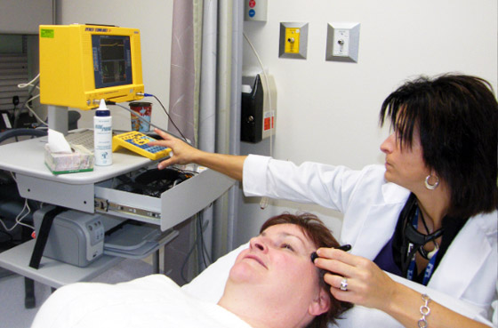
The doctor alternately brings the sensor to various areas heads:
- supraorbital;
- Temporal;
- Neck area;
- Place of transition spinal column into the occipital bone.
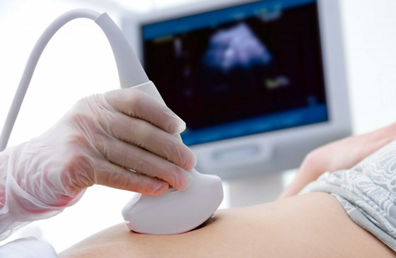
Beforehand, a special gel is applied to the examined areas, which ensures better contact of the sensor with the skin.
In addition to examining the vessels, the doctor makes functional tests (for example, a request to hold the breath) to clarify or refute the diagnosis of autonomic regulation disorders.
What does the examination say about pathologies?
- ABOUT vasculitis judged by changes in the echogenicity of the vessel, vascular wall, differentiation of the layers of the wall.
- Non-stenosing atherosclerosis can be suspected if the thickness of the vessel wall is increased, but the artery is narrowed by no more than 20%. Echogenicity at the same time changed according to an uneven type.
- Hypoechoic formations with a thin rim - this is how they are diagnosed plaques in the arteries.
- Mandatory treatment required artery narrowing by 50% or more.
Video: doctor about ultrasound of the vessels of the head
Conducting a study of cerebral vessels in children
A small child is more often comprehensive study brain called neurosonography . To be more precise, this term combines different techniques study of the state of the central nervous system: examination of the spinal column, head and spinal cord, vessels, tissues of the head. But it is customary to call neurosonography ultrasonography brain.
Using ultrasound techniques, the doctor can identify tissue pathologies, diagnose intracranial elevation pressure and other disturbances.
Today, neurosonography is performed in three different ways:
- The simplest one is transfontanellar. The name speaks for itself: the sensor is brought to the fontanel of the child and the brain is examined. The disadvantage is that the study of the brain is done in one projection.
- The second is transcranial neurosonography. It requires more serious equipment. The brain of adults is also studied in this way.
- The third method combines the other two. Transcranial-transfontanellar neurosonography is performed for young patients. Advantages - the brain is considered in different projections. Requires more time to conduct and serious equipment.
The main indications for the study:
- prematurity;
- Suspicion of pathology of the central nervous system;
- Inflammatory diseases of the CNS.
Special preparation for the examination is not needed. The procedure is absolutely safe and painless.
Examination of the brachiocephalic arteries (main vessels of the neck)
The brain is nourished not only by intracranial, but also peripheral vessels. Important role in process belongs brachiocephalic arteries (brachiocephalic trunk, carotid artery, subclavian and vertebral arteries). Atherosclerotic changes in them can become a serious problem. When plaques appear in the arterial vessels that feed the skin, bone, muscle tissues, there will be no big trouble. The brain is a completely different matter. Responsible for its blood supply are several large vessels- arteries. A blockage in any of these can eventually lead to a stroke.
most frequent complaint people with BCA pathology is dizziness. It mainly appears when sharp turn heads. Sometimes - as a result of a decrease in pressure. Of course, one-time dizziness is not a problem. It can occur with overwork, change atmospheric pressure or for other reasons, but relapses of this condition cannot be discounted.
A BCA study is carried out in the case of:
- Frequent headaches for unknown reasons;
- When manifested clinical signs stroke or;
- The presence of a disease in the patient that provokes violations cerebral circulation(vasculitis, diabetes, arterial hypertension and others);
- Clinical signs of BCA;
- The presence of pathology of the surrounding tissues (if there is a possibility of compression of the BCA).
- Upcoming operations on the heart and blood vessels.
Diagnostics
Usually, to make a diagnosis, BCA is examined using, as a result of which blood flow characteristics are determined.
To clarify the diagnosis, duplex scanning of the brachiocephalic arteries is used. The doctor can visually assess all the shortcomings in the anatomy of the arterial vessels. This undoubtedly increases the diagnostic possibilities. Using this technique, the doctor determines deformities, blood clots, plaques and other pathological changes.
Video: doctor about ultrasound of BCA
Ultrasound examination of the vessels of the kidneys
Ultrasound of the kidney vessels is performed to obtain Additional information about renal blood flow. This study is more informative than conventional ultrasound. The interstitial blood flow and vessels of the kidney pedicle are studied.
When is a Doppler examination of the kidneys prescribed?
- In man for a long time diagnosed with hypertension of unknown origin.
- Conventional ultrasound revealed a difference in the size of both kidneys.
- There are signs of renal failure.
- Traumatic injury to the kidney.
- Thrombosis is suspected.
- A violation of the movement of blood in the kidneys is predicted. (More often this situation is observed with anomalies of blood vessels, tumors, atherosclerosis, thrombophlebitis, nephroptosis).
Is preparation necessary?
To doppler ultrasound of the kidneys, you need to properly prepare. Moreover, the success of the study is directly related to the level of training. Excess intestinal gases may invalidate the test result. That's why preparation should begin a few days before the procedure. From your diet, you must exclude fruits, legumes, sauerkraut, carbonated drinks, sweets, fruit juices, Rye bread. It is useful to take enterosorbents.

It is advisable to conduct an examination on an empty stomach in morning time. If the ultrasound is performed in the afternoon, you can have a little snack in the morning. The main thing is that at least 6 hours pass from the meal to the procedure.
Important! It is impossible to carry out Dopplerography of the vessels of the kidneys after such diagnostic examinations like fibrogastroscopy and colonoscopy. This is explained big amount air entering the intestines as a result of these procedures.
There is no doubt that timely diagnosis vascular disease is essential. Thanks to her, it is possible to help many people live not only long, but (importantly) active life. Therefore, the advantages of dopplerography and duplex scanning as the main methods are indisputable. early diagnosis. Taking care of your body is the key to staying healthy.
Video: a neurologist performs dopplerography (using the example of brain vessels)
Normal operation circulatory system is an integral part in the work of all human organs and systems, each of its cells.
Blood acts as a link, saturating cells and organs nutrients and oxygen, which is vital for human health.
The slightest disruption in the work of the circulatory system provokes the development of general malaise, deterioration of well-being. When a person ignores similar phenomena, neglecting such diagnostics as dopplerography of cerebral vessels, in the future there may be serious pathologies vessels and brain.
Dopplerographic examination of the vessels of the brain and neck will help determine their structure, condition, evaluate work and determine possible deviations in functioning. Thus, the doctor can exact information and determine the tactics of subsequent treatment.
Dopplerography (USDG) of the vessels of the neck and head: what is it?
If you do not go into details, then the study of ultrasound implies ultrasound diagnostics with simultaneous use of dopplerography. This combination helps to identify pathological changes in the vessels and is essential in making a diagnosis. This method is considered the most informative for the study of blood vessels in the human body. Color coding is also used, which can clearly show the blood flow and the area with a defect.
The method allows not only to detect the problem, but also to identify the cause that provoked the development of the pathology. Ultrasound is recommended for early dates disease, which will significantly reduce the risk of complications, prevent its acute course.
There are several diagnostic modes that can be used together (for a more informative study), as well as separately. Note that duplex scanning of blood vessels differs from the ultrasound method.
Ultrasound examination makes it possible to study the structure of the vessel itself, as well as closely located tissues. And the use of duplex scanning allows you to determine the condition of the arteries and veins of the blood circulation.
Dopplerography of the vessels of the head and neck is a non-invasive diagnostic method based on reflection sound waves from moving objects (Doppler effect), after which the computer processes the information and recreates a two-dimensional image blood vessels, where the main problems in the bloodstream will be visible.
Ultrasound is modern, effective and exact method research. Due to the absence of radiation exposure this method diagnostics has a minimum of contraindications. The advantage of the method is considered to be its non-invasiveness, which eliminates the need to violate skin for research.
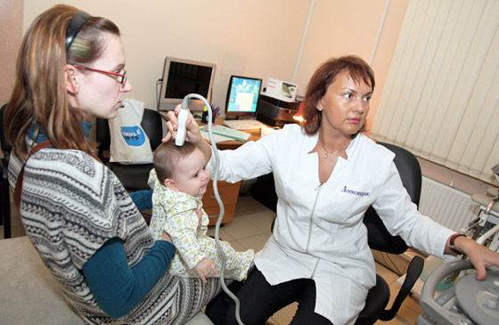
Since dopplerography is the safest, most informative and painless method for diagnosing blood vessels, it is recommended even for infants.
Vascular ultrasound: what does it show?
As already mentioned, the main task of this diagnostic method- is to evaluate the work of blood vessels and identify pathological changes in them. This study allows you to assess the state of:
- veins, vertebral arteries, subclavian;
- paired large arteries of the neck (carotid);
- main artery;
- jugular veins.
The high information content of the method allows the specialist:
- assess hemodynamics (blood movement in the vessels);
- detect aneurysms, protrusion (expansion) of the vessel wall by more than 2 times;
- to establish the cause of headaches, dizziness, ICP and spasms;
- analyze the integrity of the vessels, their thickness, density (echogenicity);
- obtain information regarding vasoconstriction, the extent of the pathology;
- identify violations of the vascular geometry of the arteries;
- makes it possible to identify vascular disorders on early stage development, as well as their defeat in the course of trauma or birth defects;
- additionally, it is possible to study the tissues that surround the vessels.

Ultrasound scanning using duplex scanning has greatly simplified the work of many specialists, since now accurate diagnosis does not require a long time. It should be noted that earlier it could take up to several months to diagnose a particular disease associated with the vessels of the head or neck, which delayed the process of therapy and recovery.
Today, the ultrasound method, if necessary, for more deep research can use additional effects such as light flashes, various sounds, etc.
Ultrasound: how is it performed?
There are a number of symptoms that require this study, in particular, it is tinnitus, causeless headaches, dizziness, disruption speech apparatus also can be used for cervical osteochondrosis. Diagnosis does not require special preparatory measures from the patient.
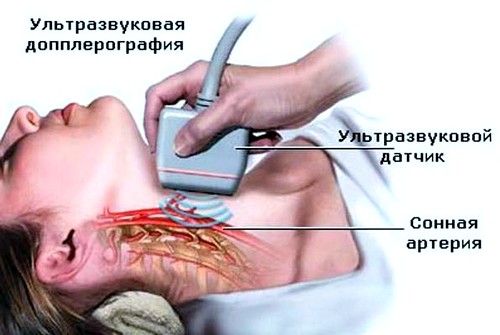
The patient needs to free the head and neck area from excess clothing, remove jewelry and lie down on the couch. For closer contact, a special gel is applied to the neck area, used in ultrasound practice. It should be taken into account that this study may take more than half an hour.
To assess blood flow through the intracranial vessels, the sensor is applied to the area temporal bone. Such a scan can be found under the name transcranial study.
The duration of such a diagnosis is due to the need to study each site separately, study the parameters and compare with the norms of the ultrasound study.

As already mentioned above, this diagnosis does not require specific methods of preparation. However, there are guidelines that should be followed for the most accurate result. So, in a few hours, the patient needs to exclude the use of food, tea, coffee and cigarettes so that there is no effect on the tone of the arteries.
Dopplerography is allowed to study the vessels of the neck, head and brain in infants. Most often, this procedure is preventive measures. The examination of newborns is carried out at the time of their sleep, so it is possible to get the most accurate results. An hour before the start of the diagnosis, the baby should be fed.
Previously, such a diagnosis of children without suspected disease was not carried out. However, experts now recommend that all parents, without exception, sign up for an ultrasound scan to prevent and prevent pathologies of cerebral vessels.
Lesions diagnosed by ultrasound
In the course of the study, the study of blood vessels, their structure and work, it becomes possible to find out pathologies, as well as determine the scheme further treatment. All possible violations that diagnostics can determine is divided into 2 groups:
- pathologies with a certain ultrasound symptom(aneurysm, thrombosis);
- pathologies without characteristic ultrasound accompaniment (vasculitis, angiopathy).
Ultrasound procedure: where can I go?
On this moment the ultrasound procedure is quite common, therefore, it is not uncommon to find it in local clinics. A referral for such a diagnosis can be obtained from a general practitioner after contacting and describing the problem.
With the help of a referral from the attending physician, dopplerography can be done at the place of residence free of charge. However, there are a number of certain disadvantages: the patient cannot choose the most convenient time for yourself, you will additionally have to wait in line, which in some cases may take more than 1 hour. Moreover, there is no individual approach, which often leads to discomfort from visiting the ultrasound room. It should also be noted that the equipment public clinics often not a modern sample, which means that the effectiveness of diagnostics is reduced.
For these reasons, research is best done in private centers. Clinics conduct a paid examination, but the patient receives highly accurate results, as it is used latest equipment, individual approach and save time as much as possible. Price policy conducting ultrasound in private clinics from 2000 rubles.
