The structure of female intimate organs diagram. Cheers! Health problems are solved here
Structure and age characteristics female reproductive system
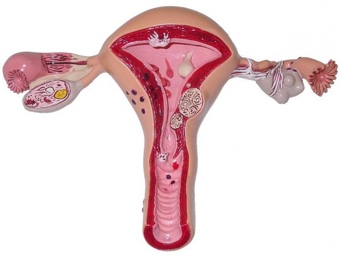
In women, the internal genital organs consist of the gonads (ovaries), uterus, fallopian tubes and vagina, and the external genitalia - from the labia majora and minora and the clitoris (Fig. 9.4).
Rice. 9.4.
Ovary – a paired gland, shaped like an oval, laterally flattened body weighing 5–6 g. It is located in the pelvic cavity on the sides of the uterus. In a newborn girl, the ovary has a cylindrical shape, in 8–12 years old it is ovoid. The length of the ovary varies from 1.5–3 cm in a newborn girl to 5 cm in adolescence, and the weight is from 0.16 to 6 g. In women after 40 years, the weight of the ovaries decreases, and after 60–70 years, their atrophy occurs. The ovaries of a newborn are located outside the pelvic cavity, above the pubic symphysis, and are strongly inclined forward. By 3–5 years they assume a transverse position, and by 4–7 years they descend into the pelvic cavity. In the ovary, there is an upper (tubal) end facing the fallopian tube, and a lower (uterine) end connected to the uterus through a ligament. The ovary has a free and mesenteric edge. The latter is attached to the mesentery, here the vessels and nerves enter the organ, which is why it is called the hilum of the ovary. The ovary is covered with a membrane consisting of connective tissue and epithelium. On a section in the ovary, the medulla and cortex are distinguished. Brain matter consists of loose connective tissue in which blood vessels and nerves pass. Present in the ovarian cortex a large number of follicles (bubbles). The follicle is shaped like a sac containing the female sex cell. In a sexually mature woman, the follicles are in varying degrees ripening and have different sizes. A newborn girl's ovary contains from 40,000 to 200,000 primary immature follicles. Their maturation begins at the time of puberty (12–15 years). However, over the course of a woman’s entire life, no more than 500 follicles mature, the rest are resolved.
In a newborn girl, the surface of the ovaries is smooth; in adolescence, irregularities and bumps appear on the surface due to swollen follicles and the presence of corpus luteum in the ovarian tissue.
The fallopian tubes serve to transport the egg from the ovary to the uterus. They have a cylindrical shape, their length in a mature woman is 8–18 cm, the lumen diameter is 2–4 mm. They are located in the upper lumen of the broad ligament of the uterus (Fig. 9.5).
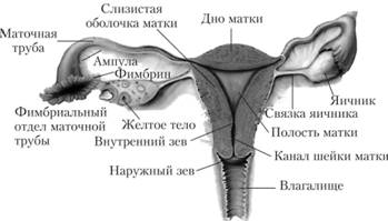
Rice. 9.5.
In the wall of the fallopian tube there is a mucous membrane covered with single-layer cylindrical ciliated epithelium, muscle layer, consisting of smooth muscle tissue, and the serous layer, represented by the peritoneum. The fallopian tube has two openings: one of them opens into the uterine cavity, the other into the peritoneal cavity, near the ovary. At this point, the end of the fallopian tube has funnels and ends in projections called fimbriae. Through these fimbriae, the egg, after leaving the ovary, enters the fallopian tube. This is where fertilization occurs. The fertilized egg divides and moves through the fallopian tube to the uterus. This movement is facilitated by vibrations of the cilia of the ciliated epithelium and contraction of the walls of the fallopian tubes. A newborn girl's fallopian tubes are curved and do not touch the ovaries. In adolescence, they lose their tortuosity, descend downwards and approach the ovaries. The length of the fallopian tube in a newborn is 3.5 cm; during puberty it quickly increases. In old age, the walls of the fallopian tubes become thinner due to atrophy of the muscular layer, and the folds of the mucous membrane are smoothed out.
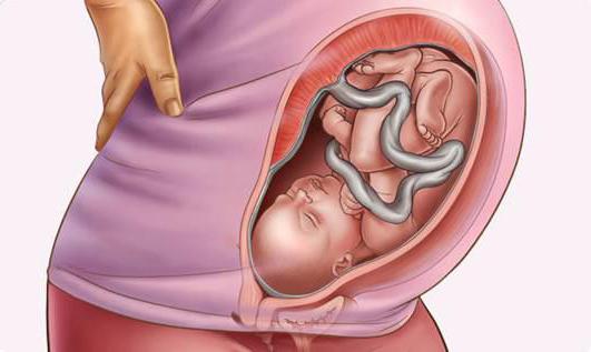
Uterus - a muscular organ that serves for the maturation and gestation of the fetus and is located in the pelvic cavity. The bladder lies in front of the uterus, and the rectum lies behind it. Up to 3 years, the uterus has a cylindrical shape and is flattened in the anteroposterior direction. By the age of 7, the uterus becomes rounded, its bottom expands, adolescence it takes on a pear shape. The length of the uterus in a newborn girl is 3.5 cm, about 2/3 of it falls on the cervix. By the age of 10, the length of the uterus increases to 5 cm, and adult woman reaches 6–8 cm. The weight of the uterus in a newborn is 3–6 g, at 15 years old – 16 g, at 20 years old – 20–25 g. The uterus has its maximum weight (45–80 g) at the age of 30–40 years, after Over the course of 50 years, its mass decreases.
The cervical canal in a newborn is wide and contains a mucus plug. The mucous membrane forms folds that disappear by 6–7 years. The uterine glands develop only during puberty. The muscular layer thickens after 5–6 years. In newborn girls, the uterus is tilted forward, located high above the pubic symphysis. The cervix is directed downward and posteriorly. The ligaments are poorly developed, the uterus is easily displaced. After 7 years, a lot of connective and fatty tissue appears around it. As the size of the pelvis increases, the uterus descends into the pelvis. In old age, due to a decrease in adipose tissue in the pelvic cavity, the mobility of the uterus increases again.
The wall of the uterus consists of inner, middle and outer layers. Inner layer ( endometrium ) is a mucous membrane lined with columnar epithelium. Its surface in the uterine cavity is smooth, in the cervical canal it has small folds. In the thickness of the mucous membrane there are glands that secrete secretions into the uterine cavity. With the onset of puberty, the mucous membrane of the uterus undergoes changes associated with the processes occurring in the ovary (ovulation, formation corpus luteum). At the time when the developing embryo from the fallopian tube is about to enter the uterus, its mucous membrane grows and swells. The embryo is immersed in such a loosened mucous membrane. If fertilization of the egg does not occur, then most of the uterine mucosa is rejected, and the blood vessels rupture, bleeding from the uterus occurs - menstruation, which lasts 3-5 days. After this, the uterine mucosa is restored and the entire cycle of its changes is repeated after 28–30 days. Middle layer (myometrium ) – the most powerful, consists of an outer longitudinal, middle circular and inner longitudinal layer. During pregnancy, smooth muscle fibers increase 5–10 times in length and 3–4 times in width. The size of the uterus and the number of blood capillaries. After birth, the weight of the uterus reaches 1 kg, and then its reverse development occurs, which ends after 6–8 weeks. Thanks to the muscular contractions of the uterus during childbirth, the fetus comes out of its cavity. Outer layer of the uterus ( perimetry ) presented serosa- the peritoneum, which covers the entire uterus, with the exception of the cervix. From the uterus, the peritoneum passes to other organs and the walls of the pelvis.
Vagina is a tube about 8–10 cm long that connects the uterine cavity with the external genitalia. The vaginal wall consists of mucous, muscular and connective tissue membranes. The mucous membrane on the anterior and posterior walls of the vagina has folds, is covered with stratified squamous epithelium and is abundantly supplied with blood vessels and elastic fibers. The outer shell consists of loose connective tissue. Before the onset of sexual activity, the outlet is covered with a fold of the mucous membrane - hymen.
External genitalia. The labia majora are paired folds of skin containing a large amount of adipose tissue. They limit a space called the genital slit. The posterior and anterior ends of the labia are connected by posterior and anterior commissures (see Fig. 9.4).
The labia minora is also a paired fold of skin. The gap between the labia minora is called vestibule of the vagina. The external opening of the urethra and the opening of the vagina open into it. At the base of the labia minora there are two glands of the vestibule - Bartpolinium glands , the ducts of which open onto the surface of the labia minora in the vestibule of the vagina. Bartholin's glands secrete a thick mucous secretion that moisturizes the vestibule of the vagina.
The clitoris is located in the vestibule of the vagina and has the shape of a small elevation (see Fig. 9.4). It consists of two cavernous bodies, similar in structure to cavernous bodies male penis. On top, the clitoris is covered with stratified squamous epithelium and contains a large number of sensitive nerve endings.
A newborn girl's labia majora are loose, the labia minora are not completely covered by the labia majora. The vestibule of the vagina is deep, with poorly developed glands. The hymen is dense. The vagina is short (2.5–3.5 cm), arched, narrow, the anterior wall is shorter than the posterior one, up to 10 years the vagina changes little, it grows in adolescence.
Before puberty, the vaginal mucosa is squamous epithelium, which is replaced by a cylindrical one during puberty. Therefore, in girls before puberty protective functions The mucous membrane of the external genitalia is poorly developed, it is thin, easily vulnerable and easily susceptible to allergic and bacterial inflammation. This is due to low level estrogens (female sex hormones) and the alkaline environment of the vagina due to the absence of Daudelein's bacillus, which secretes lactic acid and promotes self-cleaning of the vagina.
We are pleased to present you the first one on the Russian-language Internet social network supporters of a healthy lifestyle and a full-fledged platform for the exchange of experience and knowledge in everything related to the words “health” and “medicine”.
Our task is to create an atmosphere of positivity, kindness and health on the site, which will lift your spirits, improve your health and prevent you, because information and thoughts are transformed into material events! ;-)
We strive to create a highly moral portal in which it will be pleasant for the most different people. This is facilitated by the fact that we monitor the actions of all users. At the same time, we want the site to be fairly objective, open and democratic. Here everyone has the right to express a personal opinion, to make their own assessment and comment on any information. In addition, anyone can submit an article, news or any other material to most sections of the site.
Project “To your health!” is positioned as a portal about health, not medicine. In our opinion, medicine is the science of how to recover from a particular disease, and health is the result of a lifestyle in which you do not get sick. The healthier you are, the less likely you are to get sick. Our body is designed in such a way that when in the right way In life we shouldn’t get sick at all. So let's improve our health instead of studying diseases. There are quite a lot of websites about medicine, but in our opinion, they are intended more for medical professionals than for ordinary people. We strive to talk to you about health. We don’t want to write a lot about diseases and methods of treating them - enough has already been written about this. Instead, we'll focus on how to avoid getting sick.
We are interested healthy image life, and we want to live happily ever after. We believe that you are also not indifferent to the topic. healthy longevity. Therefore, if you want to be surrounded healthy people and those who strive for this, this site will help you solve this problem. Our plans include creating an active community of people leading a healthy lifestyle, and in this regard, we are pleased to offer you the following opportunities:
- create your own page with personal photos, blog, forum, calendar and other sections
Do what you like, and we will try to provide you with everything you need for this. We strive to make this site as comfortable as possible for you. There is still a lot of new and interesting things ahead.
Register yourself and invite your colleagues, friends and loved ones to the site for constant contact with them and exchange experiences. Stay in touch, discussing all the news and interesting things in the field of health.
Stay with us!
The reproductive system is necessary for the production of new living organisms. The ability to reproduce is a fundamental characteristic of life. When two people produce offspring that have genetic features both parents. The main function of the reproductive system is to create male and female (sex cells) and ensure the growth and development of offspring. The reproductive system consists of male and female reproductive organs and structures. The growth and activity of these organs and structures is regulated by hormones. Reproductive system is closely related to other organ systems, especially the endocrine and urinary systems.
Reproductive organs
Male and female reproductive organs have internal and external structures. The reproductive organs are considered either primary or secondary. The main reproductive organs are (testes and ovaries), which are responsible for the production (sperm and eggs) and hormonal production. Other reproductive organs belong to secondary reproductive structures. Secondary organs help in the growth and maturation of gametes, as well as the development of offspring.
Organs of the female reproductive system
Organs of the female reproductive system include:
- The labia majora are outer folds of skin that cover and protect internal structures genitals.
- The labia minora are smaller, spongy folds located inside the labia majora. They provide protection for the clitoris, as well as the urethra and vaginal opening.
- The clitoris is a very sensitive sexual organ located in front of the vaginal opening. It contains thousands of nerve endings and responds to sexual stimulation.
- The vagina is a fibrous, muscular canal that leads from the cervix (the opening of the uterus) to the outside of the genital canal.
- The uterus is a muscular internal organ that nourishes female gametes after fertilization. The uterus is also the place where the fetus develops during pregnancy.
- Fallopian tubes are tubular organs that carry eggs from the ovaries to the uterus. This is where fertilization usually occurs.
- The ovaries are the female primary reproductive glands that produce gametes and sex hormones. There are two ovaries in total, one on each side of the uterus.
Organs of the male reproductive system
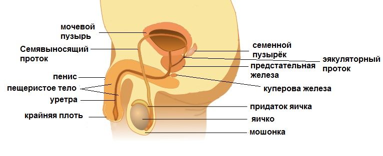
The male reproductive system consists of the reproductive organs, accessory glands, and a series of canals that provide a pathway for sperm to exit the body. The major male reproductive structures include the penis, testicles, epididymis, seminal vesicles, and prostate gland.
- Penis - main body involved in sexual intercourse. This organ consists of erectile tissue, connective tissue and skin. The urethra extends along the length of the penis, allowing urine and sperm to pass through.
- Testes are male primary reproductive structures that produce male gametes(spermatozoa) and sex hormones.
- The scrotum is the outer pouch of skin containing the testicles. Since the scrotum is located outside abdominal cavity, it can reach temperatures that are lower than those internal organs bodies. For proper development sperm require lower temperatures.
- Epididymis (epididymis) is a system of ducts that serve for the accumulation and maturation of sperm.
- The vas deferens is a fibrous, muscular tube that is a continuation of the epididymis and ensures the movement of sperm from the epididymis to the urethra.
- The ejaculatory duct is a canal formed from the connection of the vas deferens and the seminal vesicles. Each of the two ejaculatory ducts empty into the urethra.
- The urethra is a tubular structure that extends from Bladder through the penis. This channel allows reproductive fluids (sperm) and urine to be released from the body. Sphincters prevent urine from entering the urethra when the sperm passes through.
- Seminal vesicles are glands that produce fluid for sperm maturation and provide them with energy. The ducts leading from the seminal vesicles join the vas deferens to form the ejaculatory duct.
- The prostate gland is a gland that produces an alkaline milky fluid that increases sperm motility.
- Bulbourethral glands (Cooper's glands) - pair small glands located at the base of the penis. In response to sexual stimulation, these glands secrete an alkaline fluid that helps neutralize acidity from the urine and vagina.
Likewise, the female reproductive system contains organs and structures that help produce, support, grow and develop female gametes(ovum) and the growing fetus.
Diseases of the reproductive system
The functioning of the human reproductive system can be affected by a number of diseases and disorders, which also include cancer that develops in the reproductive organs, such as the uterus, ovaries, testicles or prostate. Disorders of the female reproductive system include endometriosis (endometrial tissue develops outside the uterus), ovarian cysts, uterine polyps, and uterine prolapse. Disorders of the male reproductive system include testicular torsion, hypogonadism (insufficient testicular activity leading to decreased testosterone production), increased prostate, hydrocele (swelling in the scrotum) and inflammation of the epididymis.
In all world cultures, the function of reproduction, procreation, is considered one of the main ones. The male and female reproductive systems have different structure, but performs one task: to form sex cells - gametes, the fusion of which at the moment of fertilization will become possible development future human body. This article is devoted to the study of the structure and function of the female reproductive system.
General characteristics of a woman’s reproductive organs
The female reproductive system includes the external and internal genital organs, which are also called reproductive organs.
The external genitalia, called the vulva, are visually sufficiently pronounced - these are the pubis, labia majora and minora, clitoris and the entrance to the vagina (vagina), closed by an elastic hymen called the hymen. Let's study the external organs of the female reproductive system in more detail.
The structure of the pubis
The lower abdomen at the level of the pubis (pubic bone) forms the pubis. The bone itself is anatomically correct position hangs over the entrance to the vagina and looks like an arch. Externally, the pubis has a roll-like shape, forming an elevation. A layer of fat forms under his skin. Outside it will form hairline. It has a clearly defined horizontal border. If a woman’s body produces an excess amount of androgens - male sex hormones, the hairline increases and rises upward at an acute angle to the navel. Pathology of pubic hair is a sign of sexual development.
Labia majora and labia minora
From the pubis to the anus there are two folds of skin - the labia majora, which have outer hair and contain a layer of subcutaneous fatty tissue. Their connective tissue contains the ducts of the Bartholin gland. It secretes a fluid that moisturizes the female genital organs. In case of poor hygiene harmful microorganisms penetrate the gland tissue and cause inflammation in the form of painful seals.
Under the labia minora are the labia minora, densely intertwined with blood vessels and nerves. In their upper part there is an organ homologous to the male penis - the clitoris. Its growth is inhibited by the hormones of the female reproductive system - estrogens. The clitoris contains a large number of nerves and blood vessels, which means it has high sensitivity. If a girl or woman has greatly enlarged clitoris, this may be a clear sign of hormonal pathology.

Entrance to the vagina
The vulva, in addition to the pubis, labia majora and minora, and clitoris, includes the entrance to the vagina. At a distance of up to 2 centimeters deep from it there is the hymen. It consists of connective tissue and has several holes through which blood flows during menstruation.
Internal reproductive organs of a woman
These include the vagina (vagina), uterus, ovaries and fallopian tubes. All of them are located in the pelvic cavity. Their functions are the maturation and entry of fertilized female gamete eggs into the uterine cavity. It is from the zygote that the embryo will develop.
Structure of the vagina
The vagina is an elastic tube consisting of muscle and connective tissue. It is located from the genital slit towards the uterus and has a length of 8 to 10 cm. Located in the pelvis, the vagina enters the cervix. It has a front and back wall, as well as the arch - upper section deeper than the front one.
The vagina is located at an angle of 90 degrees to the surface of the uterus itself. Thus, the internal female genital organs, which include the vagina, are densely intertwined with arterial and venous vessels, as well as nerve fibers. The vagina is separated by a thin connective tissue wall from the bladder. It is called the vesicovaginal septum. Bottom part The vaginal wall is separated from the back lower section large intestine by the perineal body.
Cervix: structure and functions
The vagina enters into a canal called the cervical canal, and the junction itself is the external os. Its shape differs between those who have given birth and nulliparous women: if the pharynx is dotted-oval, the uterus did not bear the fetus, and the appearance of the gap is characteristic of those who have given birth. The uterus itself is an unpaired hollow muscular organ, consisting of a body and a neck and located in the pelvis. Considering the structure of the female reproductive system and its functions, it becomes clear that it is responsible for the formation and development of the embryo, as well as for the process of expulsion of the fetus as a result labor activity. Let's return to the structure of its lower section - the neck. It is connected to top part vagina and has the shape of a cone (in nulliparous women) or a cylinder. The vaginal portion of the cervix is up to three centimeters long and is also anatomically divided into anterior and posterior lips. The cervix and pharynx transform as a woman ages.
Inside the cervix is cervical canal, ending internal throat. It is lined with secretory glands that secrete mucus. If its secretion is disrupted, blockage and cyst formation can occur. Mucus has bactericidal properties and prevents infection of the uterine cavity. 4-6 days before the release of the egg from the ovary, the mucus becomes less concentrated, so sperm can easily penetrate through it into the uterus, and from there into the fallopian tubes.
After ovulation, cervical secretion increases its concentration, and its pH decreases from neutral to acidic. Pregnant uterus is completely closed by a clot cervical mucus in the cervical area. IN menstrual period The cervical canal opens slightly so that the rejected endometrial layer can come out. This may be accompanied aching pain lower abdomen. During labor, the cervical canal can open up to 10 cm in diameter. This promotes the birth of a child.
Among the most common diseases of the cervix is its erosion. It appears as a consequence of damage to the mucous layer caused by infections or injuries (abortion, complicated childbirth). Erosion that is not detected and treated in time can cause inflammatory processes and even cancer.
Fallopian tubes
The fallopian tubes, also called oviducts or fallopian tubes, are 2 elastic tubes located in the abdominal cavity and entering the fundus of the uterus. The free edge of the oviduct has fimbriae. Their beating ensures the advancement of the egg released from the ovary into the lumen of the tube itself. The length of each oviduct is from 10 to 12 cm. It is divided into sections: a funnel, which has an expansion and is equipped with fimbriae, an ampulla, an isthmus, and part of the canal entering the uterine wall. For normal development pregnancy requires such a condition as complete patency of the oviducts, otherwise the woman will face infertility. The most common pathologies of the fallopian tubes are adhesions, salpingitis and hydrosalpinx.
All of the above diseases cause tubal infertility. They are complications of chlamydia, gonorrhea, trichomoniasis, genital herpes, causing a narrowing of the lumen of the fallopian tubes. Frequent abortions can provoke the appearance of adhesions that are located across the tube. Hormonal disorders cause a decrease in the mobility of the ciliated epithelium lining the oviducts, which leads to a deterioration in the motor properties of the egg.

Most dangerous complication resulting from tubal pathologies - ectopic pregnancy. In this case, the zygote stops in the oviduct without reaching the uterus. It begins to fragment and grow, stretching the pipe wall, which eventually bursts. As a result, there is a strong internal bleeding life-threatening.
Ovaries in women
They are a paired gonad and weigh 6-8 grams. The ovaries are the production of sex hormones - estrogens, controlled by the pituitary gland and hypothalamus - this is an intrasecretory function. As exocrine glands, they form sex cells - gametes, called eggs. Biochemical composition and the mechanism of action of estrogens will be studied by us later. Let's return to the structure of the female gonads - the ovaries. It must be taken into account that the structure of the female reproductive system (as well as the male) is directly related to the urinary system.
It is from the mesonephros ( primary kidney) the stroma of the female gonads develops. The precursors of eggs, oogonia, are formed from mesenchyme. The ovary has a tunica albuginea, and under it there are two layers: the cortex and the medulla. The first layer contains follicles, which, when maturing, form first and second order oocytes, and then mature eggs. The medulla of the gland consists of connective tissue and performs a supporting and trophic function. It is in the ovaries that ovogenesis occurs - the process of reproduction, growth and maturation of female reproductive gametes - eggs.
Specifics for a woman
The structure of the reproductive system of the female and male individual is controlled by special biological active substances- hormones. They are produced by the sex glands: the testes in men and the ovaries in women. Entering the blood, they specifically influence both the development of the reproductive organs and the formation of secondary sexual characteristics: body hair, development of the mammary glands, pitch and timbre of the voice. The development of the female reproductive system occurs under the influence of estradiol and its derivatives: estriol and estrone. They are produced by special ovarian cells - follicles. Female hormones- estrogens lead to an increase in the volume and size of the uterus, as well as to muscle contractions of the fallopian tubes and the uterus itself, that is, preparation occurs reproductive organ to acceptance of the zygote.
The corpus luteum of the uterus produces progesterone, a hormone that stimulates development children's place- placenta, as well as an increase glandular epithelium mammary glands during pregnancy. Violation hormonal levels female body leads to diseases such as uterine fibroids, endometriosis, and polycystic disease.
Anatomical features of the female uterus
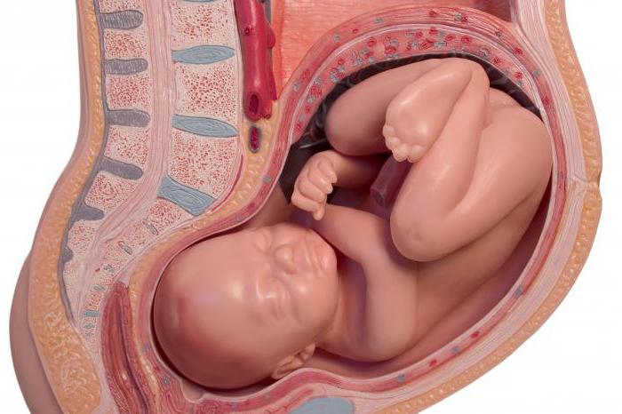
The reproductive system of the female body contains an organ that is unique in structure and function. It is located in the pelvic cavity between bladder and rectum and has a cavity. This body called the uterus. To understand the mechanism of fertilization, remember that the genital organs - the ovaries in women - are associated with fallopian tubes. The egg, entering the oviduct, then penetrates the uterus, which serves as the organ responsible for the development of the embryo (embryogenesis). It consists of three parts: the neck, which was studied earlier, as well as the body and bottom. The body of the uterus has the shape of an inverted pear, the expanded part of which includes two fallopian tubes.
The reproductive organ is covered with a connective tissue membrane and has two layers: muscular (myometrium) and mucous (endometrium). The latter is built from squamous and columnar epithelial cells. The endometrium changes the thickness of its layer: during ovulation it thickens, and if fertilization does not occur, this layer is rejected along with a portion of blood from the walls of the uterus - menstruation occurs. During pregnancy, the volume increases greatly (about 8-10 times). In the pelvic cavity, the uterus is suspended by three ligaments and entwined with a dense network of nerves and blood vessels. Her main function- development and nutrition of the embryo and fetus until the moment of physiological birth.
Pathologies of the uterus
The structure of the female reproductive system may not always be ideal and function correctly. One of the pathologies of the reproductive system associated with the structure of the reproductive organ may be a bicornuate uterus. It has two bodies, each of which is connected to one oviduct. If the pathology of the female reproductive system concerns the structure of the endometrium, we speak of hypoplasia and aplasia of the uterus. The consequence of all the above pathologies is termination of pregnancy or infertility.
This article examined the anatomical and physiological characteristics female reproductive system.
