Removal of the pericardium. Methods of surgical intervention on the pericardium
Eat various diseases, capable of exerting a strong negative impact for the work of the main body circulatory system- heart. This article will discuss one of the common problems, which is called pericarditis. This disease is dangerous and deserves attention.
What disease are we talking about?
If you try to determine scientific language, what is cardiac pericarditis, the symptoms of which worry many people, we can say the following: it is an aseptic or infectious inflammation of the parietal and visceral layers of the pericardium. But to put it simply, we are talking about a chronic or acute form of inflammation of the outer lining of the heart (pericardium), the cause of which is rheumatic lesion, the influence of infection or other types of influences.
In order to understand the essence of this disease, it makes sense to pay attention to general information.
The pericardium is the pericardial sac in which such important organ like a heart. Moreover, the very fact of active contraction in such a bag is possible due to its special structure, which prevents the occurrence of strong friction.
Symptoms of pericarditis in adults make themselves felt provided that the functioning and structure are impaired. In this case, accumulation of effusion (secret) in the pericardium, which is serous or purulent in nature, is possible. This liquid is defined as exudate. It is under its influence that the heart is compressed, which disrupts the performance of its direct functions. Obviously, such a condition is dangerous and cannot be ignored.
If excessive accumulation of fluid occurs in the pericardium, there will be a direct threat to human life. This condition can only be avoided with immediate intervention.
It is worth noting that this disease occurs most often in women, less often in men. As for children, early age It is extremely rare for such a diagnosis to be made, although it does sometimes occur.
Causes
Before considering the symptoms of pericarditis in humans, it is worth finding out what precedes the appearance of such a complex disease.
You need to understand the following fact - fluid accumulation may be a consequence various processes in organism:
Complication of pathology internal organs;
A sign of diseases directly of the heart itself;
The result of the injury;
Manifestation infectious diseases general;
A sign of systemic diseases.
But if we talk about the most common causes, then first of all attention should be paid to the impact of infection. In this case, the symptoms of pericarditis can be a manifestation of its different forms:
Infectious-allergic;
Infectious;
Non-infectious (non-purulent, aseptic).
In fact, we are talking about problems with the condition of the pericardial sac in the following diseases:
Such viral infections like influenza and measles;
Tuberculosis, provided that the infection spreads from an extrapulmonary tuberculous or primary pulmonary focus;
Microbial diseases (septic processes, scarlet fever, tonsillitis);
Fungal infections.
In addition to the effects of these diseases, symptoms of pericarditis can result from drug allergies.
A separate group of problems of the pericardial sac includes those forms of pericarditis that develop under the influence of developing pericardial defects with the formation of diverticula and cysts.
Types of disease
No matter what the symptoms of pericarditis look like, they will always indicate the development of one of two key types of this disease: acute or chronic.
If speak about chronic form, then you need to pay attention to the fact that it develops gradually and may not make itself felt for several years. In this case, such pericarditis has several common forms:
Adhesive, or adhesive, during which scars and soldering are formed;
Mixed appearance of adhesions, fluid and scars;
Exudative, or exudative form, characterized by a large accumulation of fluid in the pericardial sac.
Acute pericarditis, the symptoms of which appear much more quickly, also has several key forms:
Effusive, with the formation of a significant volume of fluid (pus, bloody contents or blood plasma);

Fibrinous, or dry, characterized by a large accumulation of an adhesive substance from blood plasma (fibrin) in the pericardial cavity.
Constrictive and exudative pericarditis: symptoms
In order to respond to the disease in a timely manner and begin timely treatment, you need to know at least general signs Problems. And if we talk about the exudative form of the disease of the pericardial sac, then you need to pay attention to this: with such a problem, the fact of fluid accumulation is often detected during a fluorographic examination, as well as when using echocardiography.
If a tumor of the chest or lungs has been detected, it also makes sense to suspect the development of pericarditis. A similar diagnosis can be made in patients with uremia, during which cardiomegaly develops without obvious reasons, and an increase in venous pressure is also observed.
The situation is somewhat different with such a problem as constrictive pericarditis, the symptoms of which are early stage often virtually invisible. Tangible signs of the disease make themselves felt already when enough fluid accumulates in the pericardial sac to complicate the work of the heart. Obvious signs appear over time after physical exertion in the form of severe shortness of breath and increased fatigue. People with this problem may experience sudden weight loss and a noticeable decrease in appetite.
There are other signs that indicate constrictive pericarditis of the heart. Over time, symptoms may increasingly resemble those of right ventricular heart failure. We are talking about ascites, pain and heaviness in the right hypochondrium.
How does dry pericarditis manifest?
Speaking about this form of diseases of the pericardial sac, you need to pay attention to the fact that this problem is often preceded by myalgia or fever. But the difficulties caused by dry pericarditis are not limited to this. Symptoms eventually manifest as pain, which is localized in the chest area.
Such symptoms can persist for several days and even weeks, expressing themselves quite clearly behind the sternum. Possible painful sensations in both hands or one upper limb, as well as in the trapezius muscles. The pain becomes especially acute when trying to change body position. When coughing, swallowing and breathing, unpleasant sensations can also make themselves felt.

If a problem such as dry pericarditis develops, the symptoms are slightly relieved if the person is in a sitting position. But if the patient lies on his back, the effect will be the opposite - his health will worsen.
In some cases, the pain syndrome can bother you around the clock. In this case, irradiation will be observed in one or both hands. This condition can stimulate myocardial infarction.
In this case, it is possible that the pain sensations described above will not occur during the gradual development of the process of fluid accumulation. Most often, this condition is caused by the formation of a problem with the pericardium against the background of tuberculosis, uremia, tumor lesions and after radiation sessions.
But pain is not the only symptom that accompanies dry pericarditis. Dyspnea and signs of dysphagia may occur.
Acute pericarditis
With this form of the disease, the earliest and most obvious symptom is pain in the heart area. At the same time, the location and strength of these unpleasant sensations may change.
Most often, pain is observed in the lower part of the sternum or at the apex of the heart. Possible irradiation to the neck, left shoulder blade and arm.
When there is an exacerbation of pericarditis, the symptoms manifest in the form of very severe pain. This condition may resemble myocardial infarction or pleurisy. In some cases, the pain is aching and dull. Sometimes patients begin to feel heaviness in the heart area.
If it is felt during an exacerbation, then most likely you have to deal with dry pericarditis.
During an exacerbation, the appearance of shortness of breath cannot be ruled out, which will indicate the presence of effusion in the pericardial sac. Moreover, the more fluid that has accumulated in the pericardium, the worse the breathing problems will be. In order to temporarily improve your well-being, you need to sit down. In this case, shortness of breath will noticeably decrease due to the concentration of exudate in the lower part of the pericardial sac and, as a result, the pressure on the heart itself will decrease. Blood circulation eventually improves, and the patient feels relief.
The shortness of breath itself may be accompanied by a cough, and in some cases even vomiting.
How does pericarditis manifest in children?
As stated above, similar problems This is a rare occurrence with heart function at an early age. However, the risk of developing such a disease before adulthood is possible.
So, what does pericarditis look like in children? Symptoms in such young patients, as in adults, are often signs of the development of another, underlying disease. Most often we are talking about the impact various viruses. Much less often the disease develops against the background of tuberculosis or rheumatoid arthritis. The cause of pain in the heart may also be the effect on children's body diseases connective tissue, for example, systemic lupus erythematosus.
Another reason why the pericardial sac is damaged at an early age is severe staphylococcal and, less commonly, septic processes. Immunological genesis is characteristic of most childhood pericarditis. In the case of the development of so-called uremic pericarditis with severe renal failure the disease may be toxic.
Problems with the pericardial sac in children during purulent processes are most often metastatic. This means that they develop due to either a breakthrough of a purulent focus into the pericardium from the myocardium, or due to hematogenous spread.
As for the general symptoms, they include moderate cyanosis, shortness of breath, stabbing pain in the heart and fever.

In the case of the development of dry pericarditis, a pericardial friction rub will be heard above the heart. Often the dry form is followed by an exudative one and you need to be prepared for this. In this state, pain and friction noises disappear, but the size of cardiac dullness expands and the condition as a whole worsens.
This is how childhood pericarditis manifests itself. Symptoms and treatment in this case are solely the responsibility of the doctor. It is better for parents not to try to influence the body of their beloved child on their own. A quick visit to the hospital will be the best solution.
Diagnostics
Of course, not all ordinary people are such sophisticated medical experts as to independently determine obvious signs dry pericarditis without the help of a doctor. That is why it is important to remember that in conditions modern medicine There is always the opportunity to undergo diagnostics with the participation of qualified doctors.
So, if your health worsens and sensations appear that are even vaguely reminiscent of the symptoms of pericarditis, you should immediately go to the doctor, who will be able to determine the presence of external signs underlying illness and fever as well. With the help of palpation in the case of pericarditis, you can detect a friction noise of the pericardial sac over the area of cardiac dullness.
As a rule, such noise is also listened to in order to obtain a more accurate picture of the patient’s condition. For this, a phonendoscope is used, which is pressed firmly against the chest. The patient should be in an upright position at this time, holding his breath as he exhales.
For more accurate diagnosis Laboratory data as well as ECG readings may be used.
Symptoms of pericarditis on the ECG, especially in its acute dry form, are expressed in the form of signs characteristic of subepicardial myocardial damage. If there is a significant accumulation of fluid, then the signs of the disease with this type of diagnosis will be expressed by reducing the voltage of the QRS complexes.
Treatment
With a disease such as pericarditis, symptoms and treatment require a competent assessment and approach. First of all, you need to ensure bed rest. This rule is especially relevant in the case of diagnosing the exudative form of the disease. On average, such a regime lasts about a month and can be expanded only if there are noticeable significant improvements in a sick condition.
If dry pericarditis has been recorded, then there is no urgent need for constant stay in bed.
But, returning to the exudative form, it should be noted that when it worsens, immediate hospitalization in the intensive care unit is necessary. The patient will need to be examined. As for nutrition during a disease such as pericarditis, this issue is regulated taking into account the underlying disease.
If the cause of fluid accumulation in the pericardium is an infection, antibiotic treatment may be prescribed. In the case of tuberculosis, appropriate drugs are also used, but such treatment will require much more time.
Often, for problems with the pericardial sac, anti-inflammatory drugs are prescribed. To reduce the severity of pericarditis and neutralize pain, the following are relevant: non-steroidal drugs, like “Voltaren”, “Indomethacin”, etc.

Glucocorticosteroids, in addition to the effects described above, can have an immunosuppressive and antiallergic effect. For this reason, they are defined as effective means of pathogenetic therapy.
Such drugs are indicated in case of diagnosing the following types of pericarditis:
With myocardial infarction, also known as Dressler's syndrome;
In case of systemic connective tissue diseases;
If there is an active rheumatic process;
For persistent tuberculous pericarditis;
An exudative form with an unclear cause and accompanied by a severe course.
In most cases, a drug such as Prednisolone is prescribed. This course usually lasts several weeks with a gradual abolition of the use of this remedy.
Attention should also be paid to pericardial puncture. We are talking about puncturing the cavity of the pericardial sac and evacuating the effusion that creates pressure on the heart. Such a puncture is performed urgently if there is a rapid accumulation of exudate, leading to the risk of cardiac tamponade.
The puncture can also be performed when purulent form diseases, in this case, after removing the fluid, antibiotics and other medications that are relevant in the case of a particular patient are injected into the pericardium through a needle.
Rapid accumulation of exudate can also lead to the development of constrictive pericarditis. In this condition, it is necessary to limit consumption table salt up to 2 g per day and significantly reduce the amount of fluid the patient consumes. Prescribing diuretics will be relevant.
In particularly severe cases, surgery may be performed. It is used mainly in the absence of the desired result after drug treatment during constrictive pericarditis. When the patient's condition improves, the surgeon performs a pericardiectomy to free the left ventricle of the heart from constant compression.
Folk remedies
There are a number alternative techniques to treat a problem such as pericarditis. Symptoms and folk remedies treatment of manifestations of this disease are actual topic. But it is worth understanding that replacing the main one with traditional methods healing process- this is a big mistake. Fluid accumulation in the pericardial sac is a problem accompanied by rapid complications, which without competent diagnosis and qualified treatment it will not be possible to neutralize it.
Folk remedies are permissible only in the recovery period, as an accompanying effect. But in any case, amateur activity is not encouraged; it is better to take all actions after consultation with your doctor.
As for topical medicines, decoctions of rose hips, strawberries, hawthorn and St. John's wort are acceptable.

For those who have pets, the topic will be relevant: “Pericarditis in dogs: symptoms and treatment.” In principle, other animals can also have problems with the pericardial sac. Symptoms for this problem boil down to the appearance of fever, pain in the heart area, as well as general depression of the animal that is sick. In general, the symptoms are similar to the course of the disease in humans, so if your pet is depressed, it is better to take it to the veterinarian, otherwise you may face serious complications.
Symptoms of pericarditis in animals can also appear after injuries. various kinds. Therefore, if your beloved dog is injured, it is simply necessary to organize a check of his condition over time.
Results
Pericarditis is enough serious illness, so it cannot be ignored. For this reason, ordinary people should familiarize themselves with at least the general symptoms. This will allow you to recognize in time dangerous illness and consult a doctor. With this approach, there is every chance of receiving effective treatment by preventing possible complications. Do not forget that if the fact of fluid accumulation near the heart is not influenced in any way, it may occur. death. So it is better to find time for an additional visit to the doctor.
Pericardial pathology that requires surgical intervention is usually divided into two categories - pericardial effusion and constrictive pericarditis. Until recently, surgical access to the pericardium traditionally required a left thoracotomy, median sternotomy, or subxiphoid approach. Development is minimal invasive methods made it possible to successfully use video-assisted thoracic surgery for pericardial diseases. As with the open approach, thoracoscopic evaluation of the pericardium provides diagnostic information regarding the etiology of pericardial disease and alleviates the hemodynamic consequences of pericardial effusion and constrictive pericarditis.
Anatomy
The parietal layer of the pericardium consists of dense collagen and elastin fibers with an internal serous lining of single-layer mesothelium. The parietal layer of the pericardium is a sac-like formation that surrounds the heart and merges with the adventitia of the proximal parts of the large vessels. The visceral layer of the pericardium covers the surface of the heart and consists of a thin layer of fibrous tissue covered with mesothelium. The parietal and visceral layers are fused at the points of attachment to the proximal parts of large vessels. Ligaments fix the pericardium to the sternum in front, spinal column behind and the diaphragm below. The phrenic nerve and pericardiophrenic artery pass along the lateral surface of the pericardium on both sides. Normally, the pericardial cavity contains up to 50 ml of serous fluid, which serves as a lubricant that promotes the movement of the heart. The pericardium reduces friction between the heart and surrounding tissues and anchors the heart in the mediastinum. Experimental data have shown that the pericardium performs an important physiological function in equalizing hydrostatic forces, limiting cardiac elongation and diastolic hemodynamic coupling.
Pathophysiology
Pericardial effusion may occur after acute pericarditis or trauma. The most common types of pericardial effusion are: neoplastic, idiopathic, infectious and traumatic. A fluid volume of only 150-250 ml can cause acute pericardial tamponade. Increased intrapericardial pressure reduces ventricular filling and systolic volume cardiac output and thus reduces cardiac output. The decrease in systolic volume is compensated by an increase in heart rate and sympathetic tone. If compensatory mechanisms are ineffective, systemic perfusion decreases and cardiogenic shock occurs.
Clinical picture and differential diagnosis pericarditis
Acute pericarditis is characterized by chest pain, pericardial friction rub, and electrocardiogram (ECG) changes. in the chest has different localization and can intensify in the supine position and when deep breathing. The classic pericardial friction murmur has 3 components, which correspond to atrial systole, ventricular systole, and ventricular filling during diastole. Four stages of ECG changes in acute pericarditis are described. Acute pericarditis usually resolves without consequences. However, hemodynamic complications may occur with cardiac tamponade due to pericardial effusion, constriction due to fibrosis, or both.
The onset of cardiac tamponade may be sudden and unnoticeable. Tamponade is characterized by dilation of the neck veins, distantly audible heartbeats, and hypotension. This triad of symptoms is known as Beck's triad. With tamponade, a paradoxical pulse is observed, which is characterized by a drop in blood pressure during inspiration by more than 10 mmHg. Cyanosis, tachycardia, and tachypnea may also be present. In general, symptoms of cardiogenic shock dominate. On the ECG, a decrease in the voltage of the waves is possible. A chest x-ray may reveal an enlarged heart shadow. Invasive monitoring shows an increase in central venous pressure with a decrease in cardiac output and mean arterial pressure.
Echocardiography is the most sensitive method for diagnosing pericardial effusion. It can detect signs of early cardiac tamponade. Increased respiratory variations in valvular blood flow, right ventricular diastolic collapse, and loss of normal inspiratory inferior vena cava collapse are accurate indicators of cardiac tamponade. Cardiac tamponade is differentiated from other serious pathologies of the chest, causing shock and hypotension. These diseases include acute myocardial infarction, congestive heart failure, inferior vena cava syndrome and constrictive pericarditis.
Pericardial windowing can be performed using a subxiphoid approach under local anesthesia and is usually well tolerated by patients. This surgery is an excellent therapeutic option for many patients, but it should not be used when the underlying disease causing the effusion may lead to constrictive pericarditis (eg, in patients with tuberculosis, Haemofilus influenzae infection, or radiation pericarditis). This is primarily due to the limited extent of pericardial resection that the subxiphoid approach allows, which leads to recurrences in 10-18% of cases. Thoracotomy allows for a more thorough resection of the pericardium and is characterized by a decrease in the incidence of recurrent effusion. However, this approach is more invasive and comes with additional morbidity due to the need for general anesthesia.
Video-assisted thoracoscopic access allows for extended resection of the pericardium with simultaneous assessment of the pathology of the lungs and pleura, avoiding thoracotomy. Postoperative pain is less severe than after thoracotomy, although general anesthesia and separate ventilation of one lung are still necessary. If symptoms of tamponade are present, pericardiocentesis should be performed before general anesthesia.
To summarize, it should be noted that the subxiphoid approach has advantages because it allows the use local anesthesia, does not require ventilation of one lung and turning the patient on his side (this maneuver is poorly tolerated by patients with severe clinical symptoms of tamponade). The disadvantage of the subxiphoid approach is that other chest pathology will not be detected with this approach, and in addition, it is characterized by a higher recurrence rate of pericardial effusion than with video-assisted thoracoscopy. The advantages of the video-assisted thoracoscopic approach include improved access and visualization of the pericardium, which allows for more extensive resection of the pericardium and associated diagnostic and therapeutic procedures (drainage of pleural effusion, decortication, lung or pleural biopsy).
Disadvantages of video-assisted thoracoscopy include the need for general anesthesia, separate ventilation, lateral positioning of the patient, and the need for decompression of the pericardial cavity before induction of anesthesia in unstable patients.
Technique of thoracoscopic surgery on the pericardium
The patient is intubated with a double-lumen endotracheal tube for separate ventilation of the lungs. A nasogastric tube and Foley catheter are placed. If tamponade is significant, pericardiocentesis should be performed before initiating anesthesia. The patient is placed in the left lateral position and lung ventilation. Right-sided access is preferred, except in cases where there is concomitant pathology of the left lung and pleura. Access to the pleural cavity is carried out using blunt dissection above the VIII rib (seventh intercostal space) along the posterior midscapular line. A 10-mm port is installed and a thoracoscope is inserted. Examine the pleural cavity. Two 5-mm ports are inserted one intercostal space above (sixth) along the midscapular and anterior axillary line. Next, the clamp and scissors are inserted. The pericardium and phrenic nerve are found. The pericardium is grasped anterior to the phrenic nerve and dissected. Care must be taken not to damage the heart underlying the pericardium. The anterior surface of the pericardium is widely excised. If there is an encysted fluid collection along the posterior surface, posterior pericardial resection can be performed. A pleural 28 or 32 is installed and directed to the pericardium. The reliability of hemostasis at the sites where the ports are inserted is checked and the thoracoscope is removed.
Results of thoracoscopic operations on the pericardium
The results of video-assisted thoracoscopic resection of the pericardium are encouraging. Hazlerrigg et al. reported thoracoscopic pericardiectomy in 35 patients. Malignant effusion was observed in more than half (52%) of patients. There were no deaths during the operation; the duration of hospitalization averaged 4.6 days. There were no recurrences of effusion observed during a 9-month follow-up. Liu et al. found similar results in a group of 28 patients who underwent thoracoscopic pericardiectomy. In 60% of them, pleuropulmonary pathology was established, which would have been impossible to detect in the case of a subxiphoid approach. The authors believe that the ability to simultaneously correct pleuropulmonary pathology was a significant advantage for patients with both diseases.
Data on thoracoscopic treatment of pericardial effusion in 230 patients, obtained in, where the authors of the chapter work, confirm the safety and therapeutic effectiveness this surgical approach. Compared with traditional subxiphoid and thoracotomy approaches, patients undergoing video-assisted thoracoscopic pericardiectomy have fewer recurrences and lower mortality (unpublished data). Other studies have also shown greater effectiveness of video-assisted thoracoscopic pericardiectomy compared with the subxiphoid approach. The use of video-assisted thoracoscopic pericardiectomy was further supported in a recent study of patients with stale hemopericardium or effusion after cardiac surgery. Thoracoscopic pericardiectomy, in addition, can serve as an effective therapeutic option in hemodynamically stable patients with penetrating pericardiectomy.
Videothoracoscopic creation of a “window” in the pericardium is a new alternative to thoracotomy and subxiphoid approach. This technique allows for wide resection of the pericardium, avoiding the morbidity that accompanies open thoracotomy. Early reports are encouraging and show a low rate of recurrence of effusions in early follow-up.
The article was prepared and edited by: surgeonIt is a disease that requires therapeutic and at the same time - perhaps from the very beginning - surgical intervention. Establishing this fact is important because it serves to prevent those severe complications that in the future pose an increasingly difficult problem for the therapist and surgeon.
Diseases of the pericardium of inflammatory origin, for which there are indications for surgical treatment, are usually divided into four groups:
1. Serous and serous-hemorrhagic pericarditis.
2. Fibrinous pericarditis.
3. Purulent pericarditis.
4. Chronic fibrous, calcifying, constrictive pericarditis.
It is typical for the first and third groups that fluid accumulates in the pericardial cavity in the form of transudate or exudate, and this accumulation of fluid can be so significant that it threatens cardiac tamponade. The goal of the operation is to release the fluid and create an easy path for local treatment.
With fibrinous pericarditis, fibrin is secreted intrapericardially, which in some cases can occur in such large quantities that it gives a picture of the pathologically well-known “hairy heart”, “cor villosum”.
Surgical opening of the pericardium is indicated:
1. if due fast education exudate poses a risk of cardiac tamponade;
2. if the exudate exists for a long time and does not show a tendency to be absorbed;
3. if the exudate is purulent, as a result of which its pumping and continuous local antibiotic treatment are indicated;
4. if removal of the pericardium is indicated to prevent the occurrence of constrictive pericarditis.
Constrictive pericarditis is a late complication of advanced pericarditis.
In the treatment of pericarditis latest development Heart surgery led to results that, by necessity, changed the old conservative views towards radicalism. Before the discovery of antibiotics and chemotherapeutic drugs, conservative views rightfully dominated in the treatment of pericarditis of tuberculous origin. However, having these medicines, conservatism was replaced by a very successful active look treatment, based on objective criticism of which the old indications and contraindications for surgery needed significant revision.
Puncture treatment of pericarditis is unsafe. Here we are less referring to heart damage due to incorrect technique, but rather to pleural complications (pleurisy, empyema, pneumothorax, etc.), which can undoubtedly be serious and undesirable consequences punctures.
Surgical opening of the pericardium is a long-known, very simple and completely safe intervention, which should always be preferred to puncture treatment. In old textbooks of surgery, the operation is known as inferior longitudinal pericardiotomy or inferior oblique pericardiotomy. The essence of the operation is that by cutting or removing the base of the xiphoid process or resection of a small sternal part of the VIIth costal cartilage, the area of the diaphragm adjacent to the pericardium is prepared without opening the peritoneum and pleura. Having made a small hole in it, the exudate is gradually released. Donaldson attaches a thin rubber tube inserted into the hole with a catgut suture and closes the several centimeter wound. Through the drainage, the outflow or suction of the accumulating pericardial fluid is ensured, and it is possible local treatment, washing the pericardial cavity with antibiotics and medications.
This method is suitable for the treatment of acute pericarditis, regardless of its specific or nonspecific nature, and it plays a very important role in the prevention of later developing constrictive pericarditis. Holman recommends after graduation acute stage pericarditis, pericardiectomy. We believe that this proposal should be considered, and we are certainly more willing to perform the operation then than in the late, advanced constrictive stage.
In chronic pericarditis, larger or smaller fusions of the pericardial sheets or cords, as well as fixation of the pericardium to surrounding organs (sternum, diaphragm, mediastinum, spine) sometimes lead only to minor, but very often to severe morphological and functional disorders.
The type of these adhesions and the harm they cause determine the indications and contraindications for the operation, as well as the appropriate method for performing it. In the treatment of pericardial adhesions, causing disorders functions, essentially two operational methods are known. One of them is pericardiectomy, based on the principle of lung decortication proposed by Sapozhnikov; Another method is Brouwer cardiolysis.
Cardiolysis had the goal, according to the views of that time, of freeing the heart from the bony chest wall. Currently, this operation is performed only very rarely. The essence is that part of the bony chest wall located in front of the heart is removed, as a result of which the heart is freed from its fixed position. This operation can be successful if diastolic expansion is primarily hampered by the fact that the heart is fixed to the chest wall.
With pericardial constriction, results can only be expected from pericardiectomy. Known for penetration different kinds access. We have very good access with the lower mediastinotomy proposed by Holman, at the middle of the lower part of the sternum. Other surgeons open both pleural cavities with a transverse incision in the sternum. We typically use the method proposed by Holman, which provides excellent access to complete pericardiectomy, including the inferior vena cava area. The hemodynamic rule for pericardiectomy is that cardiac release and dissection should begin at the surface of the left ventricle.
When removing the armor, serious difficulties sometimes arise for the surgeon, because the scars can be closely adhered to the muscles, and the thin muscles can easily rupture. Sewing it up is sometimes a very difficult or even hopeless task. It is usually not recommended to peel off the atrium, nor is it particularly necessary. The thin walls of the atria are very easily ruptured. All authors consider the release of the vena cava area to be theoretically correct. I myself have never observed their narrowing in a fairly large amount of material.
From a disease perspective, pericardiectomy is certainly a more radical and desirable solution to the issue. Still, there may be cases in which - even if not definitively - one has to be content with cardiolysis. In postoperative treatment, we have good experience also with the use of hibernation and hypothermia. By reducing the work of the heart, they have a very beneficial effect. The heart, freed from its shell, becomes sick as a result of compression, and the surgical load certainly means additional work.
The development of operating technology is unlikely to significantly reduce the high mortality rate during pericardiectomy, but early identification of indications for the operation can do this. Unfortunately, this operation in most cases was used only as a last resort, when the patient undergoing surgery was already in a hopeless condition, and the operation is hopeless, when the consequences of a long-existing constriction (ascites, cirrhosis) have already gone very far. The results will be good only if we do not delay in establishing indications for surgery and if we take into account that pericarditis should be considered from the very beginning not only therapeutic, but also surgical disease. After successful operation The improvement in general condition is striking. Blood pressure increases, the difference between systolic and diastolic pressure levels out, venous pressure reaches normal, and the formation of ascites stops. The condition, naturally, improves even more as the heart strengthens.
Constriction that occurs in childhood leads to cardiac infantilism, which, however, gradually disappears after successful surgery. As interesting case Let us point out one of our patients in whom constriction was caused by a large accumulation of fluid located outside the pericardial cavity, in a separate connective tissue sac. The pericardium itself was also thickened, but only on the surface of the right ventricle, in the area of the conus arteriosus, there was a fusion in an area the size of a palm infant. In this area there was scarring of the myocardium.
Out of 1000 operations performed for mitral stenosis, in 8 cases we encountered complete cicatricial fusion of the pericardial layers. However, this change did not cause symptoms of compression in any of the patients. We consider it necessary to point out that pericardiectomy performed simultaneously with commissurotomy almost always led to cardiac arrest. After bringing the patient to life, we reconnected the prepared edges of the pericardium and saw that in these cases the supporting role of the pericardium is very large. Thus, in our experience, a prerequisite for successful resumption of cardiac activity is the preservation of the pericardium.
When setting contraindications, it is necessary to take into account simultaneously existing valve defects, congenital anomalies development of the heart and large vessels. The operation is contraindicated in cases of severe changes in the myocardium and lungs, in cases
– inflammation of the pericardial sac (the outer shell of the heart - the pericardium), most often of an infectious, rheumatic or post-infarction nature. Manifested by weakness, constant pain in the chest, worsening with inspiration, cough (dry pericarditis). It may occur with the exudation of fluid between the layers of the pericardium (exudative pericarditis) and be accompanied by severe shortness of breath. Effusive pericarditis is dangerous due to suppuration and the development of cardiac tamponade (compression of the heart and blood vessels by accumulated fluid) and may require emergency surgical intervention.

General information
– inflammation of the pericardial sac (the outer shell of the heart - the pericardium), most often of an infectious, rheumatic or post-infarction nature. Manifested by weakness, constant pain in the chest, worsening with inspiration, cough (dry pericarditis). It may occur with the exudation of fluid between the layers of the pericardium (exudative pericarditis) and be accompanied by severe shortness of breath. Effusive pericarditis is dangerous due to suppuration and the development of cardiac tamponade (compression of the heart and blood vessels by accumulated fluid) and may require emergency surgical intervention.
Pericarditis can manifest itself as a symptom of any disease (systemic, infectious or cardiac), or be a complication of various pathologies of internal organs or injuries. Sometimes in the clinical picture of the disease it is pericarditis that acquires paramount importance, while other manifestations of the disease fade into the background. Pericarditis is not always diagnosed during the patient’s lifetime; in approximately 3–6% of cases, signs of previously suffered pericarditis are determined only at autopsy. Pericarditis occurs at any age, but is more common among the adult and elderly population, and the incidence of pericarditis in women is higher than in men.
With pericarditis, the inflammatory process affects the serous tissue membrane of the heart - the serous pericardium (parietal, visceral plate and pericardial cavity). Changes in the pericardium are characterized by increased permeability and expansion blood vessels, infiltration of leukocytes, fibrin deposition, adhesions and scar formation, calcification of the pericardial layers and compression of the heart.
Causes of pericarditis development
Inflammation in the pericardium can be infectious and non-infectious (aseptic). The most common causes of pericarditis are rheumatism and tuberculosis. In rheumatism, pericarditis is usually accompanied by damage to other layers of the heart: the endocardium and myocardium. Pericarditis of rheumatic and in most cases tuberculous etiology is a manifestation of an infectious-allergic process. Sometimes tuberculous lesions of the pericardium occur when the infection migrates along lymphatic ducts from foci in the lungs and lymph nodes.
There are primary and secondary pericarditis (as a complication of diseases of the myocardium, lungs and other internal organs). Pericarditis can be limited (at the base of the heart), partial, or involve the entire serous membrane (general diffuse).
Depending on the clinical features There are acute and chronic pericarditis.
Acute pericarditis
Acute pericarditis develops quickly, lasts no more than 6 months and includes:
1. Dry or fibrinous - the result of increased blood supply serous membrane hearts with fibrin leaking into the pericardial cavity; liquid exudate is present in small quantities.
2. Exudative or exudative - the release and accumulation of liquid or semi-liquid exudate in the cavity between the parietal and visceral layers of the pericardium. Exudative exudate can be of various types:
- serous-fibrinous (a mixture of liquid and plastic exudate, in small quantities can be completely absorbed)
- hemorrhagic (bloody exudate) with tuberculous and scurvy inflammation of the pericardium.
- with cardiac tamponade - accumulation of excess fluid in the pericardial cavity can cause increased pressure in the pericardial cleft and disruption of the normal functioning of the heart
- without cardiac tamponade
- purulent (putrefactive)
Blood elements (leukocytes, lymphocytes, erythrocytes, etc.) in different quantities are necessarily present in the exudate in each case of pericarditis.
Chronic pericarditis
Chronic pericarditis develops slowly, over 6 months and is divided into:
1. Exudative or exudative.
2. Adhesive (sticky) - represents residual effects of pericarditis of various etiologies. During the transition of the inflammatory process from the exudative stage to the productive stage, the formation of granulation and then scar tissue occurs in the pericardial cavity; the pericardial layers stick together to form adhesions among themselves, or with neighboring tissues (diaphragm, pleura, sternum):
- asymptomatic (without persistent circulatory disorders)
- With functional disorders cardiac activity
- with deposition of calcium salts in the altered pericardium (“armored” heart)
- with extracardial adhesions (pericardial and pleurocardial)
- constrictive - with germination of pericardial layers fibrous tissue and their calcification. As a result of pericardial compaction, there is limited filling of the heart chambers with blood during diastole and venous stagnation develops.
- with dissemination of inflammatory granulomas (“pearl oyster”) along the pericardium, for example, with tuberculous pericarditis
3. Exudative-adhesive.
Non-inflammatory pericarditis also occurs:
- Hydropericardium is an accumulation of serous fluid in the pericardial cavity in diseases that are complicated by chronic heart failure.
- Hemopericardium is an accumulation of blood in the pericardial space as a result of a ruptured aneurysm or injury to the heart.
- Chylopericardium is an accumulation of chylous lymph in the pericardial cavity.
- Pneumopericardium - the presence of gases or air in the pericardial cavity when the chest and pericardium are injured.
- Effusion due to myxedema, uremia, gout.
Various neoplasms can occur in the pericardium:
- Primary tumors: benign - fibromas, teratomas, angiomas and malignant - sarcomas, mesotheliomas.
- Secondary – damage to the pericardium as a result of the spread of metastases malignant tumor from other organs (lungs, mammary gland, esophagus, etc.).
- Paraneoplastic syndrome is a lesion of the pericardium that occurs when a malignant tumor affects the body as a whole.
Cysts (pericardial, coelomic) are a rare pathology of the pericardium. Their wall is represented by fibrous tissue and, similar to the pericardium, is lined with mesothelium. Pericardial cysts can be congenital or acquired (a consequence of pericarditis). Pericardial cysts can be constant in volume or progressive.
Symptoms of pericarditis
Manifestations of pericarditis depend on its form, stage of the inflammatory process, the nature of the exudate and the rate of its accumulation in the pericardial cavity, severity adhesive process. In acute inflammation of the pericardium, fibrinous (dry) pericarditis is usually observed, the manifestations of which change in the process of release and accumulation of exudate.
Dry pericarditis
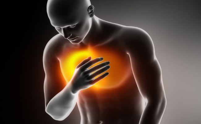
Manifested by pain in the heart area and pericardial friction rub. Pain in the chest is dull and pressing, sometimes radiating to the left shoulder blade, neck, and both shoulders. More often, moderate pain occurs, but it can be severe and painful, reminiscent of an angina attack. Unlike pain in the heart with angina, pericarditis is characterized by its gradual increase, duration from several hours to several days, lack of response when taking nitroglycerin, temporary subsidence from taking narcotic analgesics. Patients may simultaneously feel shortness of breath, palpitations, general malaise, dry cough, chills, which brings the symptoms of the disease closer to those of dry pleurisy. A characteristic sign of pain with pericarditis is its intensification with deep breathing, swallowing, coughing, changing body position (decreased in a sitting position and increased in a supine position), shallow and frequent breathing.
A pericardial friction rub is detected by listening to the patient's heart and lungs. Dry pericarditis may heal in 2-3 weeks or become exudative or adhesive.
Exudative pericarditis
Exudative (effusion) pericarditis develops as a consequence of dry pericarditis or independently with rapidly beginning allergic, tuberculous or tumor pericarditis.
There are complaints of pain in the heart area, a feeling of tightness in the chest. When exudate accumulates, blood circulation through the vena cava, hepatic and portal veins is disrupted, shortness of breath develops, the esophagus is compressed (the passage of food is disrupted - dysphagia), and the phrenic nerve (hiccups appear). Almost all patients have a fever. For appearance Patients are characterized by a swollen face, neck, anterior surface of the chest, swelling of the neck veins (“Stokes collar”), pale skin with cyanosis. On examination, smoothing of the intercostal spaces is noted.
Complications of pericarditis
When exudative pericarditis the development of acute cardiac tamponade is possible, in the case of constrictive pericarditis - the occurrence of circulatory failure: compression of the vena cava and hepatic veins, the right atrium by exudate, which complicates ventricular diastole; development of false cirrhosis of the liver.
Pericarditis causes inflammatory and degenerative changes in the myocardial layers adjacent to the effusion (myopericarditis). Due to the development of scar tissue, fusion of the myocardium with nearby authorities, chest and spine (mediastino-pericarditis).
Diagnosis of pericarditis
It is very important to diagnose pericardial inflammation in a timely manner, as it can pose a threat to the patient’s life. Such cases include compressive pericarditis, exudative pericarditis with acute cardiac tamponade, purulent and tumor pericarditis. It is necessary to differentiate the diagnosis from other diseases, mainly acute myocardial infarction and acute myocarditis, identify the cause of pericarditis.
Diagnosis of pericarditis includes taking a medical history, examining the patient (listening and percussing the heart), laboratory research. General, immunological and biochemical (total protein, protein fractions, sialic acids, creatine kinase, fibrinogen, seromucoid, CRP, urea, LE cells) blood tests are performed to clarify the cause and nature of pericarditis.
ECG has great importance in the diagnosis of acute dry pericarditis, the initial stage of exudative pericarditis and adhesive pericarditis (with compression of the cavities of the heart). In the case of exudative and chronic inflammation of the pericardium, a decrease in the electrical activity of the myocardium is observed. FCG (phonocardiography) notes systolic and diastolic murmurs not associated with the functional cardiac cycle, and periodically occurring high-frequency oscillations.
X-ray of the lungs is informative for the diagnosis of exudative pericarditis (an increase in the size and a change in the silhouette of the heart is observed: a spherical shadow is characteristic of an acute process, triangular - for a chronic one). When up to 250 ml of exudate accumulates in the pericardial cavity, the size of the heart shadow does not change. There is a weakened pulsation of the heart shadow contour. The shadow of the heart is poorly visible behind the shadow of the pericardial sac filled with exudate. With constrictive pericarditis, visible fuzzy outlines heart due to pleuropericardial adhesions. A large number of adhesions can cause a “stationary” heart that does not change shape and position when breathing and changing body position. With an “armored” heart, calcareous deposits are noted in the pericardium.
With a rapid increase in the accumulation of exudate (threat of cardiac tamponade), a pericardial puncture (pericardiocentesis) is performed to remove the effusion. Pericardial puncture is also used in case of prolonged resorption of effusion (with treatment for more than 2 weeks) to identify its nature and nature (tumor, tuberculosis, fungal, etc.).
Patients with constrictive pericarditis in case of chronic venous stagnation and compression of the heart, operations on the pericardium are performed: resection of scarred areas of the pericardium and adhesions (subtotal pericardectomy).
Prognosis and prevention
The prognosis in most cases is favorable; with correct treatment started in a timely manner, patients’ ability to work is almost completely restored. When purulent pericarditis in the absence of urgent therapeutic measures the disease can be life-threatening. Adhesive (adhesive) pericarditis leaves permanent changes, because surgical intervention is not effective enough.
Only secondary prevention of pericarditis is possible, which consists of clinical observation with a cardiologist, rheumatologist, regular monitoring of electrocardiography and echocardiography, sanitation of lesions chronic infection, healthy way life, moderate physical activity.
Extended combined pulmonary resections of the vascular-atrial type include surgical interventions performed: with resection of the pericardium, pulmonary veins with the wall of the left atrium, superior vena cava, extrapulmonary parts pulmonary arteries and the walls of their common trunk, the aorta.
Resections of this type are most often performed during combined operations for lung cancer. Thus, out of 605 patients operated on in the clinic, they were performed on 424, which amounted to 70.1%. Only in 168 (42%) they were single, and in the majority of patients they were multiple. Moreover, only in 82 cases they included other resections of the same type, and more often they were combined with resections of other types (mediastinal-esophageal, tracheobronchial, parietal-diaphragmatic). Of the 424 patients, 401 (94.6%) underwent pneumonectomies and 23 (5.4%) underwent partial pulmonary resections.
In all patients who underwent resections of the vascular-atrial type, multiple metastases were determined in the lymph nodes of the root of the lung and mediastinum. In only 31 patients they were limited to damage to the lymph nodes of the lung root; in all other patients they affected the lymph nodes of the mediastinum.
Pericardial resection is the most frequent sight resections of extrapulmonary formations and organs chest cavity in patients with advanced stages of lung cancer. Resection of the pericardium was performed in 362 patients, which was 85.4% among all who underwent resections of the vascular-atrial type and 59.8% among those who underwent combined operations. The need to perform it arises when various localizations tumors, equally often on both the right and left. In our observations, it was less often performed in isolation, more often it was combined with other resections of various extrapulmonary formations and organs of the chest cavity. Resection of the pericardium is, as a rule, a mandatory element during resection of the heart wall and its vessels, and is often combined with resections of the mediastinal-esophageal and tracheobronchial type.
Invasion of the cardiac membrane by a tumor or its metastases requires its wide excision. The resulting defect in the pericardial wall, especially after pneumonectomy, creates the prerequisites for the development of a severe complication - “dislocation” of the heart into the pleural cavity with a sharp disruption of its activity (Vishnevsky A.A. et al., 1978; Ivchenko Yu.B., Volotsenko M.A., 1990). After extensive resections of the pericardium, it is rarely possible to close the defect.
More often you have to resort to plastic closure. Many methods of pericardial plastic surgery have been proposed. The most commonly used flap of the parietal pleura, taken on a pedicle or freely along with the intrathoracic fascia, pericardial adipose tissue. However, they are mechanically fragile and it is not always possible after extensive surgical interventions to cut out a sufficiently large area of parietal pleura or adipose tissue to close the resulting pericardial defect. It is much more convenient and reliable to use alloplastic materials for this purpose.
Since 1981, the clinic has been using the Bulgarian antibacterial polycaproamide mesh “Ampoxen” (BAPP) for pericardial alloplasty after resection, created in 1976 at the suggestion of Professor N. Vasilev by a team of employees led by Professor K. Dimov. The mesh is knitted from polyfilament fibers with a thickness of 20 microns, cell size 1 mm. The antibacterial effect is achieved by creating a special chemical bond between the polymer and medicinal substance, which may contain various antibiotics and antiseptics. BAPP has strength, optimal elasticity, does not allergize the body, does not have a blastomogenic effect, has chemical and biological inertness, and hemostatic properties (Vasilev N. et al., 1982; Penchev R. et al., 1984).
To study the fate of the implant and the reaction of surrounding tissues to it, we carried out 29 experimental research on dogs for pericardial plasty of BAPP. After pneumonectomy was performed in 14 animals on the right and 15 on the left, a section of the pericardium with an area of 10 cm was resected and the resulting defect in the BAPP was repaired. Animals were removed from the experiment at 1, 6, 8, 11, 14 days, 1 and 2 months, 1 year, followed by macroscopic and histological examination of the preparations.
It has been established that in the tissues directly adjacent to the mesh, a natural change in the phases of inflammation is detected: unexpressed alterative phenomena in the adjacent epicardium on the 1st day, accompanied by the loss of fibrin on the mesh, are replaced by a picture of alterative-infiltrative inflammation in the subepicardial layers of the myocardium with the formation of adhesions on the 3rd day. day. Subsequently, proliferative changes progress at the site of plastic surgery and tissue contact with the implant, manifested by the formation of a connective tissue scar. In the long term (up to 1 year), complete resorption of the mesh does not occur.
Thus, the Bulgarian antibacterial polycaproamide mesh "Ampoxen" is a good plastic material for closing defects when performing extended combined operations for lung cancer with resection of the pericardium. Emerging inflammatory reaction tissue on the implant is not pronounced and is local in nature, does not cause progressive pericarditis. Subsequently it happens partial resorption mesh with the formation of a soft connective tissue scar at the point of contact of the heart with plastic material, which does not impede the work of the heart.
BAPP "Ampoxen" was used for plastic surgery of pericardial defects in 61 patients during extended combined resections for lung cancer. We did not note the development of complications in any patient. postoperative period, which could be connected with the use of a grid. In our opinion, the Ampoxen BAPP is a convenient, reliable and safe material for alloplasty of pericardial defects after extensive resection.
The second most common type of vascular-atrial resection is resection of the pulmonary veins with a section of the left atrium. Among our patients, they were performed in 64 patients, which amounted to 15.1% of patients who underwent resections of the vascular-atrial type and 10.6% of all operated patients who underwent combined operations.
Resections of the pulmonary veins with the atrium equally often have to be performed for right- and left-sided lung lesions. The need to perform them arises in patients with advanced local spread of the tumor, characterized by the extensiveness and multiplicity of damage to various extrapulmonary anatomical structures and organs of the chest cavity. Therefore, as a rule, they are multiple, often combined. Such surgical interventions are traumatic, increased danger development of severe intraoperative complications.
Resection of the pulmonary veins with a section of the left atrium with a right-sided tumor localization is much more technically complex and dangerous, due to the peculiarities of their anatomical structure. Short, inactive, especially when germinated by tumor tissue, deeply located, flowing into the atrium on the posterior surface, they are, as a rule, inaccessible to typical treatment with separate ligation of vessels. Ligation of a wide, thin-walled common venous trunk poses a significant risk due to the possibility of cutting through it with a ligature.
It is preferable to bring the mechanical suturing device of the cardiac auricle directly to the atrium and apply stapled sutures here. If the local spread of the tumor allows, in order to increase the reliability of the sutures, even before cutting off the vessel, a second one is applied somewhat proximally, at a distance of 3 mm from the first line.
During such resection, a double mechanical hardware suture does not require additional reinforcement and is quite reliable. When performing resection of the atrium from the right-sided approach using mechanical suturing devices, it is necessary, when applying the device, to control the location of the interatrial groove of the heart and suturing the left atrium posterior to it. Involvement in the seam interatrial septum and a section of the anterior wall of the right atrium can lead to severe disorders heart rate and even mechanical narrowing of the mouth of the superior vena cava (Volodos N.L., 1987).
Suturing the left atrium wall during right-sided resections using hand sutures is difficult and dangerous. Performing a well-adapted manual suture requires prior application of a vascular clamp (such as a Satinsky clamp) and intersection of the atrial wall distal to it. With constant traction on the clamp while making a suture, the thin and mechanically fragile posterior wall of the atrium may rupture or slip out of the jaws of the clamp, which leads to massive, very difficult to stop bleeding.
If such a complication occurs, it seems advisable for us to abandon attempts to capture the damaged atrium deep in the wound filled with blood using a vascular clamp, as this can lead to an increase in the rupture and increased bleeding. It is necessary to press the atrium wall to the spine with a tuffer to temporarily stop or at least reduce the bleeding, drain the surgical field, and then suture the atrial defect with a continuous suture or furrier's suture, be sure to include the section of the dissected posterior pericardial layer in the suture along with the posterior wall of the atrium. The pericardium, acting as a gasket, avoids the cutting of sutures. In such a situation, leading to back wall the atrium of other autologous or alloplastic materials is extremely difficult.
Bisenkov L.N., Grishakov S.V., Shalaev S.A.
