Anatomical structure and functions of the ear. The structure and functions of the ear Human ear canal
There are a lot of diseases that signal their development with pain in the ears. To determine what specific disease affected the organ of hearing, you need to understand how the human ear is arranged.
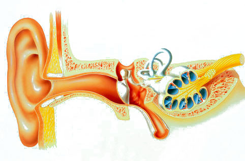
Diagram of the auditory organ
First of all, let's understand what an ear is. This is an auditory-vestibular paired organ that performs only 2 functions: the perception of sound impulses and responsibility for the position of the human body in space, as well as for maintaining balance. If you look at the human ear from the inside, its structure suggests the presence of 3 parts:
- external (external);
- average;
- internal.
Each of them has its own no less intricate device. Connecting, they are a long pipe penetrating into the depths of the head. Let us consider the structure and functions of the ear in more detail (the diagram of the human ear demonstrates them best).
What is the outer ear
The structure of the human ear (its outer part) is represented by 2 components:
- ear shell;
- external ear canal.

The shell is an elastic cartilage that completely covers the skin. It has a complex shape. In its lower segment there is a lobe - this is a small skin fold filled inside with a fatty layer. By the way, it is the outer part that has the highest sensitivity to various kinds of injuries. For example, for fighters in the ring, it often has a form that is very far from its original form.
The auricle serves as a kind of receiver for sound waves, which, falling into it, penetrate deep into the organ of hearing. Since it has a folded structure, the sound enters the passage with little distortion. The degree of error depends, in particular, on the place where the sound comes from. Its location is horizontal or vertical.
 It turns out that more accurate information about where the sound source is located enters the brain. So, it can be argued that the main function of the shell is to catch sounds that should enter the human ear.
If you look a little deeper, you can see that the shell extends the cartilage of the external ear canal. Its length is 25-30 mm. Next, the cartilage zone is replaced by bone. The outer ear completely lines the skin, which contains 2 types of glands:
- sulfuric;
- greasy.
The outer ear, the device of which we have already described, is separated from the middle part of the hearing organ by a membrane (it is also called the tympanic membrane).
How is the middle ear
If we consider the middle ear, its anatomy is:
- tympanic cavity;
- eustachian tube;
- mastoid process.
All of them are interconnected. The tympanic cavity is a space outlined by the membrane and the region of the inner ear. Its location is the temporal bone. The structure of the ear here looks like this: in the anterior part, there is a union of the tympanic cavity with the nasopharynx (the function of the connector is performed by the Eustachian tube), and in its posterior part - with the mastoid process through the entrance to its cavity. Air is present in the tympanic cavity, which enters there through the Eustachian tube.

The anatomy of the ear of a person (child) up to 3 years old has a significant difference from how the ear of an adult is arranged. Babies do not have a bone passage, and the mastoid process has not yet grown. The children's middle ear is represented by only one bone ring. Its inner edge has the shape of a groove. It just houses the tympanic membrane. In the upper zones of the middle ear (where there is no this ring), the membrane is connected to the lower edge of the scales of the temporal bone.
When the baby reaches the age of 3, the formation of his ear canal is completed - the structure of the ear becomes the same as in adults.
Anatomical features of the internal department
 The inner ear is the most difficult part of it. The anatomy in this part is very complex, so she was given a second name - "webbed labyrinth of the ear." It is located in the stony zone of the temporal bone. It is attached to the middle ear with windows - round and oval. Comprises:
- vestibule;
- snails with the organ of Corti;
- semicircular canals (filled with fluid).
In addition, the inner ear, the structure of which provides for the presence of the vestibular system (apparatus), is responsible for constantly keeping the body in a state of balance by a person, as well as for the possibility of accelerating in space. The vibrations that occur in the oval window are transmitted to the fluid that fills the semicircular canals. The latter serves as an irritant for the receptors located in the cochlea, and this already becomes the cause of the launch of nerve impulses.
It should be noted that the vestibular apparatus has receptors in the form of hairs (stereocilia and kinocilia), which are located on special elevations - maculae. These hairs are located one opposite the other. By shifting, stereocilia provoke the occurrence of excitation, and kinocilia help inhibition.
Summing up
In order to more accurately imagine the structure of the human ear, the diagram of the organ of hearing should be in front of the eyes. It usually depicts a detailed structure of the human ear.
 Obviously, the human ear is a rather complex system, consisting of many different formations, each of which performs a number of important and truly irreplaceable functions. The diagram of the ear demonstrates this clearly.
Regarding the structure of the outer part of the ear, it should be noted that each person has individual genetically determined features that in no way affect the main function of the hearing organ.
Ears need regular hygienic care. If you neglect this need, you can partially or completely lose your hearing. Also, lack of hygiene can lead to the development of diseases affecting all parts of the ear.
The ear is a paired organ located deep in the temporal bone. The structure of the human ear allows you to receive mechanical vibrations of the air, transmit them through internal media, transform and transmit them to the brain.
The most important functions of the ear include the analysis of body position, coordination of movements.
In the anatomical structure of the human ear, three sections are conventionally distinguished:
- external;
- average;
- internal.
ear shell
It consists of cartilage up to 1 mm thick, over which there are layers of perichondrium and skin. The earlobe is devoid of cartilage, consists of adipose tissue covered with skin. The shell is concave, along the edge there is a roller - a curl.
Inside it is an antihelix, separated from the curl by an elongated recess - a rook. From the antihelix to the ear canal there is a recess called the cavity of the auricle. The tragus protrudes in front of the ear canal.
ear canal
Reflecting from the folds of the ear shell, the sound moves into the auditory 2.5 cm in length, with a diameter of 0.9 cm. The basis of the ear canal in the initial section is cartilage. It resembles the shape of a gutter, open up. In the cartilaginous region, there are santorian fissures bordering the salivary gland.
The initial cartilaginous part of the ear canal passes into the bone part. The passage is bent in a horizontal direction, to inspect the ear, the shell is pulled back and up. In children - back and down.
The ear passage is lined with skin with sebaceous, sulfuric glands. Sulfur glands are modified sebaceous glands that produce. It is removed during chewing due to vibrations of the walls of the ear canal.
It ends with the tympanic membrane, blindly closing the ear canal, bordering:
- with the joint of the lower jaw, when chewing, the movement is transmitted to the cartilaginous part of the passage;
- with cells of the mastoid process, facial nerve;
- with salivary gland.
 The membrane between the outer ear and the middle ear is an oval translucent fibrous plate, 10 mm long, 8-9 mm wide, 0.1 mm thick. The membrane area is about 60 mm 2 .
The plane of the membrane is inclined to the axis of the auditory canal at an angle, drawn funnel-shaped into the cavity. The maximum tension of the membrane is in the center. Behind the tympanic membrane is the cavity of the middle ear.
Distinguish:
- middle ear cavity (tympanic);
- auditory tube (Eustachian);
- auditory ossicles.
tympanic cavity
The cavity is located in the temporal bone, its volume is 1 cm 3. It houses the auditory ossicles, articulated with the eardrum.
Above the cavity is placed the mastoid process, consisting of air cells. It houses a cave - an air cell that serves as the most characteristic landmark in the anatomy of the human ear when performing any ear surgery.
auditory trumpet
 The formation is 3.5 cm long, with a lumen diameter of up to 2 mm. Its upper mouth is located in the tympanic cavity, the lower pharyngeal mouth opens in the nasopharynx at the level of the hard palate.
The auditory tube consists of two sections, separated by its narrowest point - the isthmus. The bony part departs from the tympanic cavity, below the isthmus - membranous-cartilaginous.
The walls of the tube in the cartilaginous section are usually closed, slightly open when chewing, swallowing, yawning. The expansion of the lumen of the tube is provided by two muscles associated with the palatine curtain. The mucous membrane is lined with epithelium, the cilia of which move towards the pharyngeal mouth, providing the drainage function of the tube.
 The smallest bones in the human anatomy - the auditory ossicles of the ear, are intended for conducting sound vibrations. In the middle ear there is a chain: hammer, stirrup, anvil.
The malleus is attached to the tympanic membrane, its head articulates with the incus. The process of the incus is connected to the stirrup attached by its base to the window of the vestibule located on the labyrinth wall between the middle and inner ear.

The structure is a labyrinth consisting of a bone capsule and a membranous formation that repeats the shape of the capsule.
In the bony labyrinth, there are:
- vestibule;
- snail;
- 3 semicircular canals.
Snail

The bone formation is a three-dimensional spiral of 2.5 turns around the bone rod. The width of the base of the cochlear cone is 9 mm, the height is 5 mm, and the length of the bone spiral is 32 mm. A spiral plate extends from the bone rod into the labyrinth, which divides the bone labyrinth into two channels.
At the base of the spiral lamina are the auditory neurons of the spiral ganglion. The bony labyrinth contains perilymph and a membranous labyrinth filled with endolymph. The membranous labyrinth is suspended in the bony labyrinth with the help of strands.
Perilymph and endolymph are functionally related.
- Perilymph - in ionic composition close to blood plasma;
- endolymph - similar to the intracellular fluid.
 Violation of this balance leads to an increase in pressure in the labyrinth.
The cochlea is an organ in which the physical vibrations of the perilymph fluid are converted into electrical impulses from the nerve endings of the cranial centers, which are transmitted to the auditory nerve and to the brain. At the top of the cochlea is the auditory analyzer - the organ of Corti.
threshold
The most ancient anatomically the middle part of the inner ear is a cavity bordering the scala cochlea through a spherical sac and semicircular canals. On the wall of the vestibule leading to the tympanic cavity, there are two windows - oval, covered with a stirrup and round, which is a secondary tympanic membrane.
Features of the structure of the semicircular canals
All three mutually perpendicular bony semicircular canals have a similar structure: they consist of an expanded and simple pedicle. Inside the bone there are membranous canals that repeat their shape. The semicircular canals and sacs of the vestibule make up the vestibular apparatus, are responsible for balance, coordination, and determining the position of the body in space.
In a newborn, the organ is not formed; it differs from an adult in a number of structural features.
Auricle
- The shell is soft;
- the lobe and curl are poorly expressed, are formed by 4 years.
ear canal
- The bone part is not developed;
- the walls of the passage are located almost close;
- the tympanic membrane lies almost horizontally.
- Almost the size of adults;
- in children, the eardrum is thicker than in adults;
- covered with mucous membrane.
tympanic cavity
 In the upper part of the cavity there is an open gap through which, in acute otitis media, the infection can penetrate the brain, causing meningism. In an adult, this gap is overgrown.
The mastoid process in children is not developed, it is a cavity (atrium). The development of the process begins at the age of 2 years, ends by 6 years.
auditory trumpet
In children, the auditory tube is wider, shorter than in adults, and is located horizontally.
A complex paired organ receives sound vibrations of 16 Hz - 20,000 Hz. Injuries, infectious diseases reduce the threshold of sensitivity, lead to a gradual loss of hearing. Advances in medicine in the treatment of ear diseases and hearing aids make it possible to restore hearing in the most difficult cases of hearing loss.
Video about the structure of the auditory analyzer
The ear is a complex organ with two functions: listening, through which we perceive sounds and interpret them, thus communicating with the environment; and maintaining body balance.
Auricle- captures and directs sound waves into the internal auditory canal;
rear labyrinth, or semicircular canals - directs movements to the head and brain to regulate the balance of the body;
front labyrinth, or cochlea - contains sensory cells, which, capturing the vibrations of sound waves, transform mechanical impulses into nerve impulses;
Auditory nerve- directs general nerve impulses to the brain;
Bones of the middle ear: hammer, anvil, stirrup - receive vibrations from auditory waves, amplify them and transmit them to the inner ear;
external ear canal- picks up sound waves coming from outside and sends them to the middle ear;
Eardrum- a membrane that vibrates when sound waves hit it and transmits vibrations along the chain of bones in the middle ear;
Eustachian tube canal that connects the tympanic membrane to the pharynx
in equilibrium the pressure created in the middle ear with the pressure of the environment.
The ear is divided into three sections, the functions of which are different.
; the outer ear consists of the auricle and the external auditory canal, its purpose is to capture sounds;
; the middle ear is located in the temporal bone, separated from the inner ear by a movable membrane - the tympanic membrane - and contains three articular bones: the hammer, anvil and stirrup, which are involved in the transmission of sounds to the cochlea;
; the inner ear, also called the labyrinth, is formed from two sections that perform different functions: the anterior labyrinth, or cochlea, where the organ of Corti is located, is responsible for hearing, and the posterior labyrinth, or semicircular canals, in which impulses are generated that take part in maintaining balance body (article "Balance and hearing")

The inner ear, or labyrinth, consists of a very strong bony skeleton, the ear capsule, or bony labyrinth, within which is a membranous mechanism with a bone-like structure, but consisting of membranous tissue. The inner ear is hollow but filled with fluid: between the bony labyrinth and the membrane is perilymph, while the labyrinth itself is filled with endolymph. The anterior labyrinth, whose bony form is called the cochlea, contains structures that generate auditory impulses. The posterior labyrinth, which takes part in the regulation of the balance of the body, has a bone skeleton, consisting of a cubic part, a vestibule and three channels in the form of an arc - semicircular, each of which includes a space with a flat plane.
The cochlea, so named because of its spiral shape, contains a membrane composed of fluid-filled channels: a triangular central canal and a whorl containing endolymph, which is located between the scala vestibuli and the scala tympani. These two scalas are partly separated, leading to large canals of the cochlea covered with thin membranes separating the inner ear from the middle ear: the scala tympani begins at the oval fenestra, while the scala vestibuli reaches the round fenestra. The cochlea, which has a triangular shape, consists of three faces: the upper one, which is separated from the scala vestibule by the Reissner membrane, the lower one, separated from the scala tympani by the main membrane, and the side, which is attached to the shell and is a vascular groove that produces endolymph. Inside the cochlea there is a special auditory organ - Corti (the mechanism of sound perception is described in detail in the article "
Hearing is one of the important sense organs. It is with the help of it that we perceive the slightest changes in the world around us, we hear alarm signals warning of danger. is very important for all living organisms, although there are those who do without it.
In humans, the auditory analyzer includes the external, middle, and from them, along the auditory nerve, information goes to the brain, where it is processed. In the article we will dwell in more detail on the structure, functions and diseases of the outer ear.
The structure of the outer ear
The human ear consists of several sections:
- External.
- Middle ear.
- Internal.
The outer ear includes:
Starting with the most primitive vertebrates, which developed hearing, the structure of the ear gradually became more complicated. This is due to the general increase in the organization of animals. For the first time, the outer ear appears in mammals. In nature, there are some species of birds with an auricle, for example, a long-eared owl.
Auricle
The outer ear of a person begins with the auricle. It consists almost entirely of cartilaginous tissue with a thickness of about 1 mm. It does not have cartilage in its structure, only it consists of adipose tissue and is covered with skin.
The outer ear is concave with a curl at the edge. It is separated by a small depression from the internal antihelix, from which the auricle cavity extends towards the ear canal. A tragus is located at the entrance to the ear canal.
ear canal
The next department, which has the outer ear, - ear canal. It is a tube 2.5 centimeters long and 0.9 cm in diameter. It is based on cartilage, resembling a gutter in shape, opening up. There are santorian fissures in the cartilaginous tissue, which border on the salivary gland.

Cartilage is present only in the initial section of the passage, then it passes into bone tissue. The ear canal itself is slightly curved in a horizontal direction, so when examining a doctor, the auricle is pulled back and up in adults, and back and down in children.
Inside the ear canal there are sebaceous and sulfuric glands, which produce its removal is facilitated by the process of chewing, during which the walls of the passage vibrate.
The ear canal ends with the tympanic membrane, which blindly closes it.
Eardrum
The tympanic membrane connects the outer and middle ear. It is a translucent plate with a thickness of only 0.1 mm, its area is about 60 mm 2.

The tympanic membrane is located slightly obliquely relative to the auditory canal and is drawn in the form of a funnel into the cavity. It has the greatest tension in the center. Behind her is already
Features of the structure of the outer ear in infants
When a baby is born, his hearing organ is not yet fully formed, and the structure of the outer ear has a number of distinctive features:
- The auricle is soft.
- The earlobe and curl are practically not expressed, they are formed only by 4 years.
- There is no bony part in the ear canal.
- The walls of the passage are located almost nearby.
- The tympanic membrane is located horizontally.
- The size of the tympanic membrane does not differ from that of adults, but it is much thicker and covered with a mucous membrane.
The child grows, and with it the additional development of the organ of hearing occurs. Gradually, he acquires all the features of an adult auditory analyzer.
Functions of the outer ear
Each department of the auditory analyzer performs its function. The outer ear is intended primarily for the following purposes:

Thus, the functions of the outer ear are quite diverse, and the auricle serves us not only for beauty.
Inflammatory process in the outer ear
Quite often, colds end with an inflammatory process inside the ear. This problem is especially relevant in children, since the auditory tube is short in size, and the infection can quickly penetrate the ear from the nasal cavity or throat.
For everyone, inflammation in the ears can manifest itself in different ways, it all depends on the form of the disease. There are several types:

You can cope at home only with the first two varieties, but internal otitis media requires inpatient treatment.
If we consider otitis externa, then it can also be of two forms:
- Limited.
- diffuse.
The first form occurs, as a rule, as a result of inflammation of the hair follicle in the ear canal. In a way, this is a common boil, but only in the ear.
The diffuse form of the inflammatory process covers the entire passage.
Causes of otitis media
There are a lot of reasons that can provoke an inflammatory process in the outer ear, but among them the following are often found:
- bacterial infection.
- Fungal disease.
- Allergic problems.
- Improper hygiene of the ear canal.
- Self attempt to remove ear plugs.
- Entry of foreign bodies.
- Viral nature, although this happens very rarely.
Cause of outer ear pain in healthy people
It is not at all necessary that if there is pain in the ear, a diagnosis of otitis media is made. Often such pain can occur for other reasons:
- Walking in windy weather without a hat can cause ear pain. The wind exerts pressure on the auricle and a bruise forms, the skin becomes cyanotic. This condition passes quickly enough after getting into a warm room, treatment is not required.
- Swimmers also have a frequent companion. Because during exercise, water enters the ears and irritates the skin, it can lead to swelling or otitis externa.
- Excessive accumulation of sulfur in the ear canal can cause not only a feeling of congestion, but also pain.
- Insufficient excretion of sulfur by the sulfur glands, on the contrary, is accompanied by a feeling of dryness, which can also cause pain.
As a rule, if otitis media does not develop, all discomfort in the ear disappears on its own and does not require additional treatment.
Symptoms of otitis externa
If the doctor diagnoses damage to the ear canal and auricle, the diagnosis is otitis externa. Its manifestations may be as follows:
- Pain can vary in intensity, from very subtle to disturbing sleep at night.
- This condition can last for several days, and then subside.
- In the ears there is a feeling of congestion, itching, noise.
- During the inflammatory process, hearing acuity may decrease.
- Since otitis media is an inflammatory disease, body temperature may rise.
- The skin near the ear may acquire a reddish tint.
- When pressing on the ear, the pain intensifies.
Inflammation of the external ear should be treated by an ENT doctor. After examining the patient and determining the stage and severity of the disease, medications are prescribed.
Therapy of limited otitis media
This form of the disease is usually treated with surgery. After the introduction of an anesthetic drug, the boil is opened and the pus is removed. After this procedure, the patient's condition improves significantly.
For some time, you will have to take antibacterial medicines in the form of drops or ointments, for example:
- Normax.
- "Candibiotic".
- "Levomekol".
- "Celestoderm-V".
Usually, after a course of antibiotics, everything returns to normal, and the patient recovers completely.
Therapy for diffuse otitis media
Treatment of this form of the disease is carried out only conservatively. All medications are prescribed by a doctor. Usually the course includes a set of measures:
- Taking antibacterial drops, for example, Ofloxacin, Neomycin.
- Anti-inflammatory drops "Otipaks" or "Otirelax".
- Antihistamines ("Citrine", "Claritin") help relieve swelling.
- To relieve pain, NPS are prescribed, for example, Diclofenac, Nurofen.
- To increase immunity, the intake of vitamin-mineral complexes is indicated.
During treatment, it must be remembered that any warming procedures are contraindicated, they can only be prescribed by a doctor at the stage of recovery. If all the doctor's recommendations are followed and the full course of therapy is completed, then you can be sure that the outer ear will be healthy.
Treatment of otitis media in children
In babies, the physiology is such that the inflammatory process very quickly spreads from the nasal cavity to the ear. If you notice in time that the child is worried about the ear, then the treatment will be short and uncomplicated.

The doctor usually does not prescribe antibiotics. All therapy consists in taking antipyretic drugs and painkillers. Parents can be advised not to self-medicate, but to adhere to the doctor's recommendations.
Drops that are bought on the recommendation of friends can only harm your child. When a baby is sick, the appetite usually decreases. You can’t force him to eat by force, it’s better to give him more to drink so that toxins are eliminated from the body.
If the child is too often over ear infections, there is reason to talk to the pediatrician about vaccination. In many countries, such a vaccination is already being done, it will protect the outer ear from inflammatory processes that are caused by bacteria.
Prevention of inflammatory diseases of the external ear
Any inflammation of the external ear can be prevented. To do this, you need to follow only a few simple recommendations:

If the pain in the ear does not cause much concern, this does not mean that you should not see a doctor. Running inflammation can turn into much more serious problems. Timely treatment will allow you to quickly cope with otitis externa and relieve suffering.
The ear is an important organ in the human body that provides hearing, balance and orientation in space. It is both an organ of hearing and a vestibular analyzer. The human ear has a rather complex structure. It can be divided into three main sections: outer, middle and inner. This division is associated with the characteristics of the functioning and defeat of each of them in various diseases.
outer ear
The human ear includes the outer, middle and inner ear. Each part performs its functions.This section of the auditory analyzer consists of the external auditory meatus and the auricle. The latter is located between the temporomandibular joint and the mastoid process. It is based on elastic type cartilage tissue, which has a complex relief, covered with perichondrium and skin on both sides. Only one section of the auricle (lobe) is represented by adipose tissue and is devoid of cartilage. The size of the auricle can vary slightly from person to person. However, normally its height should correspond to the length of the back of the nose. Deviations from this size can be regarded as macro- and microotia.
The auricle, forming a constriction in the form of a funnel, gradually passes into the ear canal. It has the form of a curved tube of various diameters about 25 mm long, which consists of a cartilaginous and bone section. From above, the external auditory meatus borders on the middle cranial fossa, below - on the salivary gland, in front - on the temporomandibular joint and behind - on the mastoid cells. It ends at the entrance to the middle ear cavity, closed by the tympanic membrane.
Data on this neighborhood are important for understanding the spread of the pathological process to adjacent structures. So, with inflammation of the anterior wall of the auditory canal, the patient may experience severe pain during chewing due to the involvement of the temporomandibular joint in the pathological process. The back wall of this passage is affected by (inflammation of the mastoid process).
The skin covering the structures of the outer ear is heterogeneous. In the depths of it, it is thin and vulnerable, and in the outer sections it contains a large number of hairs and glands that produce earwax.
Middle ear
The middle ear is represented by several air-bearing formations that communicate with each other: the tympanic cavity, the mastoid cavern and the Eustachian tube. With the help of the latter, the middle ear communicates with the pharynx and the external environment. It has the appearance of a triangular canal about 35 mm long, which opens only when swallowing.
The tympanic cavity is a small, irregularly shaped space resembling a cube. From the inside, it is covered with a mucous membrane, which is a continuation of the nasopharyngeal mucosa and has a number of folds and pockets. It is here that the chain of auditory ossicles is located, consisting of the anvil, malleus and stirrup. Between themselves, they form a mobile connection with the help of joints and ligaments.
The tympanic cavity has six walls, each of which plays an important role in the functioning of the middle ear.
- The tympanic membrane, which separates the middle ear from the environment, is its outer wall. This membrane is very thin, but elastic and low-elastic anatomical structure. It is funnel-shaped drawn in in the center and consists of two parts (stretched and loose). In the stretched part, there are two layers (epidermal and mucous), and in the loose part, a middle (fibrous) layer is added. The handle of the malleus is woven into this layer, which repeats all the movements of the eardrum under the influence of sound waves.
- The inner wall of this cavity is at the same time the wall of the labyrinth of the inner ear; it contains the window of the vestibule and the window of the cochlea.
- The upper wall separates the middle ear from the cranial cavity, it has small holes through which blood vessels penetrate there.
- The bottom of the tympanic cavity borders on the jugular fossa with the bulb of the jugular vein located in it.
- Its back wall communicates with the cave and other cells of the mastoid process.
- The mouth of the auditory tube is located on the anterior wall of the tympanic cavity, and the carotid artery passes outward from it.
The mastoid process in different people has an unequal structure. It can have a lot of air cells or be made of spongy tissue, or it can be very dense. However, regardless of the type of structure, there is always a large cavity in it - a cave, which communicates with the middle ear.
inner ear
 Schematic representation of the ear.
 The inner ear consists of the membranous and bony labyrinths and is located in the pyramid of the temporal bone.
The membranous labyrinth is located inside the bone labyrinth and exactly repeats its curves. All its departments communicate with each other. Inside it is a liquid - endolymph, and between the membranous and bony labyrinth - perilymph. These fluids differ in biochemical and electrolyte composition, but they are closely related to each other and participate in the formation of electrical potentials.
The labyrinth includes the vestibule, cochlea, and semicircular canals.
- The cochlea belongs to the auditory analyzer and has the appearance of a curled canal making two and a half turns around the bone tissue rod. A plate extends from it into the canal, which divides the cochlear cavity into two spiral corridors - the scala tympani and the scala vestibule. In the latter, the cochlear duct is formed, inside which there is a sound-perceiving apparatus or the organ of Corti. It consists of hair cells (which are receptors), as well as supporting and nourishing cells.
- The bony vestibule is a small cavity resembling a sphere in shape, its outer wall is occupied by the vestibule window, the anterior one by the cochlear window, and on the back wall there are openings leading to the semicircular canals. In the membranous vestibule there are two sacs with otolithic apparatus embedded in them.
- The semicircular canals are three curved tubes located in mutually perpendicular planes. And accordingly, they have names - anterior, posterior and lateral. Inside each of them are vestibular sensory cells.
Functions and physiology of the ear
The human body picks up sounds and determines their direction with the help of the auricle. The structure of the ear canal increases the pressure of the sound wave on the eardrum. Together with it, the middle ear system, through the auditory ossicles, ensures the delivery of sound vibrations to the inner ear, where they are perceived by the receptor cells of the organ of Corti and transmitted along the nerve fibers to the central nervous system.
The sacs of the vestibule and the semicircular canals act as a vestibular analyzer. The sensory cells located in them perceive various accelerations. Under their influence, various vestibular reactions occur in the body (redistribution of muscle tone, nystagmus, increased blood pressure, nausea, vomiting).
Conclusion
In conclusion, I would like to note that knowledge about the structure and functioning of the ear is extremely important for otolaryngol doctors, as well as therapists and pediatricians. This helps specialists to correctly diagnose, prescribe treatment, perform surgical interventions, as well as predict the course of the disease and the possible development of complications. But a general idea of \u200b\u200bthis can also be useful to an ordinary person who is not directly related to medicine.
Informative videos on the topic "Anatomy of the human ear":
