How to check the blood vessels of the body, indications for such studies. Where is ultrasound of the vessels of the legs done, and what are the indications for ultrasound diagnostics of the arteries of the lower and upper extremities
Ultrasound of the vessels of the upper and lower limbs- one of the most informative, safe, quick ways diagnostics that allow assessing the degree vascular pathology and reveal it on early stages development of the disease. Initial method examinations are of the upper and lower extremities. Reinforced, this technique has no analogues in terms of studying the pathology of the vessels of the upper and lower extremities.
Principles of vascular ultrasound
The ultrasound method is based on the ability of low ultrafrequencies to be reflected from objects in motion. Analyzing the data obtained through ultrasound sensors, a specially designed algorithm (computer program) builds a graphic display of the characteristics of blood flow and vascular structure. A number of devices allow you to see a color image of the recorded processes. The movement of blood and pulsation of the veins and arteries of the upper and lower extremities can not only be seen, but also heard.
The ultrasound method allows you to see the blood flow system and a graphical representation of active processes. Some devices are capable of providing color images of structures
Ultrasound of the vessels of the lower extremities, and, if necessary, the upper extremities, is performed to study blood flow in the arteries and veins. In this case, it is possible to assess how passable the vessel under study is, its diameter, the lumen of the narrowing, etc. Unlike X-ray angiography, the image of arterial and venous structures during ultrasonic method It is done non-invasively (without damaging the skin) and without the introduction of contrast agents.
It should be said that the advantages of ultrasound of the vessels of the legs include harmlessness to the patient, since there is no radiation exposure. Therefore, it can be performed frequently (if necessary) on pregnant women and children.
Doppler ultrasound of blood vessels lower limbs
Indications for ultrasound scanning of the legs
Echography is performed if changes in arterial or venous circulation caused by varicose veins, trophic disorders or atherosclerotic process.
Indications for performing ultrasound scanning of the vessels of the lower extremities:
- painful sensations in the calf area (which appear at the end of the day; to relieve them, you can pour cool water on your legs or lift them up);
- the appearance of heaviness in the legs (especially in the evening);
- the appearance of pain in the leg, which occurs when walking (more than three to four kilometers), exercise (cycling, roller skating, etc.);
- if you suspect varicose veins of the legs, as well as nodular vascular formations An ultrasound scan of the veins of the lower extremities is performed;
- the appearance of swelling of the legs of unknown origin (in the evening);
- Constantly cold skin on the feet forces an examination;
- numbness, cramps in them;
- Ultrasound of the vessels of the legs is performed when the leg increases in volume;
- Availability trophic lesions skin of the lower leg (for example, ulcers);
- the study is done when it is detected in the patient the following diseases: hypertension, diabetes (mellitus), as these processes can lead to atherosclerosis;
- this examination is done when smoking, since this bad habit can lead to the appearance of atherosclerotic plaques on the walls of blood vessels.
Atherosclerotic plaque
Along with this, ultrasound of the vessels of the lower extremities, and, if necessary, the upper extremities, can be done for preventive purposes (as screening). In order to diagnose the pathology of the vascular bed as early as possible, especially in persons with a predisposition to this (persons of the following professions: truck drivers, surgeons, athletes).
Ultrasound examination of veins and arteries has no contraindications (practically). The only thing, ultrasound of the lower veins, if necessary - upper limbs, it will be difficult to do for a patient who is in in serious condition, so in this case it is not done.
Ultrasound scanning capabilities
Ultrasound of the veins of the lower extremities allows you to visualize vessels of various diameters, and you can identify the following manifestations of varicose veins:
- the cause of the disease;
- severity pathological changes;
- incompetence of venous valves;
- thrombotic changes (thrombus size, structure, flotation).
Along with this, ultrasound of the vessels of the lower extremities, in some cases, is carried out to study the arteries. In this case, the following arterial pathology can be identified:
- atherosclerotic plaques in the arteries;
- changes in the arteries caused by blood clots;
- blood flow disorders;
- the presence of arterial stenosis, the degree of this change;
- arterial aneurysms.
It should be noted that ultrasound of the vessels of the lower extremities is an integral method that can identify pathological changes leading to circulatory disorders. It is worth adding that modern vascular surgery cannot exist without this research.
Preparing the patient for an ultrasound scan
It should be said that preparation for this examination is not required (there are no dietary restrictions, taking medicines don't stop). It is not recommended to take stimulating drinks on the day of the study: tea, coffee; You should not smoke for several hours before the procedure.
Immediately before diagnostic manipulation, you should not use ointments, or undergo physical activity.
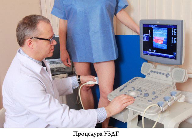
How is ultrasound scanning of blood vessels performed?
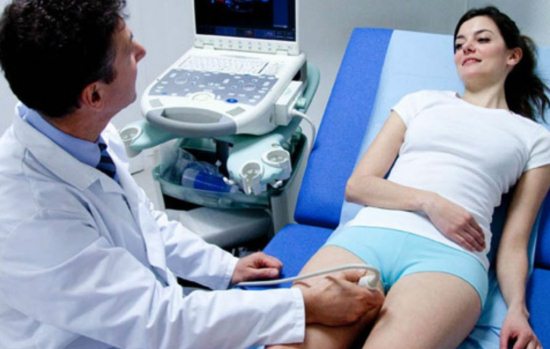
So, before starting this procedure, the patient needs to remove clothing from the area being examined (also compression underwear). Before the procedure begins, the doctor interviews the patient about complaints, the presence and duration of the disease.
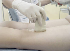
It should be added that the examination is usually carried out in horizontal position(lying) the patient on the couch, if necessary - standing. Before the examination, a special gel is applied to the patient’s skin to promote closer contact between the patient’s skin and the ultrasound sensor.
Application of special gel
The study is usually done within: thirty to thirty-five minutes. Immediately after the examination, the doctor interprets the results and gives a conclusion.
It should be noted that patients may experience numbness in the feet. This may be due to poor circulation in the leg. Therefore, in order to identify the cause pathological disorders Of course, an ultrasound of the veins of the lower extremities should be performed.
The condition of the deep veins, the speed and direction of blood flow in them, is shown by ultrasound of the veins of the lower (if necessary, also the upper extremities).
Advantages of ultrasound scanning
So to positive aspects this beam method relate:
- painlessness;
- non-invasive (no damage to the skin surface, no injections);
- ease of examination;
- relative cheapness (compared to MR angiography, X-ray angiography);
- absence of ionizing radiation;
- the research is done in real time;
- you can do a biopsy (if pathological formations are identified);
- good visualization of soft tissues (compared to X-ray angiography).
Ultrasound scanning of the blood vessels of the legs is completely painless and safe procedure
Disadvantages of Ultrasound Scanning
It can be said that vascular examination limbs (upper and lower) has the following negative aspects:
- Ultrasound alone is not enough to make a diagnosis;
- it is not always possible to assess the condition of small-caliber arteries and veins;
- the presence of atherosclerotic changes interferes with the passage of the ultrasound wave;
- cannot replace angiography (including computer or magnetic resonance imaging);
- if the study is done on an old device, then diagnostic value method is low.
It should be said that ultrasound doctors recommend periodically examining blood vessels: arteries, veins in patients with a predisposition to pathology ( occupational hazards: hairdressers, taxi drivers, overweight, smoking). This will help reduce the risk of developing diseases of the lower, and in some cases, upper extremities.
Thus, an ultrasound scan to identify vascular pathology of the legs is done to identify circulatory disorders. No patient preparation is required for this study. Given the need complex diagnostics vascular pathology, ultrasound examination done in combination with other radiation and functional methods. Attention! The ultrasound specialist does not make a diagnosis.
Ultrasound of the vessels of the lower extremities is a method using ultrasonic waves that allows you to show the vessels graphically and evaluate the parameters of their condition. In order to analyze the characteristics of blood flow, the property of an ultrasonic wave is used to visualize the picture when reflected from moving shaped elements blood. This technique is called Doppler ultrasound or Doppler ultrasound.
Types of ultrasound of blood vessels of the lower extremities
1.USDG (two-dimensional Doppler ultrasound)
- This study is now used quite rarely due to the emergence of other types of ultrasound of the vessels of the legs, as well as devices of higher diagnostic level. This type of examination can be used to assess the patency of the veins, the condition of the valves that exist in the superficial, deep and perforating veins (provided that the valves have a typical location).
2.USDS or USAS (duplex angioscanning)
- This combination Doppler study and energy mapping. To put it simply, when this study is performed, areas of different blood flow rates are highlighted in different colors.
Duplex ultrasound of the vessels of the lower extremities is the “gold standard”. Only on the basis of this study (except for his own examination) will the doctor be able to make a reasonable diagnosis and prescribe treatment.
It allows you to evaluate:
- condition of the walls of arterial and venous vessels
- patency of veins, both deep and superficial
- the nature of the damage to the vein valves, the degree of their insufficiency at any location of the veins
- the presence of blood clots, their type and size, the degree of narrowing of blood vessels by them
- and also determine the cause of relapse varicose veins after surgery or sclerotherapy.
3. Triplex ultrasound of vessels and veins of the lower extremities
- This is a volumetric (3D) study of blood vessels in color. This - optimal method examinations for those patients who have problems with the arteries or veins of the legs.
- It is especially informative in cases where you need to most clearly draw up a plan for the upcoming surgical intervention to avoid relapse or complications after surgery.
Who needs to undergo this examination?
You need to examine the arteries of the legs:
- for diabetes
- if you suffer from hypertension blood pressure, no matter what the reason is
- if walking is accompanied by pain in the legs
- for night pain in the legs, especially when it becomes easier when lowering them from the bed
- if you are suffering overweight body
- when smoking
- feet get cold quickly even with normal temperature environment
- history of myocardial infarction
- previous operations on the blood vessels of the legs
- increased blood cholesterol levels
Dopplerography of the venous collectors of the lower extremities is performed in the following cases
- swelling of the legs, especially occurring in the evening
- varicose veins visible to the eye
- leg cramps
- if pregnancy is complicated by varicose veins in the legs, swelling, pain
- pain in the leg, especially if they are accompanied by an increase in local or general temperature
- change in skin color on legs
- trophic ulcers.
How to prepare for the examination
Doppler ultrasound of the lower extremities is done without any preliminary preparation. She doesn't require anything special diet, nor stopping medications that treat your arteries or veins. If you are wearing compression garments, you will need to remove them during the examination.How is an ultrasound of the legs performed?
- Inspection of the vessels is carried out first in a lying position, with legs bent at the knees.
- Then the doctor must examine the vessels in vertical position patient.
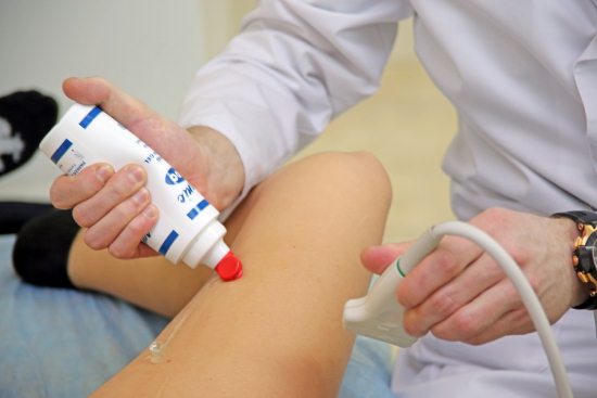
- Before starting the inspection, a small amount of special gel is applied to the legs, which serves to eliminate interference associated with air entering directly under the sensor.
During the procedure, the doctor manually selects the frequency of ultrasound radiation depending on the depth of the vessels and necessary degree its detail. The most commonly used frequency is 6-12 megahertz. Deep veins inspected with low-frequency sensors.
How is ultrasound decoding performed?
It is best to have an ultrasound scan of the vessels of the legs where you will not just be given a protocol with numbers and a brief conclusion, but where the vessels will be examined directly by a phlebologist or vascular surgeon. Based on the data obtained, he will assess the characteristics of blood movement through the vessels, and then describe further treatment tactics.
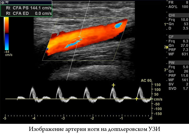
To assess blood flow through the arteries, the following indicators are used:
- Vmax – maximum speed blood flow in the artery, which is recorded in systole
- Vmin – minimum blood velocity recorded in diastole
- RI - vascular resistance: this is the ratio of the difference between the systolic and minimum blood flow speed to the maximum speed
- PI – pulsatility index, defined almost like RI, but is a more sensitive indicator of changes in the lumen of the vessel
- thickness of the inner and middle layers of the vessel (IMT).

For femoral artery IMT should be less than 1.1 mm. When it does not reach 1.2 mm, it is still a borderline value. Atherosclerosis is indicated by this indicator of more than 1.3 mm, or an increase in this value by 50% of the same indicator in a nearby area.
- ABI: attitude systolic pressure on the tibial artery to the same pressure on brachial artery. It should be approximately 1.0 or slightly more.
There are no such digital indicators for veins. During ultrasound examination of the vessels of the extremities, prices for which differ in various clinics, the doctor evaluates the patency of the venous collectors on the lower leg - deep and superficial, as well as the largest veins passing into abdominal cavity and the pelvic cavity - the iliac and inferior vena cava.
All possible blood clots are visualized, the valve system and the condition of the communicating veins are assessed. Tests are also carried out with raising the leg, squeezing the veins with a tourniquet, which allows one to get an idea of the direction and nature of blood movement, the presence or absence of pathological discharges in the opposite direction.
Who to trust to carry out the research
Where can I get an ultrasound of the vessels of the lower extremities? This is best done in specialized phlebological centers or at hospitals that have a department vascular surgery. First of all, about where to go for an ultrasound of your legs, consult a phlebologist, and not a general surgeon or therapist.It is not the centers that establish high price on this study 3000 rubles and above, and those in which the examination and this study are performed by a vascular surgeon or phlebologist. On average, the price for
For really effective treatment Vascular disorders and their diagnosis must be at the proper level. One of the most popular screening methods for detecting disorders of all types of blood circulation in arteries and veins is ultrasound of the vessels of the lower extremities. This type diagnostics has a fairly indicative result, is absolutely safe and can be used in all cases if indicated.
Basics of ultrasound examination of vascular structures
The diagnostic principles of the ultrasound method are based on the ability of low-frequency ultrasound waves to be reflected from moving objects. With the help of special sensors, these oscillations are recorded and, based on the difference in their sum, special computer programs build a graphic image of the blood flow and show the vessels being studied. Today, there are ultrasound devices that can convert received signals into a color image that can be seen on a monitor screen. If necessary, pulse filling with blood can not only be seen, but also heard in the form of pulsating or smooth uniform noises, which depends on the arterial or venous vessel being examined.
Alarm signals in determining indications for diagnosis
Despite its absolute harmlessness, ultrasound of the blood vessels of the legs, like other diagnostic methods, must be performed according to strict indications. They can be determined not only by doctors, but also by patients themselves. But it is better if everything happens under the supervision of a specialized specialist who will compare clinical and instrumental data.
Signal about vascular disorders and the need to do research, the following complaints may occur:
- The appearance of dilated veins or stars of small vessels on the skin of the legs.
- Swelling of the legs and feet, especially unilateral.
- Darkening of the skin of the legs, thickening or non-healing for a long time trophic disorders and ulcers.
- Feeling of coldness in the legs and their rapid freezing, despite adequate ambient temperature.
- Numbness and crawling sensation.
- Pain in the legs when walking, any exercise and at rest. Most often they force you to do an ultrasound.
- Pale feet.
- Reduction in volume of the lower leg with a violation of its trophic indicators (hair growth, muscle tone and strength).
- Weakness of the lower extremities in relation to loads.
- Darkening and blueness of the toes or the entire foot.
- Muscle cramps on the back of the leg.
Types of ultrasound angioscanning
Regarding the terminology of ultrasound diagnostics of blood vessels, there are specific names that often raise a lot of questions. Any ultrasound examination vascular structures called Dopplerography. Among its methods for diagnosing circulatory disorders, there are two basic studies that are fundamentally different from each other in their diagnostic capabilities.
- Standard Doppler ultrasound is a graphical recording or audio recording of blood flow in the vessel being examined. In this case, a black and white image of the nature of the blood flow in the form of a line is obtained. The method allows you to conduct Doppler measurements (description of the characteristics of the resulting image) and draw conclusions about the characteristics of the blood supply to the studied leg segments. Mainly used for diagnostics arterial diseases lower extremities. The advantage of this ultrasound technique is that it is easy to perform and can be performed at the patient’s bedside due to the availability of portable devices.
- Duplex angioscanning – obtaining a color image of blood vessels depending on the speed and direction of blood flow. This method is more accurate and gives almost comprehensive information about his condition. Makes him relatively happy low price compared to other similar informative methods.
What does vascular ultrasound show?
Thanks to Doppler ultrasound, you can get an idea exclusively about the function of blood vessels - the intensity and nature of blood flow in them. It is impossible to obtain direct information about its structure. This has to be judged indirectly by assessing the results obtained and determining the presumed localization of pathological changes within its lumen, if any, based on the results of Doppler ultrasound.
Duplex mapping evaluates not only functional ability, but also directly shows an image of the vessel in those places where there is an obstacle to normal blood flow. With its help, you can determine the presumable cause of narrowing of the lumen: spasm, atherosclerotic plaque, thrombus, thromboembolus (thrombus that has broken off from the heart or aorta and migrated to peripheral vessels lower extremities), external compression by tumor.
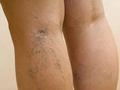
Spider veins on the legs - indication for ultrasound of the lower extremities
Ultrasound for diseases of the veins of the lower extremities
The method is indispensable for this pathology, since there are no analogues that could replace it. Ultrasound allows you to fully establish the signs:
- Varicose veins.
- Thrombophlebitis (formation of blood clots in superficial veins).
- Phlebothrombosis (thrombosis in the deep venous system).
- Chronic venous insufficiency.
- Insufficiency of the valve apparatus of the veins of the perforating and deep system, and mark them before surgery, which is only possible with ultrasound of the vessels of the legs.
Ultrasound in the diagnosis of arterial pathology of the legs
In all cases of violation arterial circulation An ultrasound scan of the lower extremities is necessary. Primary examination performed using Doppler sonography. Its only competitor is arteriography, which gives even more full information O vascular system legs But, if we take into account its invasiveness and complexity of implementation, Dopplerography turns out to be irreplaceable, especially duplex study. It is impossible to overestimate its importance in diagnosis:
- Obliterating atherosclerosis and endarteritis.
- Aortic diseases.
- Thrombosis and thromboembolism of the arteries of the lower extremities.
- Chronic arterial insufficiency.
- Peripheral aneurysms arterial vessels legs
- Raynaud's disease.
From this article you will learn how ultrasound examination of the vessels of the lower extremities is performed and who is prescribed the procedure. What can be diagnosed using ultrasound.
Ultrasound Dopplerography is Doppler ultrasound. This diagnostic method, unlike other methods of examining blood vessels, is able to show the speed of blood flow, which makes it possible to accurately diagnose the severity of the disease that impairs blood circulation.
For any vessels, this procedure is carried out according to the same principle - using an ultrasound sensor, like any ultrasound. More often, this procedure is required to examine veins; it is used less often to examine arteries.
Various doctors can refer you for this examination: therapist, phlebologist, angiologist. A specialist performs the procedure ultrasound examination.
Indications
Ultrasound scanning of leg vessels is prescribed for the diagnosis of the following diseases:

For what symptoms is ultrasound ultrasound prescribed?
Patients are referred for this diagnostic procedure if they suspect vascular diseases legs Your doctor may order an ultrasound scan if you experience the following symptoms:
- swelling of the legs;
- heaviness in the legs;
- frequent paleness, redness, blue discoloration of the legs;
- “goosebumps”, numbness in the legs;
- pain when walking less than 1000 meters;
- cramps in the calf muscles;
- spider veins, webs, protruding veins;
- tendency to freeze feet, cold feet even when warm;
- the appearance of bruises on the legs even after the slightest blow or for no reason at all.
When is preventive Doppler ultrasound needed?
Get a Doppler ultrasound of the blood vessels in your legs for preventive purposes every six months to a year if you are at risk. The following are prone to vascular diseases of the lower extremities:
- overweight people;
- busy physical labor(loaders, athletes);
- those who constantly stand or walk a lot at work (teachers, security guards, couriers, waiters, bartenders);
- those who have already been diagnosed with atherosclerosis of other vessels;
- people whose direct relatives suffered from vascular diseases;
- those with diabetes;
- smokers;
- people over 45 years old;
- women during pregnancy and menopause;
- women taking oral contraceptives for a long time.
Preparation
The procedure does not require any complex preparation.
The only thing is that your feet must be clean. If you mean individual characteristics thick hairline on the legs, it is advisable to shave it off to make it easier for the doctor to work.
On the day of the procedure, do not drink alcohol, stimulating drinks (coffee, strong tea, energy drinks), do not expose your legs to physical activity (do not run, do not lift weights, do not go to workouts). 2 hours before ultrasound examination of the vessels of the lower extremities (and other vessels too), do not smoke. It is better to go for examination in the morning.
Bring napkins or a towel with you to the procedure to dry your feet later. Also bring a referral for an ultrasound scan from your doctor and the results of previous vascular examinations.
How the research is carried out
First, you free your legs from clothing.
The examination will be carried out standing or lying down. The doctor applies ultrasound gel and moves the ultrasound probe along the legs.
An image of your vessels is displayed on the specialist’s monitor. Immediately during the procedure, he analyzes and records the received data.
If you are being examined lying down, the doctor will first tell you to lie on your stomach and raise your feet on your toes. Or you can place a cushion under your feet. In this position, it is most convenient for a specialist to examine the popliteal, peroneal, small saphenous and sural veins, as well as the arteries of the posterior surface of the legs. You will then be asked to roll over onto your back and bend your legs slightly. knee joints. In this position, the doctor can examine the veins and arteries of the front surface of the legs.
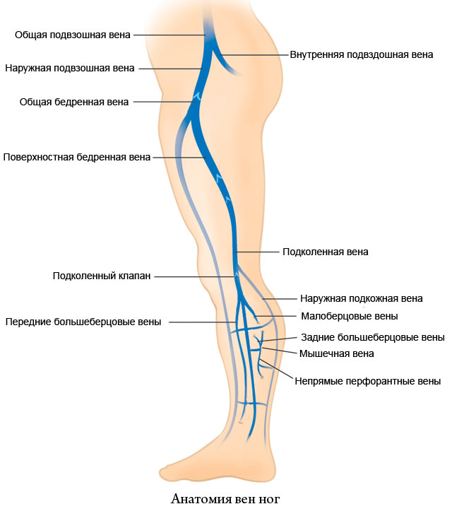 Anatomy of leg veins. Click on photo to enlarge
 During an ultrasound scan to detect reflux (reverse discharge of blood), the doctor can perform special tests:
- Compression test. The limb is compressed and the blood flow in the compressed vessels is assessed.
- Valsalva maneuver. You will be asked to inhale, pinch your nose and mouth, and strain slightly as you try to exhale. If there initial stage varicose veins, reflux may occur during this test.
Dopplerography of blood vessels takes about 10–15 minutes in total.
At the end of the examination, you wipe your feet from any remaining ultrasound gel, get dressed, pick up the result and are good to go.
What does ultrasound examination of the blood vessels of the legs show?
Using Dopplerography of the lower extremities, you can examine the following vessels of the legs:
During this diagnostic procedure the doctor can see:
- shape and location of blood vessels;
- diameter of the vessel lumen;
- condition of the vascular walls;
- condition of arterial and venous valves;
- blood flow speed in the legs;
- the presence of reflux (reverse discharge of blood, which often occurs with varicose veins veins);
- presence of blood clots;
- size, density and structure of the blood clot, if any;
- the presence of atherosclerotic plaques;
- the presence of arteriovenous malformations (connections between arteries and veins that should not normally exist).
Ultrasound ultrasound standards, conclusion with explanations
The veins must be passable, not dilated, and the walls not thickened. The lumens of the arteries are not narrowed.
All valves should be healthy, there should be no reflux.
The speed of blood flow in the femoral artery is on average 100 cm/s, in the arteries of the leg - 50 cm/s.
Atherosclerotic plaques and blood clots in the vessels should not be detected.
There are normally no pathological connections between the vessels.
An example of a normal ultrasound examination of the leg veins and explanations for it
Conclusion: all veins on both sides are passable, compressed, the walls are not thickened, the blood flow is phasic. No intraluminal structures were identified. The valves are consistent at all levels. There are no pathological refluxes when performing compression tests and the Valsalva maneuver.
| Abstracts from the conclusion | What do they mean |
|---|---|
| All veins on both sides are passable, compressed, the walls are not thickened. | All veins on both sides are patent, which means that blood can flow freely through the vessels. Compressive - that is, they have not lost their natural tone, they can shrink. The walls are not thickened - this indicates that there are no inflammatory or other pathological processes. |
| Blood flow is phasic. | The blood flow is phasic - faster when exhaling and slower when inhaling. This is how it should be normally. |
| No intraluminal structures were identified. | No intraluminal structures were identified - there are no atherosclerotic plaques, blood clots or other inclusions that should not be there. |
| The valves are consistent at all levels. | The valves are healthy - that is, they perform their functions normally and do not allow backflow of blood. |
| There are no pathological refluxes when performing compression tests and the Valsalva maneuver. | There are no pathological refluxes during the tests - under no circumstances is blood discharged in the opposite direction, which indicates healthy blood circulation. |
Contraindications
Dopplerography of the vessels of the lower extremities is an absolutely safe procedure. It has no contraindications or age restrictions.
It can be performed with any frequency and to any people, including:
- children of any age;
- elderly;
- people with chronic diseases;
- patients with acute inflammatory diseases;
- those who have a pacemaker implanted (they can direct the ultrasound sensor to their legs, and ultrasound of organs chest cavity cannot be done);
- pregnant and lactating women;
- those who are allergic to contrast agents(angiography, for example, cannot be performed in this case);
- people weighing more than 120 kg (but it is impossible to perform MRI on obese patients using most machines, since they are not designed for such dimensions).
The only limitation that can be allowed is an allergy to the ultrasound gel. She meets in isolated cases. And she isn't absolute contraindication to perform diagnostics. Allergic reaction can be avoided by choosing hypoallergenic gel, which is right for you.
 Gel for ultrasound
 Summary, advantages of the procedure
Dopplerography of the vessels of the lower extremities is an absolutely painless diagnostic method. It doesn't cause any side effects and has no contraindications (except for allergies to ultrasound gel). As scientists' research shows, ultrasonic waves do not cause any harm to the body, so ultrasound of the blood vessels of the legs can be performed with any frequency.
