Duplex scanning of the arteries of the head. Duplex scanning of brachiocephalic arteries: indications, conduct, preparation
Duplex vessels of the head and neck - what is it? This is most often a question not of idle curious, but of people who are faced with pathologies. vascular nature. This diagnostic method research based on modern achievements science and technology and allows you to effectively and quickly identify many disorders in the vessels of the head, neck, limbs. Such studies are carried out in specialized medical institutions using special equipment. They are completely safe, although they have certain limitations in use.
The essence of the technique
Duplex or duplex scanning of the vessels of the head and neck is ultrasound procedure(ultrasound) of vessels simultaneously in two modes (B-mode and doppler study). This technology shows high efficiency in the study of extracranial (cervical) and intracranial (inside the brain) vessels. It provides a clear picture of the change vascular structure(malformation, bends, patency, stenosis) and disorders of the blood supply to the brain. In addition, the technique is widely used to study peripheral blood circulation.
Duplex is based on 2 methods. When conducting a study in the B-mode, the ultrasound radiation is configured in a pulsed mode. As a result of receiving the reflected signal, a two-dimensional echogenic picture appears on the monitor, which makes it possible to assess the structure of tissues. In this way, it is possible to examine almost all vessels and nearby tissues, with the exception of those located inside the skull.
Second component duplex is based on the Doppler effect, i.e. evaluates a moving object by reflected ultrasound. As a moving element, the blood flow is studied, or rather the speed of blood movement in various vascular areas. Modern technology allows you to reproduce on the computer monitor a color sketch of the blood flow, while the color intensity corresponds to the value of the blood flow velocity. The picture has a two-dimensional display: along and across the vessels.
For getting complete information transcranial duplex scanning (TCDS) is performed, allowing you to look inside the skull. TKDS is used in the study of changes in the tissues and vessels of the brain for the primary diagnosis of volumetric lesions in the cerebral hemispheres, to identify intracranial hematomas and other brain lesions, as well as when monitoring the development of pathologies.
What problems does a duplex solve?
The combination of 2 research methods makes it possible to obtain the necessary information on anatomical changes vessels, lumen, condition vascular wall, reveal morphological changes; and to establish the extent to which such anomalies affect hemodynamics. duplex study refers to non-invasive diagnostic methods that provide the detection of pathologies such as stenosis, thrombosis, occlusion, plaques, malformations, tumor formations, injuries. This method scans
veins and arteries that provide brain activity(incl. carotid artery) and peripheral vessels extremities, and with the development of TKDS, it became possible to assess cerebral hemodynamics.
duplex scanning recognized as an informative method that has become popular in modern medicine due to their positive qualities.
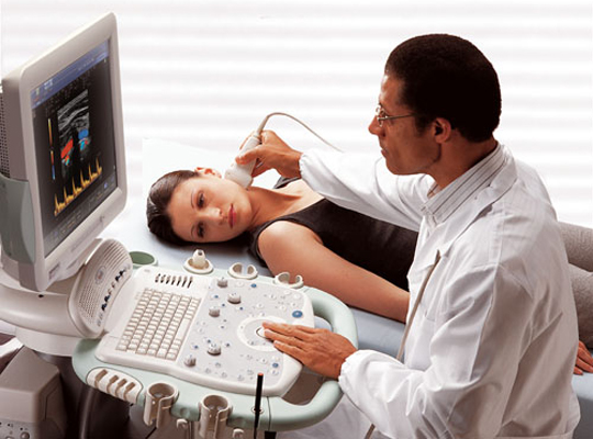
Ultrasound duplex or triplex vascular scanning is an effective, affordable, informative method diagnosing diseases of the vessels of the head. Such a study should be carried out by a qualified specialist using an ultrasound machine. To examine the vessels, a special sensor is used that transmits ultrasonic waves to the monitor in real time. Duplex head scan helps the specialist to identify pathological abnormalities and diseases of the circulatory system. Also this survey does not harm the health and well-being of the patient, since the possibility of introducing a contrast agent is completely excluded.
DS survey area
Duplex scanning helps to identify such abnormalities and diseases:
- Chronic leg artery disease (CALE).
- Progressive disease of the arteries that completely closes their gaps (PEANK).
- foot diabetes.
- Narrowing of the carotid arteries.
- Pathology of cerebral vessels.
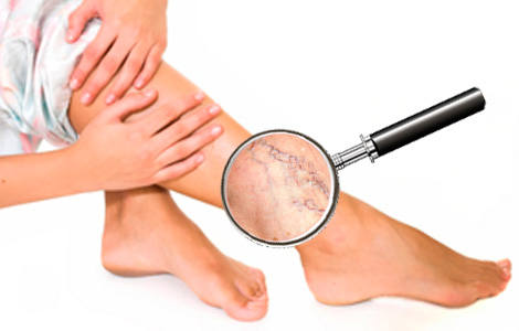
- Formation of vein clots in the lower extremities.
- Varicose veins lower extremities.
- Post-thrombotic syndrome.
Also this method examination helps to identify atherosclerotic plaques in the lumen of the arteries, narrowing of the lumen of these arteries and other pathologies associated with arteries, vessels, veins, both of the lower extremities and the brain.
Indications for vascular DS
 This examination is recommended if the patient has suspicions or progression of diseases such as:
- Varicose disease.
- cerebrovascular disease.
- Vasculitis, angiitis, arteritis.
- Thrombosis, with inflammation of the vein wall.
- The formation of a blood clot in a vein, with its complete blockage.
- PTFB.
- Chronic disease of the arteries of the elastic and musculo-elastic type.
- Chronic vascular disease with a primary lesion of the arteries of the legs.
- Trophic ulcers.
- Aortic aneurysm.
- Vascular injuries and their consequences.
- Arterial hypertension.
- Vascular control before and after surgery.
- Examination as a preventive measure.
Also duplex scanning, modern method research, helps to identify violations occurring in the blood circulation. It is also recommended to carry out DS if the first alarming symptoms were noticed:
- Constant dizziness, loss of consciousness.
- Migraines and pain in the neck.
- Sudden memory loss and distracted attention.
- Unexplained swelling.
- Cramps, pains and numbness in the limbs.
Advantages and disadvantages in the study of arteries
Duplex scanning not related to the application x-rays, so it does not cause any harm to the body. It can be done repeatedly without worrying about your well-being and health. Also, this method is non-invasive, which reduces the risk of allergic reactions to a minimum, or rather to zero. Duplex scanning is recognized as an inexpensive and most common method for diagnosing blood circulation and the condition of blood vessels, arteries and veins, both of the head and lower extremities.
The only disadvantage of such an examination is that it can not always be used, for example, in the study of heart vessels.
Preparation for DS
 Duplex scanning of the brachiocephalic arteries does not require any special and pre-training. The only thing that experts recommend is that on the day of the BCA scan, it is necessary to refrain from eating foods that can provoke expansion or narrowing of blood vessels. if you accept antihypertensive drugs and other medicines that can distort the results of the study, you must first consult with a neurologist.
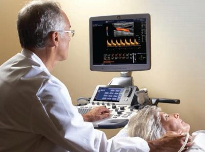
Carrying out a BCA scan of the head and neck
This examination of the BCA of the head is carried out according to generally accepted principles:
- The patient needs to take a supine position on the couch.
- Be sure to put a roller under his head. If there are any diseases cervical region, then the roller can be replaced with a solid pillow.
- The patient needs to release the neck for the BCA examination and turn the head in the opposite direction to the sensor.
- A special gel must be applied to the skin to help the sensor slide and prevent air from entering. After that, each vein and BCA is carefully examined by a specialist, be sure to take measurements in parallel.
To view the vessels of the brain, it is necessary to conduct an examination through the cranial bones. In this case, the sensor is located:
- At the temples on both sides.
- Above the eyes.
- At the junction occipital bone with the spine.
- In the region of the occipital bone.
The interpretation of the results obtained is carried out immediately after the examination, which should be dealt with exclusively by a specialist.
Identification of the disease with the help of DS
![]() This study is not only recognized as informative, but also of high quality, and thanks to duplex scanning, a qualified specialist is able to identify such diseases in a patient as:
This study is not only recognized as informative, but also of high quality, and thanks to duplex scanning, a qualified specialist is able to identify such diseases in a patient as:
- Abnormal arrangement, course or branching of blood vessels. It can also be congenital.
- Chronic arterial disease.
- Injuries to arteries or veins.
- Inflammation occurring in the walls of arteries and capillaries.
- Angiopathy of various types.
- Dyscirculatory encephalopathy.
Survey results
The interpretation of the results is carried out only qualified specialists. Never worth doing self-diagnosis and treatment. The results of the duplex scan examination indicate blood flow, any defects occurring in the vessels, and may also show the presence of intraluminal formations. As for the numbers, they are practically absent in such a survey. More detailed digital data can be obtained thanks to the Doppler. For any pathologies found in the arteries, the doctor informs you and directs you to a specialist for further monitoring and prescribing a correct and effective treatment.
To understand all the possibilities of duplex scanning of the brachiocephalic arteries and what it is, you need to understand the methodology for its implementation.
This is currently ultrasound examination is one of the most informative and exact ways determination of disorders associated with the blood circulation of the brain.
Its decoding makes it possible to identify a number of pathological conditions, including the development of a stroke.
The main purpose of the brachiocephalic arteries is to supply the brain with blood flow necessary for its normal functioning.
Crashes in normal operation brachiocephalic arteries can lead to the development of serious diseases.
The most informative way to identify all possible pathologies is a duplex scan of the neck and head, which is carried out in the direction of a doctor in any medical institution where appropriate ultrasonic equipment is available.
Physicians include three in the composition of the brachiocephalic arteries, namely: carotid, subclavian and vertebral.
These arteries have a common junction, which is called the brachiocephalic trunk. In turn, the junction of these arteries and some other veins is called the circle of Wellis.
It is here that the main distribution of blood flows throughout the brain occurs.
Each of the parts of the brain has its own specific blood norm, which it must receive for normal functioning.
The manifestation of various pathological conditions in the carotid, vertebral, main or some other arteries leads to the development of the most various diseases up to a stroke.
Duplex scanning of the neck and head is the method that timely detects malfunctions of the entire circulatory system generally.
Such an ultrasound examination is absolutely harmless to human health.
When it is carried out, the possibility of injuries on the mucous membranes is excluded - the method is non-invasive.
By itself, duplex scanning of the carotid, vertebral, or main veins can be performed frequently, as needed.
Duplex scanning of the neck and head is highly sensitive and most informative.
Due to a somewhat specific way of obtaining data on the state of the brachiocephalic arteries and veins, the accuracy of the readings allows for the correct diagnosis.
There are other methods for checking the condition of the carotid, vertebral, and main veins, but all of them are inferior to duplex scanning in informational content.
In some cases, the doctor may additionally prescribe a transcranial examination of the brachiocephalic arteries. This will allow you to get a clearer picture of the state of blood flow in the neck and head.
When conducting a Doppler scan, a blind method is used without visualization, which does not allow to identify the cause of the manifestation of pathologies.
The sensor itself, which emits ultrasonic wave, is installed approximately in the neck and head area.
When performing the procedure, you can see the vessel itself on the monitor screen, which makes it possible to accurately assess the speed of blood flow, as well as its patency at different points.
This allows you to timely notice the thickening of the walls, blood clots and many other pathologies. With the help of duplex scanning, it is possible to examine the anatomy of the veins.
In addition to the brain, in some cases duplex scanning of the arteries of the lower extremities is performed.
For each of the indicators obtained as a result of the procedure, there is a norm with which the doctor will compare the data and draw conclusions about the patient's condition.
Indications for scanning
As mentioned above, all arteries have a common junction, which is called the circle of Wellis. Malfunctions in the work of one of the vessels can lead to the development of serious pathological conditions.
To prevent Negative consequences, a duplex scan of the brachiocephalic veins is performed, aimed at examining all parts of the brain.
In this case, the study is carried out in the head and neck area. In addition to sleep and vertebral arteries, the state of extracranial and main veins is clarified.
The examination itself does not require any preparatory measures.
To undergo a duplex scan, you need to get a referral from your doctor. In this case, the reason for its implementation may be hypertonic disease or development arterial hypotension.
It is recommended to undergo an examination of the neck and head for diseases of a vascular nature, such as vegetative-vascular dystonia. Duplex scanning is mandatory for abnormal development of cardio-vascular system and at various forms etiology.
The condition of the extracranial veins and other arteries should also be checked if the patient has been diagnosed with diabetes mellitus for a long time.
During a duplex scan, the doctor carefully examines the picture on the screen, analyzes it and takes all the necessary indicators about the work of the brain vessels.
The final diagnosis is made by the attending physician after he carefully examines the final conclusion.
Diagnosis of the state of cerebral vessels is distinguished by its accessibility and maximum information content.
The main symptoms that may lead to the appointment of a duplex scan are frequent dizziness and severe headaches that occur when it is turned.
If a person feels tinnitus for a long time, which gives him some discomfort, then it will also not hurt to diagnose brachiocephalic arteries.
It is worth thinking about the state of your health and doing a duplex scan of the head and neck even in the case when fainting is regularly observed.
An examination of the veins of the brain is necessary when the limbs are constantly numb and dark dots flash before the eyes. An ultrasound examination is also recommended for sharp decline visual acuity.
Currently, if desired, the patient can also undergo a color scan of the head and neck.
This will make it possible to get a clearer image on the monitor screen, due to which the accuracy of the result will be higher compared to the conventional method.
Color research reveals pathological conditions arteries for more early dates disease development.
Duplex scanning itself is practically no different from ultrasound. When scanning the neck and head, a special sensor is used, which is the emitter of an ultrasonic wave.
For stronger contact with skin a special composition is applied to the examination area.
It should be noted that the decryption of the received data should be carried out only experienced specialist, since the correctness of the diagnosis made to the patient, and, accordingly, the prescribed course of therapeutic therapy, largely depends on this.
What metrics can you get?
The main goal of a duplex examination is to collect as much information as possible about the state of the arteries of the brain. The procedure shows the most informative picture in real time.
Using the duplex method, you can set a large number of most different pathologies arteries. The scan shows the presence of blood clots and plaques, as well as various aneurysms.
In addition, the procedure allows you to clearly observe arterial pathologies such as elongations, loops, bends and abnormal tortuosity.
Based on the data obtained, it is possible to accurately diagnose vascular stenosis, damage to their walls.
The underdevelopment of the vessels of the brain is also very clearly visible, as well as a change in their main diameter up or down.
Duplex diagnostics is good because in real time you can observe the exact location of plaques on the monitor screen, as well as the degree of vasoconstriction.
The doctor can see the thickness and uniformity of the veins, the change in their shape and the mobility of the outer and inner walls.
Diagnostics allows you to determine not only the speed blood flow but also its direction at the moment.
After decoding all the received data and comparing them with the norms, medical worker who conducted duplex examination, a final conclusion is made.
This conclusion is transmitted to the doctor for diagnosis and treatment.
The data obtained during the diagnosis can be the reason for diagnosing pathologies such as thrombosis and dystonia. In addition, the development of angiopathy and atherosclerosis in the vessels can be detected.
Duplex vascular examination is good because the patient does not feel any discomfort during it, and the doctor can get a lot of information necessary to prescribe the correct and effective treatment.
The method is distinguished by its high information content of the examination of brain regions.
For the treatment and prevention of diseases caused by Candida fungi (including nail fungus), our readers successfully use antifungal agent"Tinedol"- effective remedy from foot fungus, unpleasant odor and itching. Essential oil Mint will give coolness and eliminate unpleasant odors after a stressful labor day. As well as: Get rid of insomnia ... "
In addition, the patient who is scheduled for the examination is not required to comply with any preparatory measures.
