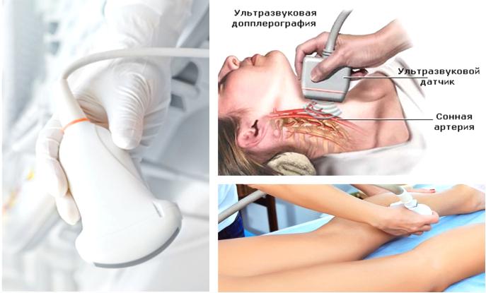
Duplex scanning of the brachiocephalic arteries. Doppler ultrasound and duplex scanning
Duplex ultrasound scanning of the brachiocephalic arteries, or abbreviated USDS BCA, is a modern ultrasound method for diagnosing vessels, including the carotid and vertebral vessels, which supply blood to the brain, and the subclavian arteries.
First of all, the person who is scheduled for this study may have a question: what are the brachiocephalic arteries and where are they located.
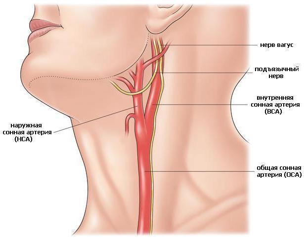
Brachiocephalic vessels are the largest arteries and veins that are responsible for blood flow to the tissues of the head, brain and upper limbs. They are also called main lines.
The brachiocephalic arteries include the carotid, subclavian, vertebral and their junction, which forms the brachiocephalic trunk. The listed vessels and some others near the base of the brain form the Circle of Wellis, which is responsible for the distribution of blood flow throughout all parts of the brain.
What is duplex scanning of the brachiocephalic arteries, and what is the method based on?
The apparatus for examining the BCA is based based on the principles of echolocation. The working surface emits and then picks up ultrasonic pulses. The information is converted into a digital signal. This is how the image appears on the monitor.
The method is based on union advantages of B-mode- visual interpretation of the state of blood vessels and adjacent tissues and Doppleroscopy - qualitative and quantitative properties of blood flow. The Doppler spectrum can also be supplemented with color mapping.
What does the ultrasound scan of the BCA show?
Ultrasonic scanning of the BCA shows:
- lumen of blood vessels;
- blood clots, plaques, detachments;
- stenosis, wall expansion;
- ruptures, deformations.
Using ultrasound BCA can be diagnosed:
- vascular pathologies;
- violation of wall tone during VSD;
- arterial aneurysms;
- fistulas between vessels;
- angiopathy;
- thrombosis;
- vascular injuries;
- varicose veins.
Brain vessels are complicated arranged system, which is capable of self-regulation and maintenance cerebral blood flow. Only comprehensive diagnostics, which includes ultrasound scanning, CT, MRI, allows you to accurately and timely select treatment, and then evaluate its effectiveness.
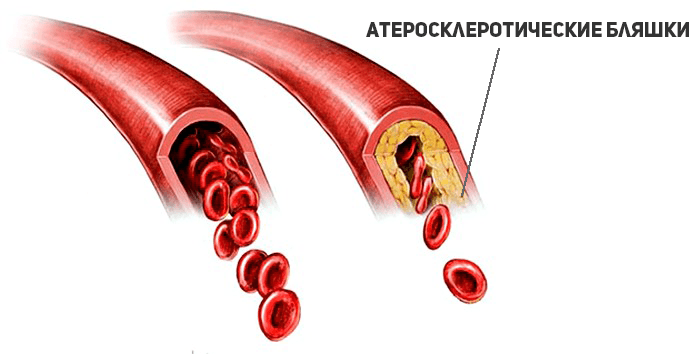
Ultrasound scanning helps to assess the anatomy of blood vessels, determine the characteristics of blood flow, and assess the condition of the walls and lumen. This way you can diagnose early stage the occurrence of atherosclerotic plaques, blood clots, tortuosity of arteries and their dissection.
Peculiarities
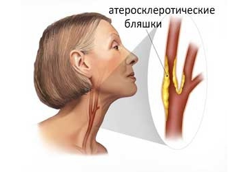
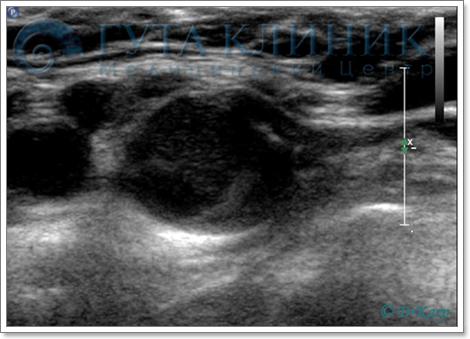
The initial sign of atherosclerosis, which an ultrasound examination can show, is not even a plaque, but thickening of the wall of the carotid artery by just a fraction of a millimeter. With duplex scanning, this indicator is well determined. The thickness of the intima-media complex (the so-called IMM) is also called. IMT is taken into account to assess the effectiveness of treatment.
 An increase in IMT greater than 1 mm is most often associated with with risk factors such as: smoking, arterial hypertension, diabetes, etc.
As the disease progresses, plaques begin to form. Usually they are localized in the so-called. Carotid bifurcation is the site of division of the common carotid artery into internal and external. The presence of a plaque in this segment is a serious risk factor for stroke and myocardial infarction. Therefore, it is very important to promptly identify atherosclerotic changes in early stages.
Duplex scanning reveals the location of the plaque, as well as its shape, size, structure and degree of stenosis (narrowing of the lumen). When the lumen is already completely closed - this is occlusion.
During examination, BCA is often detected tortuosity of the arteries due to their lengthening. Arteries lengthen due to atherosclerosis and increased blood pressure. Tortuosity of the vertebral arteries usually occurs due to defects cervical region spine. If tortuosity leads to compression of the lumen, this can cause disruption of cerebral blood flow.
Ultrasound scanning is also used for examination of patients with traumatic injury vessels: Wall delamination or similar. The main symptom of this disease is severe headache, which cannot be reduced with conventional painkillers.
The advantages of BCA ultrasound are:
- high information content;
- efficiency of research;
- safety and possibility of repeated implementation;
- painlessness of the procedure.
During the examination on the monitor an image similar to a conventional ultrasound is formed, but the vessel is clearly visible against its background, in which blood flow is formed. Due to the advantages of ultrasound scanning, BCA is considered the gold standard for diagnosing pathologies. A timely vascular ultrasound can save lives and prevent possible disability.
Indications for use
The indications for prescribing duplex scanning of the BCA are:
- headache;
- dizziness;
- violation of movement coordination;
- blood pressure problems;
- fainting;
- elevated cholesterol levels;
- impaired sensitivity (numbness) of the limbs;
- blurred vision;
- flickering spots in the eyes;
- memory impairment and decreased concentration;
- preoperative examination.
Direct indications for the study are the following pathologies:
- atherosclerosis;
- hypertension;
- heart pathologies;
- neck injuries;
- compression of arteries and veins and other vascular injuries;
- vasculitis;
- blood diseases;
- suffered a stroke or heart attack.
Preparation
Preparation before the study consists of excluding from the menu foods and dishes that can affect the tone and filling of blood vessels, which will distort the results of the study.
On the day of the study, you should not drink tea, coffee, energy drinks, Coca-Cola, alcohol, and do not indulge in overly spicy and salty foods. Directly before ultrasound examination of the BCA, you should not be in stuffy or smoky rooms, as this can also change the blood flow to the vessels. It is better to refrain from taking vitamins and nootropics the day before the study.
The use of the device is absolutely harmless and has no effect on the body person.
How is it carried out?
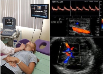
The patient lies on his back on the couch near the machine, the doctor places a cushion under his neck. The head should be turned in the direction opposite to the device. The doctor lubricates the surface of the skin with a gel that facilitates the passage of the ultrasound signal.
Using the sensor, the doctor will examine segment by segment, observing the change in signal on the monitor. He may press the sensor lightly on the vessels or ask for a short time stop breathing.
None There is no discomfort during the examination: The procedure feels no different from a regular ultrasound scan, familiar to everyone. The study lasts 20-30 minutes.
Decoding the research results
The scanner will record the necessary indicators, and the doctor will enter them into the scanning protocol. Decoding the Doppler spectrum and blood flow charts will take no more than 10 minutes, after which you will receive a transcript.
The result of the scan is a transcript of the information received, printed with a list of the examined vessels and a description of their size and condition. Decoding gives the ability to determine whether the vessels correspond to the anatomical norm, is there any pathology, etc. Based on the transcript, your attending physician, if necessary, prescribes treatment.
Decoding is carried out by comparing indicators:
- nature of blood flow;
- its speeds: systolic (max) and diastolic (min);
- wall thickness;
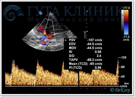
- pulsator index (so-called PI) is the ratio of the difference between max and min speeds to the average (the sum of max speed and two min, divided by three);
- resistive index (so-called RI) is the ratio of the difference between max and min speeds to min;
- systolic-diastolic ratio: max speed divided by min.
Based on the last 3 indices, the patency of the vessel is judged.
Blood flow is assessed in the external and internal carotid arteries, common (ECA and ICA, CCA), supratrochlear (SBA), main (OA), vertebral (VA) and in its segments, each of which has its own designations, for example, Vo, V1, V3 etc.
 Also in the front, back, middle cerebral arteries(ACA, PCA, MCA), subclavian (RCA), anterior and posterior communicating (ACA, PCA) arteries. Changes in indicators can also be assessed with horizontal and vertical position bodies.
It can be summarized that ultrasound scanning of the BCA is a special type of ultrasound diagnostics of vessels that provide nutrition to the brain and other organs of the head, neck, and upper limb girdle.
This is an accessible, safe, detailed and informative study, which within ten minutes can show the condition of the blood vessels and identify the cause of some unpleasant symptoms. An annual examination will allow you to predict the development of a cerebral stroke by 90%.
The study of extracranial (neck) and intracranial (intracerebral) vessels is the most informative modern method diagnosis of disorders cerebral circulation, allowing you to evaluate not only functional indicators blood flow, but also anatomical changes vessel (patency, wall condition, bends, malformations, etc.). Duplex scanning also used for peripheral circulation studies.
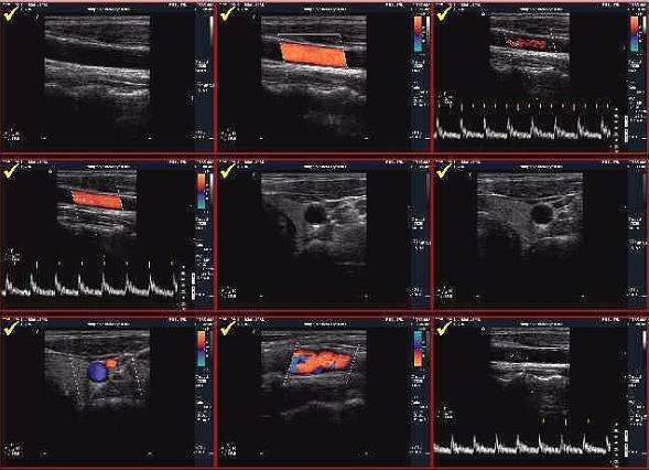
 The term "duplex" means a combination of two ultrasound modes: B-mode and Doppler. When examining in B-mode, the device's sensor emits ultrasound of a certain frequency, which penetrates tissue. At the border of tissues with different densities, ultrasound is reflected and returned to the sensor. The sensor operates in the so-called pulse mode, emitting ultrasound and picking up the reflected signal at various intervals. The farther the reflective structure is located from the sensor (it is also called echogenic), the more time passes between the moment of emission and reception of the signal. Multiple ultrasonic probe (sensor) crystals allow signals to be emitted at different angles with a variable time delay. Thus, modern powerful systems make it possible to almost instantly scan and reconstruct a two-dimensional image of the organ under study. The Doppler mode is based on the “Doppler” effect - when colliding with a moving object, ultrasound is not only reflected, but also changes frequency (“Doppler frequency shift”), the value of which is directly proportional to the speed of the object. In research blood vessels The “moving object” is red blood cells. In this way, the speed of blood flow is measured (more precisely, the spectrum of speeds, since different flows in the vessel move with at different speeds). Modern systems They also allow you to build a color cartogram of the flow in the vessel of interest - where the direction and intensity of blood flow in the veins is color coded. This method is called color Doppler mapping(CDC).
The combination of two modes allows you to get important information how to evaluate the anatomy of the vessels, their lumen, wall condition morphological changes, and evaluate the impact of these changes on circulatory function and hemodynamics. Ultrasound duplex scanning of veins and arteries is a non-invasive way to assess the condition of blood vessels, allows you to identify various pathologies, for example, stenoses, occlusions, atherosclerotic plaques, vascular malformations, etc.
 The duplex scanning technique has received wide use, especially for the assessment of veins and arteries, as well as brachiocephalic and large vessels, supplying blood to the brain (in particular, carotid arteries) And peripheral vessels limbs. In the last decade, thanks to the development of technology, it has been possible to introduce clinical practice and transcranial duplex scanning, all these methods are available to people by affordable prices. Before that the only way assessment of intracranial cerebral circulation was transcranial Dopplerography, which, despite the emergence of transcranial duplex, retained its importance as a method for assessing cerebral circulatory function and monitoring hemodynamic parameters.
Purposes of duplex scanning of cerebral vessels
- identification of early (preclinical) signs vascular pathology
- detection of stenotic and occlusive pathology of cerebral vessels
- detection of vascular development anomalies (aneurysms, arteriovenous malformations, hypoplasia, anastomosis)
- assessment of the hemodynamic significance of vascular pathology
- detection of vasospasm and venous circulation disorders
- identification of a complex of disorders associated with the presence of systemic vascular disease
- assessment of reserve capabilities of the cerebral circulatory system
- assessment of the effectiveness of treatment
- gradebrachiocephalic vessels (BCA)
Duplex Scanning System Image Gallery
If you have a question: “Where can I do a duplex scan of veins, vessels, head, brain and lower limbs at affordable prices?", the Neuro-Med clinic is always at your service. Our specialists are true professionals in their field and they will be happy to help you by providing detailed advice on issues of interest and performing the scans that you need to do.
Currently, the most common methods for studying the human cardiovascular system are ultrasound (ultrasound) with Doppler techniques:
- Ultrasound Dopplerography (USDG)
- Echocardiography (Echo-CG)
The main advantages of these methods are their absolute non-invasiveness (no trauma skin and mucous membranes), safety for the patient, high information content, sensitivity and specificity of the data obtained, the ability to conduct dynamic studies with registration of both background blood flow parameters in real time and induced parameters when using a variety of functional stress tests.
What is ultrasound with Doppler techniques?
The basis of ultrasound techniques used for vascular studies is the Doppler effect, described by Christian Doppler in 1842. Registration of blood flow during ultrasound examinations is based on a change in the frequency of the ultrasonic signal when it is reflected from moving blood particles, the bulk of which are erythrocytes, or red blood cells. Thus, they will make it possible to obtain objective information about the blood flow inside almost any vessel in the human body.
Where are Doppler techniques used?
Main directions in vascular studies, where Doppler techniques have found the most wide application, are:
Duplex scanning of the brachiocephalic arteries (DS BCA), also known as duplex scanning main arteries heads (DS MAG). Is a basic study to assess the blood supply to the brain. In this case, general, external, internal carotid and vertebral arteries on the neck.
The next stage is the study of intracranial, i.e. intracranial sections of the same arteries and their branches - TKDS.
Duplex scanning (DS) and Doppler ultrasound (USD) are currently used to study blood flow in vessels.
Duplex scanning (DS) (sometimes you can find triplex scanning). Unlike ultrasonography, the DS method is a visualization method, which significantly expands its diagnostic capabilities, since a direct assessment of the pathological process in a specific vessel of the studied vascular region becomes possible.
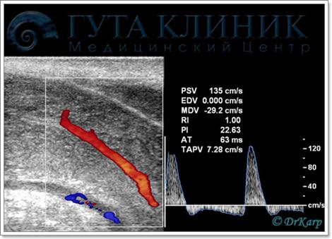
 An atherosclerotic plaque that is practically invisible in the greyscale mode
in the common carotid artery
The duplex scanning method combines visualization of vessels and tissues surrounding the vessel in B-mode with simultaneous study of blood flow in the lumen of the vessel using the Doppler effect through color Doppler coding (CDC) and (or) spectral Doppler analysis. In this case, the result of computer processing can be either a Doppler spectrum or a color flow cartogram obtained using various color coding technologies. The color flow cartogram is an “impression” obtained from the lumen of the vessel.
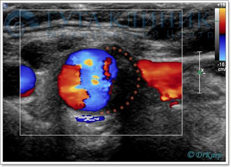 Clear visualization of the filling defect of the color flow cartogram
in color flow mode
Thus, any deviation from the normal course of the vessel (tortuosity, deformation), as well as any changes in the lumen of the vessel (plaques, blood clots, etc.) are easily determined. The Doppler spectrum characterizes the distribution of flow in the lumen of the vessel, and the calculation of a number of additional indices allows us to clarify the nature of the pathological process. The duplex scanning method allows you to visualize and assess the state of blood flow in almost all departments vascular system human, starting from large main trunks and ending with small organ and subcutaneous (subcutaneous) vessels.
In large-caliber vessels, a reliable visual assessment of all existing changes is possible vascular wall already in the early stages vascular diseases, for example, with non-stenotic atherosclerosis, diabetic angiopathy. Moreover, diagnosis is not difficult pathological processes in the presence of lesions characterized by various intraluminal changes (atherosclerotic plaques in stenotic atherosclerosis, blood clots) that impair the patency of the vessel.
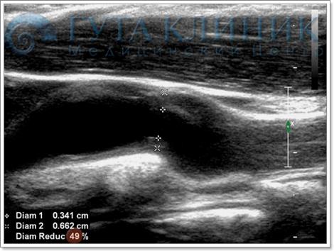 Accurate calculation of the degree of vessel stenosis
The duplex scanning method allows you to obtain direct echographic signs of various vascular processes:
The main advantages of the duplex scanning method include: the ability to identify early preclinical signs diseases with assessment vascular lesions, as well as changes in hemodynamics in real time, identifying not only organic, but also functional disorders blood flow with the possibility of studying functional state vascular system.
The main limitations and disadvantages of the duplex scanning method are: the dependence of the obtained data on the operator’s experience due to the subjective nature of obtaining and interpreting the resulting ultrasound picture, as well as resolution ultrasound scanner and on the anatomical and constitutional characteristics of the patient.
 Transcranial duplex scanning (TCDS)
Second ultrasonic method used to study the vascular system is Doppler ultrasound (USDG).
The Doppler ultrasound (USDG) method allows one to obtain only indirect information about the condition of the vessel wall and the presence of vascular pathology due to the impossibility of visualizing the vessel itself. To obtain diagnostic information using the USDG method, an ultrasound sensor is installed in the anatomical projection of a certain arterial trunk, the blood flow is located and then displayed on the display screen Doppler spectrum of blood flow from the located vessel.
The main disadvantages of the Doppler sonography method include:
- high probability of error in assessing blood flow velocity. This usually occurs due to the impossibility of correcting the position of the interrogation window and the angle of inclination of the ultrasound beam to the longitudinal axis of the vessel being examined;
- the impossibility in some cases (with anatomical variations in structure and location) of the exact location of the required arterial (or venous) trunk;
- impossibility of diagnosis initial stages vascular lesions that do not lead to hemodynamic disturbances;
- the impossibility of echographic diagnosis of various vascular processes leading to the same type of hemodynamic disorders (for example, when a vessel is occluded by an atherosclerotic plaque or thrombus or embolus).
- If there are minimum vascular disorders the information content of the method is very low, which makes it diagnostically useless for patients with similar violations. Similar restrictions has a transcranial Doppler method used to assess blood flow in large intracranial vessels.
For these reasons this study used less and less in the clinic modern medicine. Most specialists prefer duplex scanning.
 Aneurysm popliteal artery in panoramic scanning mode
Duplex scanning in the diagnosis of lesions of small vessels
For small vessels, including the distal parts of peripheral arteries and veins, due to Low quality visualization of the vascular wall due to its small thickness, as well as the peculiarities of the orientation of most small vessels qualitative assessment of the presence of changes in the vascular wall and vessel lumen is practically impossible. In this regard, the leading role in studying the state of such vessels is played by data from Doppler modes - color and spectral.
The color mode allows you to localize the vessel, thanks to the visualization of the color cartogram of the flow in its lumen, to evaluate anatomical features the location of the vessel, as well as the presence of deformations. If in the lumen of a vessel there are pathological deposits on the walls that interfere with its patency, direct visual confirmation of their presence is possible by the size of the filling defect in the color flow cartogram. However, in most cases, color mode data does not allow reliably diagnosing intraluminal pathology. In this regard, the data of the spectral Doppler mode play a decisive diagnostic role, making it possible to record all hemodynamic disturbances in the affected area according to the nature of changes in the qualitative and quantitative parameters of the Doppler spectrum.
The main limitation of the duplex scanning method when studying the condition of small vessels is the impossibility of diagnosing processes that do not lead to reliable hemodynamic disturbances in the affected area. Thus, bottom line The diagnostic resolution of the method provides for a degree of narrowing of the lumen of the vessel in diameter of more than 45-50%. The sensitivity and specificity of the DS method in diagnosing stenoses of more than 50% in diameter, as well as occlusions of large main trunks, ranges from 90 to 100% according to various authors. In the same range of values (from 95 to 100%) are the parameters of the positive and negative predictive value of ultrasound examination.
When studying changes in the microvasculature (presence of structural and functional changes vascular wall) an assessment of arterial vascular reactivity is carried out according to the nature of the blood flow reaction in large arterial trunks in response to functional load stimuli of various directions.
 Erectile function study
Arterial vascular reactivity is the ability of blood vessels to additional change its diameter in response to the use of loading stimuli (in experiment) or fluctuations in central hemodynamics to maintain a constant level of distal perfusion due to the inclusion of mechanisms for regulating vascular tone (myogenic, metabolic, neurogenic, humoral). It should be noted that muscular-type vessels (small-caliber arteries, precapillary arterioles) are capable of significant changes in diameter. Since when increasing functional activity all changes in metabolism in an organ occur at the level of the microcirculatory bed, which is accompanied by an increase in blood flow in it; peripheral vascular reactivity characterizes changes in this particular part of the vascular system.
 Functional stress tests (FST) are used to assess reactivity. Depending on the nature and method of influencing the system in question, regulatory mechanisms will strive to either return the intensity of blood flow to its original value or change it in order to adapt to new operating conditions.
To obtain reliable information, it is necessary to use as FNT influences that imitate stimuli characteristic of the circulatory regulation system. Based on the mechanism of action, stimuli can be divided into metabolic and myogenic. Stimuli can be of a chemical or physical nature.
Vascular examination using duplex scanning of any region in our clinic is carried out by the leading specialist of GUTA-CLINIC, doctor highest category, candidate medical sciences, Karpochev Maxim Viktorovich.
The introduction of the duplex scanning method into the practice of doctors made it possible to raise diagnostics by more high level. It is important that the equipment is quite affordable for urban and rural hospitals. Therefore, patients do not have to travel far for examination.
Duplex scanning of the veins of the lower extremities is widely used to study vascular patency and determine the stage of vein damage varicose veins. At the same time, not only the veins are visible, but also the arterial network.
The duplex effect is a type of response to ultrasound. In this version, ultrasound allows you to observe blood flow and quantify its parameters compared to normal ones. Different devices use black-and-white image modes (B mode) or color mode (Colorful Color Dynamics mode).
Physical basis of the method
The usual effect of ultrasound reflection from tissues, used in ultrasound diagnostics, is not suitable for duplex scanning. Because it reflects organs that are immobile or slowly changing. This method will not reveal, for example, the flow rate of venous blood.
Duplex scanning uses the Doppler wave return effect. Not only the reflected part is taken into account, but also the property of the wave to coincide with the direction of the moving particle. Even if the object of study is at an angle of up to 60 degrees to the ultrasound beam, the technique allows you to record movement and determine its speed.
Always present in the blood shaped elements, by reflecting the signal from these cells, the blood flow carrying them can be recorded. The color image is obtained by special encoding of the speed graph. Therefore, on the screen the doctor sees a bright image of the vessels against the background of a black and white picture of the surrounding tissues.
The sensor sends ultrasonic signals and reads the response
Method capabilities
The advantages of duplex scanning are:
- the ability to examine vessels in places inaccessible to regular ultrasound- for example, if it is necessary to diagnose blood flow through the brain, a “blind” study consists of approximately installing a sensor at the point of projection of the vessel and recording the reflected sound wave, although the doctor does not see the vessel itself;
- visibility of small atherosclerotic plaques, blood clots in arteries and veins of medium and small caliber;
- obtaining “online” characteristics of blood flow in a visualized vein or artery;
- identification distinctive features vascular formations with cavities and excretory ducts (for example, you can consider the vessels of the liver and gall bladder without confusing them with small and medium-sized bile ducts, renal arteries visible at the level of the intrarenal vessels, separate from the ureters).
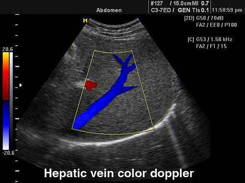
The method allows you to isolate and distinguish hepatic vein from the bile ducts
When examining blood vessels, duplex scanning is used as a independent method, and if necessary precise definition shape, consistency of the organ (for example, thyroid gland) - as an addition to ultrasound, since it clarifies the blood supply to the tissue. Often the technique is used simultaneously with ultrasound as a supplement and to obtain valuable information.
The principle of the method is preserved in echocardiography and allows for diagnostics valve defects, notice places of pathological discharge of blood flow.
Great importance is attached early detection tumors of the uterus, prostate by the nature of tortuosity vascular bundle, changes in the vascular pattern.
Duplex scanning of the veins of the lower extremities makes it possible to check the consistency of the valves in the superficial and deep vessels, the functioning of the perforating veins.
The duplex technique is part of a combination of triplex scanning, which first captures the “area of interest” and then adds a spectral pulse test.
Who is prescribed duplex scanning of extremities?
- pain in the legs during exercise (walking) and at rest;
- feeling of heaviness, unmotivated fatigue;
- swelling in the ankle area, on the legs;
- convulsive contractions calf muscles, fingers of the upper limbs;
- inability to determine pulse peripheral arteries limbs;
- appearance spider veins on the skin, visible bluish strands under the skin;
- darkening, pigmentation of the skin of the legs, paleness or redness;
- palpation painful lumps along the veins;
- detection of non-healing trophic ulcers.
Symptoms indicate vascular disease. To begin treatment, an accurate diagnosis is necessary.
How is this method convenient for diagnosing venous diseases?
The duplex scanning technique is simple and quick. Its distinctive properties:
- the patient does not need special preparation;
- does not use chemicals, no side effects;
- the patient does not experience pain or discomfort;
- not associated with damage to the skin (injections);
- has no age restrictions.
In 30–45 minutes, the doctor is able to identify the following pathology veins:
- blood clots, their stage, size, condition of surrounding tissues;
- reasons for recurrence of varicose veins after phlebectomy, sclerotherapy;
- dysfunction of perforating veins;
- decreased patency and sagging valves in deep and superficial vessels;
- change in the condition of the vascular wall.
How is the research conducted?
To make the procedure comfortable, the patient is advised to first put on clothing that will allow the desired area of the body to be quickly exposed. Before examining the veins on your arms, it is better to remove all jewelry. You need to take your own sheet and several napkins to the office to wipe off the gel at the end of the procedure.
The technique does not depend on the weight and age of the patient. The person is placed on a couch with his head elevated. The skin of the limb is lubricated with a special gel to ensure tight contact with the sensor. Without the gel, the signal clarity is lost.
The research is carried out in stages:
- starts from the groin area (sensor power up to 7 megahertz);
- the sensor moves down with a slight variable pressure, is examined deep vein hips;
- below knee joint the anterior tibial vein is scanned;
- then the patient is asked to turn over on his stomach, a cushion is placed under the knee, and the popliteal vein is examined on the screen;
- small vessels are divided into branches of the small and large tibial vein and viewed from their origins to their mouth, low-frequency sensors are used.
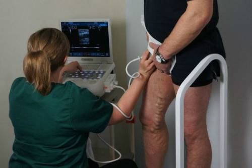
The veins of the legs are also examined in a standing position, it is enough to straighten the arm
Results for leg vein pathology
The results are deciphered immediately after the procedure. The result is given to the patient. Only a doctor can correctly assess blood circulation parameters. Blood flow is measured by the device according to the following criteria:
- maximum speed in systole;
- minimal - in diastole;
- vascular wall resistance;
- pulsation index;
- thickness of the venous wall.
The final diagnosis is made vascular surgeon or a phlebologist based on clinical manifestations and scan data.
The method allows you to identify at an early stage:
- varicose veins of superficial and deep veins;
- thrombosis and thrombophlebitis of blood vessels in the legs and arms;
- atherosclerosis of the arteries of the extremities;
- obliterating endarteritis.
Is the method safe?
The Doppler effect is accompanied by the release of a beam of energy. Modern ultrasound machines have special filters. When using high power, there is still a danger of damaging the cells in the retina. When examining pregnant women and children, use the minimum radiation power.
You will have to temporarily refuse the examination. if the patient has:
- unhealed and bleeding wounds, burns on the arms and legs;
- skin diseases in the form of rashes, ulcers;
- during the height of infectious diseases;
- with exacerbation of bronchial asthma.
After recovery, the study can be carried out in full.
Duplex scanning is not easy modern approach to diagnosis, but also most affordable way for the population.
Among the advantages of duplex ultrasound, it is worth noting high information content, safety and absence pain. In addition, such diagnostics do not require special preparation from the patient. The procedure does not have side effects and complications, therefore does not imply restrictions on the patient’s age.
Operating principle
This research method is based on the Doppler effect. Ultrasound, reflected from the vessels, changes its frequency. This allows you to determine the angle of the received signal. The Doppler shift can be encoded as a stream moving at different speeds. Each indicator is assigned its own characteristic color. It can be easily seen against the background of a monochrome picture displayed on the monitor screen. Blood flow can be collateral or main. The first is characterized by a reduced speed, the second - normal.
Indications for the study
Ultrasound scanning of the arteries of the extremities examines the diameter of the vessels and the nature of blood movement. It is often supplemented functional tests. These studies help diagnose abnormalities in the functioning of the vascular system, as well as the presence of general disorders in the body. In addition, such tests make it possible to study the mechanisms responsible for the movement of arms and legs. Thus, in a healthy limb, blood vessels tend to expand, which significantly increases the rate of blood flow. Disturbances in the mechanisms of the limbs often lead to malfunctions of blood vessels. To conduct such studies, the patient is subjected to a small amount of physical activity. The doctor then measures blood flow rates and compares them with those recorded before the procedure. The change in this data should not exceed 40%.
Sometimes tests involving tension of the limbs are used. In this case, data taken before and after the study are also recorded. Specialists also sometimes prescribe a test using nitroglycerin. In this case, the property of the substance to relax the muscles of blood vessels is used.
The first signs of arterial damage are uneven, thickened, or discontinuous inner layers. These symptoms may indicate the onset of severe abnormalities in the functioning of the arteries. They can be detected using Doppler scanning. The use of this method makes it possible to visualize vessels well and provide information about possible deviations from the norm in their work.
This study allows you to clearly examine even those vessels that could not be detected with standard ultrasound. Thus, duplex scanning of the skull is considered one of the few types of diagnostics that can detect circulatory disorders in the brain. Using it, you can accurately determine the location of the arteries and analyze their work.
