A bone grows on the leg: anatomical changes, causes and symptoms of the disease. Bone on the big toe: treatment at home
Healthy feet are the key to a full life and activity. In the event of any pain, a person experiences not only significant discomfort, he also loses the ability to lead a habitual lifestyle and move freely. One of the most common joint problems nowadays is the occurrence of pain in the big toe bone. It is noteworthy that women suffer from this problem more often, and often already at the age of 25-35 years. In this case, severe pain can be accompanied by inflammation, as well as deformity of the joint, first on the thumb, and then on others.
In the normal state, the human foot is designed to perform a shock-absorbing function. This means that it distributes the load evenly throughout the leg while walking, and also protects the spine from excessive shaking.
In the natural state, the metatarsal bones of the fingers on the foot are parallel, however, as the ligaments and muscles weaken, they begin to gradually shift to the side, pain appears. As a rule, first there is a deformation and an increase in the bone on the thumb, a kind of bump is formed. Such a diagnosis sounds like this - valgus deformity of the foot.
As a result of the deflection of the arch of the foot, flat feet develop in the transverse plane, which provokes such changes:
- loss of shock-absorbing qualities of the foot;
- offset of reference points;
- the foot is loaded incorrectly;
- there is a subluxation of the thumb;
- the head of the bone slips out of place in the joint.
If this condition is not treated, then over time, the pain in the bone will only become more intense.

Foot deformity at different stages
Visible changes in the condition of the big toe occur gradually and differ in such symptoms at different stages:
- The deviation of the metatarsal bone does not exceed 20 °, so a person experiences pain only if he puts on shoes that are too narrow.
- The deflection angle increases up to 30°. As a result, the bone on the leg near the big toe hurts after a long walk and wearing uncomfortable shoes, including high heels.
- Displacement of the metatarsal bone of the thumb by 30-50°. At the same time, the patient feels pain in the bone already quite strongly, regardless of the choice of shoes.
- Deviation of the bone by more than 50 °. In this case, the protruding bone already hurts very badly and almost continuously.
Prerequisites for the development of pathology
The reasons for which there is pain in the bone of the thumb can be very different, in particular:
- Primary joint pathologies: arthrosis, bursitis, arthritis, osteoarthritis, osteophyte, ankylosis, Ehlers-Danlos syndrome and many others. They are characterized by inflammation and degenerative-dystrophic changes, the formation of bone growths and even hemorrhages. Such pathologies can be diagnosed in both adults and children.
- Injuries - primary or recurrent. Permanent mechanical microtraumas due to uncomfortable shoes provoke the formation of edema and circulatory disorders, and as a result, the finger is displaced to the side and severe pain. A broken or cracked bone can also have this effect.
- genetic predisposition. As a rule, bulging of the bone near the thumb is diagnosed in patients whose family has already had cases of the disease. The cause of pain in the bone in this case is a violation of the production of collagen or its mutation, which is transmitted genetically.
Orthopedist Kontaev Arman Zhazitovich will tell you more about the pathology:
- Weak ligaments and transverse flat feet, acquired as a result of wearing uncomfortable shoes or a congenital predisposition.
- Excess weight has a very detrimental effect on the joints of the legs. They wear out faster, the bones in them become unstable and change position over time.
- Age. The production of collagen occurs in the human body only up to 21-25 years. In the future, the lack of this substance provokes the development of joint diseases, dislocations and fractures in the elderly.
- Hormonal imbalance. With a decrease in estrogen production, the concentration of cholesterol in the blood increases. This, in turn, provokes blockage of blood vessels with atherosclerotic plaques, impaired blood circulation and metabolism in bone tissues.
- Cartilaginous tissues in the joints wear out faster as a result of a lack of trace elements, in particular magnesium, vitamins and polyunsaturated fatty acids.

- Reinforced sports training can lead to foot deformity, a decrease in its shock-absorbing ability, so that over time the patient may find that he has a bone sticking out near his thumb.
- Autoimmune diseases (rheumatoid arthritis). If the body begins to perceive its own cells as foreign, antibodies gradually destroy the joint tissues, disrupting their performance.
- Inflammatory processes in the joints as a result of infection with gonococcus, staphylococcus, tick-borne encephalitis and other pathogens.
Whatever the reason why the bone hurts on the leg near the big toe, it is necessary to pay attention to the problem in time and seek treatment from a doctor.
Diagnostic methods
Before starting any treatment, it is worth finding out the reason why the bone is swollen. This is not always a hallux valgus deformity. In particular, acute pain can be caused, for example, by bursitis when it occurs. With gout, patients also experience long-term aching pain, as uric acid crystals accumulate in the joints due to metabolic disorders.
To determine exactly why the bone on the big toe hurts, laboratory tests of blood and urine samples are carried out. Further diagnostics is performed using instrumental methods.

Podometry - one of the methods for examining the feet
Effective diagnostic methods are recognized:
- X-ray - the leg of a standing patient is photographed in several projections. In the presence of flat feet or deformities of the joints, everything can be seen in the pictures.
- Podometry is the calculation of the ratio between the height of the instep and the long foot. Flat feet are recognized when the index value is less than 29.
- Plantography – Scanning a loaded foot allows an accurate image of any existing foot defects to be obtained.
Treatment of pain in the bone on the leg
The first thing to do if there is pain in the bone is to consult a therapist. He will prescribe an appropriate examination, and treatment will be prescribed by one of the highly specialized specialists:
- traumatologist - if the cause of the disease was a fracture or bruise;
- endocrinologist - if a hormonal imbalance is detected;
- surgeon - to remove the build-up, if it is already useless to treat it with medicines;
- orthopedist - in all other cases.
If you seek help immediately, there is no way to relieve severe pain, you should limit the mobility of the leg, rub it with a soothing infusion of calendula or a warming ointment. In addition, pain can be relieved with analgesics (Pentalgin, Tempalgin) or ibuprofen-based painkillers (Nurofen, Naklofen).

Pain medication
Treatment of the bone on the leg at the big toe by traditional methods is carried out in several directions at once:
- At the initial stages of the development of the disease, patients are recommended to use special devices, for example, insoles, linings and. They allow you to redistribute the load on the entire foot and reduce pain when walking. The overlay on the bone, in particular, prevents further exit from the joint of the head of the thumb bone and an increase in the angle of deformation.
- Massage to restore blood supply to the joints.
- Special medical gymnastics. Exercises are done daily to achieve a positive result.
- Measures aimed at reducing the load on diseased joints. Hard physical labor and intense training should be abolished. Walking daily for 30-40 minutes, swimming and light gymnastics will be useful to prevent complete relaxation or overload of the joints.

A tool designed specifically for straightening the bones on the foot
- Diet adjustment. Food is added to dishes rich in vitamins and microelements necessary for the regeneration of bone and cartilage tissues. In addition, weight loss will also have a positive effect on the joints. Alcohol, sweet and too fatty foods should be excluded.
- Pharmaceutical therapy. To relieve swelling, inflammation and pain in the bone, mainly based on indomethacin and diclofenac. At the stage of exacerbation, they are used in the form of tablets and ampoules for injection, and also supplement therapy with creams, gels and ointments.
- , for example, Chondroxide, as well as vitamins A, C, D, and E.
- Folk methods of treatment - compresses, lotions and baths.
It is worth noting that aids and procedures - plaster, baths, compresses, massage and rubbing with ointments are not the main ones in the process of treating bones at home. However, they significantly help to alleviate the patient's condition, relieve swelling and pain.
Since it is very difficult to stop the growth of a bone if the disease is already running, you can at least make sure that it does not cause so much inconvenience.
Physiotherapy
Light physical activity in the treatment of pain in the bone on the finger is still necessary. Doctors recommend performing simple preventive exercises.
These simple exercises will be useful:
- walk around the room on toes and barefoot;
- using your toes, try to pick up small objects from the floor, such as a sheet of paper;
- bend and unbend your toes, spread them apart and keep them apart for a minute;
You will see exercises for the treatment of the disease in the video below:
- try to straighten the rug on the floor using only your toes;
- roll the bottle under your feet for two to three minutes;
- hold the muscles of the feet in tension for 40 seconds;
- hold a pencil between your toes and draw figure eights with it.
Regularly doing this simple exercise will work out stiff joints and muscles and is a good way to reduce swelling and improve circulation in your legs.
Folk methods of treatment
Folk remedies for the treatment of bones are represented by many recipes that are successfully used to relieve pain if a bone grows on the leg near the big toe. Reviews about a particular tool can be contradictory, because not everyone is equally suited to them.
Popular means:
- Foot bath with the addition of iodine solution and sea salt. It will take 1 liter of warm water, 2 tablespoons of salt, 10 drops of iodine solution. You need to steam your legs in this composition for 15 minutes, performing the procedure daily for a month. After warming up, you can apply an iodine mesh to the bone.
- Propolis compress. A small amount of propolis is heated so that it rolls into a ball. The resulting plastic mass is distributed over the place where pain is felt, and wrapped with a warm cloth on top. Such a compress is done at night for 2 weeks. Helps reduce inflammation.

Foot bath with iodine and sea salt
- Compress based on honey and cabbage. After steaming the legs in the bath, a thin layer of honey is applied to the sore spot, which is covered with a cabbage leaf on top and wrapped with a bandage. Rinse your feet in the morning. Repeat the procedure for about a month.
- Mix 50 g of ammonia, camphor alcohol and red pepper, 30 g of bodyagi and 1 bottle of medical alcohol. Transfer to a glass container that does not let light through. The bone is lubricated with this composition at night and carefully insulated. Repeat the procedure for 2 weeks.
- An infusion can be prepared from iodine and dandelion flower. It will take 100 g of dried raw materials, which are poured with iodine and insisted in a dark place for 5 days. After steaming the legs, tincture is applied to the sore joint. Treatment continues for 2 weeks.
Prevention measures
To avoid the appearance of pain in the bone as a result of hallux valgus, it is worth taking elementary preventive measures:
- it is worth monitoring your weight to reduce the load on the legs and pain;
- physical activity should be moderate;
- take care of your diet - it should be balanced and include dishes rich in vitamins and trace elements;

- shoes should be comfortable and not squeeze the foot;
- massage helps relieve fatigue in the legs and improve blood circulation;
- any injury should be treated immediately.
When it comes to a painful bone on the foot, it means hallux valgus. What is a disease, and how can suffering be alleviated? Let's take a look at the causes of the disease together and find out if it is possible to quickly treat the bones on the leg at home.
What is hallux valgus
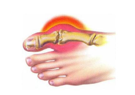
The problem of the appearance of a protruding bone is familiar mainly to female representatives. The reason for this is the lack of elasticity of the ligaments and weakness of the bone tissue. A bump appears on the inside of the foot at the base of the big toe, which causes severe aching pain, especially when wearing shoes. The appearance of a large bone on the feet can be aggravated by the inflammatory process of soft tissues, swelling and fever. The big toe noticeably deviates inside the foot, which causes a lot of trouble for a person.
Progressive disease can lead to complete which will noticeably affect walking. In addition to painful sensations that can ruin life, the curvature of the toes has an unaesthetic appearance. It becomes impossible to wear your favorite shoes, there are great difficulties with the choice of new models.
Why do bumps appear
Treatment of a large bone in the leg requires serious attention and consultation with an orthopedic doctor. Otherwise, over time, the pain will increase and, possibly, surgery will be required.
Medicine names several probable causes of the disease:
- joint pathology (bursitis, arthrosis, flat feet and others);
- genetic predisposition;
- disruption of the endocrine system;
- osteoporosis - bone fragility;
- overweight;
- improperly selected shoes;
- foot and leg injuries;
- congenital defects or complications after severe neuromuscular diseases;
- improper catering.
An x-ray in three projections will help determine the degree of deformity and the presence of concomitant diseases (arthritis, cyst, etc.).
Treatment methods

Surgical intervention is performed in complex neglected situations, when conservative measures no longer give the desired effect. Correction of the deformity occurs due to the excision of the damaged joint, followed by fixation with screws or plates. The operation, of course, solves cosmetic problems, but does not exclude the appearance of subsequent complications. In addition, the rehabilitation period can last up to six months, making life difficult for the patient.
It is recommended to start treatment of a bone on the leg at home by leveling the deformity and reducing the protruding bone with the help of various orthopedic devices: instep supports, orthopedic insoles, interdigital liners, rollers, ties. After long walks, it is useful to do warm foot baths with a massage of the metatarsal bones.
Preventive measures

To avoid a critical situation, you should start treating the bones on your toes as early as possible. First of all, you need to stop wearing tight, hard shoes and high heels. Regular gymnastics for the feet, relaxing massage, walking barefoot on uneven surfaces helps to increase the tone of the muscles of the foot. In addition, active physical activities prevent the development of arthritis and
Particular attention should be paid to the appearance of bones in children and adolescents. The cause of the bumps may be flat feet. The presence of painful symptoms can lead to circulatory disorders, and in the future to the development of early osteochondrosis. Therefore, children's shoes should be of high quality and comfortable. Preference should be given to models with a rounded toe and a stable low heel.
Preventive treatment of the bones on the leg at home also consists in a healthy diet. Alcohol, coffee, chocolate, spicy spices should be excluded from the diet. Give preference to fruits and vegetables. You should drink plenty of fluids, it is better if it is tea or still water.

Traditional medicine services
In addition to preventive measures and therapy prescribed by an orthopedist, alternative treatment of bones in the legs can be recommended. In most cases, alternative medicine relieves pain and reduces foot discomfort. The effectiveness of the impact is explained by the healing properties of natural products that underlie folk recipes.
Consider the most common and time-tested methods of dealing with hallux valgus.
salt help
Table salt has a pronounced antipyretic and anti-inflammatory effect, reliably disinfects and disinfects. This is the simplest and most affordable remedy for pain, which lends itself
Treatment consists in prescribing a two-week course of salt baths for the feet. The temperature of the water should be at the level of the body - no more than 36.6 ° C. It is better to use coarse salt for the procedure, because fine salt (such as "Extra") does not have many useful qualities due to special processing. Fifteen minutes every day is enough for the severity of pain to decrease, inflammation of the bone to decrease. After a week break, the salt treatment should be repeated again. In some cases, three or four courses of therapeutic baths may be needed.
If the disease is in the initial stage, then salt procedures are the fastest treatment for a bone in the leg at home. The folk method can not only stop the development of the disease, but also reduce the bumps in a short time.

ice salt
Gives a good effect in combination with ice. Such a remedy contributes to complete resorption Ten days of use are enough, and you will forget how the bones on your legs hurt. Treatment is carried out with a mixture of coarse salt and finely chopped ice, which must be applied to the sore spot and pressed with a towel for several minutes. In winter, pure snow can be used instead of ice.
A strong burning sensation will be felt on the skin, which is worth enduring, because a positive result will not be long in coming. As soon as the discomfort intensifies, you need to remove the ice and, without washing off the remnants of the mixture, cover the sore spot with gauze. From above, the leg is wrapped in a warm scarf. The compress can be left until the morning. If the burning sensation does not stop, then the bandage must be removed, and the bone should be lubricated with sea buckthorn oil.
Healing properties of iodine
It has long been known that iodine is a universal remedy for preventive and therapeutic properties. You can not do without it in traditional medicine, when the bone on the leg is worried. Treatment with iodine should be carried out for a long time and methodically, and then the result will be pronounced and stable.

Before the procedure, be sure to wash your feet with warm water and wipe them dry. Lubricate sore spots with camphor oil, and cover with oil on top in order to avoid possible burns. Useful procedures are best done before bedtime. After two weeks, the growth of the bones will stop.
There is another successful treatment option with iodine. To do this, you need to buy medical bile at the pharmacy. First, you need to apply a grid of iodine to the bone, and then lubricate the sore spot with a layer of bile and cover with a thin film. From above, the leg must be tied with gauze and insulated with a terry sock. The compress is left all night, and in the morning the remnants of bile are washed off with warm water without soap. If you do compresses daily for a month, then you will no longer be bothered by a bone on your leg.
Iodine treatment can also be combined with other beneficial substances. A good effect is the interaction with salt. Warm fifteen-minute baths are recommended to be done every night before going to bed. Ten drops of iodine and two tablespoons of salt dissolved in one liter of water can relieve pain and inflammation in the joint for a long time.
If you add 5 aspirin tablets to 10 drops of iodine, you get a “pharmacy cocktail”. For three days, it is useful for them to treat the bones on their legs at home. Reviews of many people who have tried the iodine remedy agree that the three-day procedure should be repeated after 10-12 days. This will consolidate the effect obtained and relieve you of painful manifestations for a long time.

On summer days, do not miss the flowering period of dandelions - they can also help a lot when a protruding bone on the leg bothers. Treatment requires preliminary preparation of the healing composition. Yellow heads of dandelions are crushed and dried in the sun. Then add a little iodine so that it completely covers the flowers. The resulting mixture must be infused for at least four days. A mesh of the resulting composition is applied to the dry, steamed skin of the legs. The procedure should be carried out every evening for two weeks.
Benefits of chicken eggs
A good result is the treatment of the bones on the leg at home with an ointment made on the basis of a chicken egg. It will take two weeks to prepare the medicine - that is how long an egg must lie, soaked in vinegar (concentration 9%) until the shell is completely dissolved. Then it is taken out of the acid and triturated with a tablespoon of unsalted pork fat and ten grams of pharmacy turpentine.
Ghee can be replaced with butter or Vaseline. The result is a healing ointment that needs to be applied every evening to sore spots. To enhance the effect, you can alternate the medicine with iodine nets.

bone tar
We suggest you consider another specific method of treatment. Some people claim that even advanced rheumatism can be healed with tar obtained from bones, not to mention such a simple thing as a bone in the leg.
Treatment, reviews of which are not too unambiguous, consists in prolonged calcination of animal bones (chicken or beef) in a clay pot. The container must be buried halfway in the ground, and then overlaid with firewood and set on fire. Tar, obtained as a result of prolonged heating, is used in the form of compresses.
The bee helps
For rubbing into the affected joints, bee honey is used, which is preheated in a water bath. A good effect also gives the use of compresses based on softened propolis. You can replace it with an alcohol tincture from a pharmacy. Soft gauze is abundantly moistened with liquid and applied to the sore spot for the whole night. From above, the compress is covered with cellophane and a warm sock is put on.

Potato peelings
Treatment of the bones on the leg at home with the help of potatoes is also an excellent remedy. Gruel from raw grated potatoes is applied to sore spots or steam foot baths are prepared. It happens in the following way.
Potato peelings are poured with a small amount of water and boiled for a quarter of an hour. The resulting decoction is added to hot water, in which sore feet are then soared. After the procedure, it is useful to apply the boiled husk directly to the bones. The procedure should be carried out for at least half an hour, after which the legs should be covered with a warm blanket. If you regularly steam your legs with potato broth for one and a half to two weeks, then the pain will soon pass, and the bones will begin to dissolve.
fish days
A very good ancient method of treatment was discovered by ancient fishermen. To relieve inflammation and heal from deformities, it is useful to apply pieces of fresh fish to sore joints. You can use any breed caught in a river or lake. Frozen fish should not be taken, because there are practically no healing properties left in it.
A compress of fresh fish pulp is applied to the sore spot, which is tightly bandaged to sore joints, and left overnight. In the morning, the leg is washed with water, removing the unpleasant odor.
Treatment should be repeated every night for a week, then take a break for two to three days. Repeating the therapeutic course fixes the effect obtained for a long time. Unfortunately, the proposed method of healing today may not be available to all people. In this case, we recommend paying attention to other methods of traditional medicine.

rice diet
In addition to various compresses and healing ointments, one should not forget about the peculiarities of nutrition. It is very useful to follow a diet that helps cleanse the connective tissue. The most common and affordable means is rice. Loose porridge is cooked from it without oil and salt. Rice of dark varieties is pre-soaked overnight in cold water. In the morning, it is thoroughly washed and boiled in plenty of water. Once a week, you need to arrange a fasting day and eat only rice porridge, washed down with a rosehip broth. The diuretic effect of the unloading menu will help to remove uric acid salts from the body and prevent the appearance of joint deposits.
Another version of the rice diet suggests eating two tablespoons of boiled rice every day on an empty stomach. For the rest of the day, you can eat in the usual way, increasing only the consumption of pure water. Rice peeling should be carried out for at least one month with periodic repetition throughout the year.
And finally
The program of alternative treatment for bones can include the use of herbal decoctions and herbal tinctures. The following plants and fruits will be good helpers: sage, chamomile, calendula, birch buds, ginger root, elderberries, lingonberries and many others.
Always remember that the beauty and health of the legs are in your hands. In order not to suffer from pain in the future and not to wonder how to cure the bones, try to take all measures today to prevent the development of hallux valgus.
Bumps, bones on the legs - this is what people call valgus deformity of the big toe. The main symptom of this disease is the appearance of a tubercle on the leg. The bump gradually increases in size, grows and causes pain. This makes it very difficult to find comfortable shoes. In advanced cases, the bone causes difficulty in walking.
If the bone grows on the big toe, then this problem must be paid attention to. A visit to the doctor is necessary. The sooner you can visit a specialist, the faster you can get rid of the problem. In the initial stages, the treatment of the disease is simple, as orthopedic agents are used. But in advanced cases, only surgery can help.
What are the bumps on the legs? Not all people suffering from hallux valgus know the answer to this question. To find it, let's remember the anatomy.
The big toe of the human foot is formed by the phalanges and the metatarsal bone. When the disease occurs in the leg, changes occur. In sick people, the phalanges of the thumb take a position at an angle to the metatarsal bone. That is why the protrusion becomes clearly visible on the leg. It is a protrusion of the metatarsal head.
With a pathological displacement, a person experiences pain. The appearance of this symptom is due to the fact that the synovial bag becomes inflamed (it is located between the phalanges of the fingers and the head of the metatarsal bone and is a bag filled with a special fluid, due to which friction over the surface of the joint is reduced). The reasons for the increase in the bone on the big toe are different. Let's talk about them.
Why does the bone grow on the leg
An enlarged bump is a sign that the foot is becoming more and more deformed, and the big toe is deviating more and more from its normal position. The bone may seem enlarged, because in this area, with hallux valgus, a swelling forms. The reasons for the increase in bumps include:
- illiterately chosen shoes: prolonged wearing of narrow shoes with high heels becomes a prerequisite for the development of the disease;
- hereditary factors: very often the bone on the leg begins to grow in those people whose close relatives suffer from valgus deformity of the big toe, complain about the growth of the bone on the leg;
- flat feet, with this disease, the weight on the foot is distributed unevenly, and as a result of this, the big toe shifts, protrudes outward;
- injuries, for example, as a result of a heavy object falling on the foot, the foot may be damaged, which will provoke the growth of the bone in the area of the thumb.
Other reasons for the appearance of a protruding bone on the leg include foot diseases, congenital bone anomalies, overweight, and a serious load on the feet.

In connection with the changes taking place in the foot, the structure of the bone tissue of the metatarsal head begins to slightly thicken. Then the articular surface is flattened. The head is covered with additional layers of bone tissue. In this regard, the size of the bump on the affected leg increases.
Treatment with folk remedies
If the bumps on the legs increase, then you can try proven traditional medicine. However, it is worth remembering that in the later stages the deformation cannot be eliminated with the help of grandmother's recipes. You will need qualified medical attention.
It is possible to stop the growth of bones at the initial stage through the use of red clay compresses, salt baths, decoctions from various medicinal plants. Before treating a protruding bone on the leg, you should familiarize yourself with the following rules that must be observed when using folk remedies:
- wear special insoles, use special orthopedic correctors that fix the metatarsal bone in the correct position;
- engage in therapeutic exercises daily (exercises can relieve pain, strengthen muscles and ligaments);
- change your diet and lose excess weight - due to a decrease in the load on the legs, deformation processes are suspended, bone growth stops;
- do therapeutic massage (its goal is to return the normal biomechanics of the foot).
The result from the use of folk remedies will be good if they are used in combination with other procedures prescribed by the attending physician. Before using this or that recipe, you should consult with a specialist. He will tell you which remedies are really effective and which do not bring the desired result.
Which specialist to contact
If a protruding bump appears in the area of the big toe, you should immediately go to the doctor. First, you should visit a therapist. He is a general practitioner. The therapist will examine the bone of the big toe, make a preliminary diagnosis and refer you to a specialist of the appropriate profile - an orthopedist, surgeon or traumatologist.

A professional will conduct the necessary diagnostic studies (visual examination, X-ray). In some cases, it turns out that the bones in the legs are a concomitant ailment of diseases such as arthritis or gout. It is important to correctly diagnose, to distinguish valgus deformity of the big toe from other diseases. Only then can you choose the most appropriate way to treat the bones on the legs and eliminate their growth.
Non-surgical methods of bone correction
If hallux valgus deformity of the big toe is detected at the initial stage, the specialist will choose non-surgical methods of correction. Thanks to them, you can both remove a protruding bone and stop its further growth.
Non-surgical methods of correction include the following.
- Use of orthopedic shoes. Professionals advise wearing special wide models made of soft materials and equipped with special pads.
- The use of special tires. These orthopedic devices hold the human foot in an anatomically correct position and fix the big toe.
- Physiotherapy: ultrasound, electrophoresis, mud therapy, therapeutic baths, magnetotherapy.
The growing bone at the base of the thumb can be treated with anti-inflammatory drugs. They should only be used as directed by your doctor. Medicines relieve swelling, eliminate pain. The inflammatory process gradually passes through the use of creams, ointments, tablets and injections.
Surgical treatment of the disease
Many people who have a bunion in their foot decide not to go to the clinic. For a long time they endure discomfort, pain and do not try to fight them, but in vain. At the moment, there are a number of effective surgical methods for treating the disease.

The goal of any surgery performed for valgus deformity of the big toe is to correct the position of the bones, restore the functions of the foot, eliminate existing symptoms and improve the quality of life. During surgery, doctors do the following:
- remove excess bone
- fix the big toe in the correct position;
- reconstruction and stabilization of the joint.
The most popular surgical treatment for a protruding bone in the leg is an osteotomy. During the operation, doctors make a Z-shaped incision in the bone that supports the thumb. This method of treatment allows you to return the head of the bone to an anatomically correct position.
Thus, only a doctor can tell how to remove a bump on the leg after making an accurate diagnosis and determining the stage of the disease. Self-medication is undesirable, as the disease progresses rapidly. Even the use of folk remedies should be agreed with your doctor.
Illness with a history
Bumps in the area of the thumbs are an unpleasant problem that mankind has known for a long time. The first mention of this disease dates back to the III century BC. e. The symptoms of the disease were first described by Hippocrates. The famous ancient Greek physician found that the bone on the big toe basically begins to grow in women during menopause. Men, on the other hand, faced this problem earlier after reaching puberty. It is noted that the growth of cones at the bases of the thumbs is affected by heredity.
Currently, there are quite a few different methods of dealing with cones. However, any modern doctor will tell you that the help of specialists may not be needed at all if certain preventive measures are followed:
The emerging and gradually growing bone near the big toe most likely indicates that the patient has transverse flat feet. This pathology is the most common deformity of the musculoskeletal system and accounts for about 10% of all orthopedic diseases. The disease is characterized by a progressive course (the bone on the leg at the big toe continues to grow). Transverse flat feet are recorded mainly in women (55.2% versus 38.1% in men). The formation of a bone or bump (the so-called hallux valgus) is an early symptom of an existing disease and indicates damage to the 1st metatarsophalangeal joint.
- predisposing - anatomical, constitutional (congenital weakness of the ligamentous and tendon apparatus of the foot, aggravated with age) and supporting features of the foot;
- producing - static and dynamic overloads (long standing on the feet and peculiarities of gait lead to a weakening of the muscular apparatus, which ultimately leads to changes in the muscles and ligaments of the foot), the use of irrational shoes.
- 1. hereditary-constitutional predisposition;
- congenital dysplasia of the osteoarticular apparatus of the foot, the most likely signs of which are the following:
- significant lengthening or shortening of the 1st metatarsal bone;
- excessive bevelling of the gap of the first metatarsal-sphenoid joint;
- the presence of additional sesamoid bones.
- 3. primary weakness of the ligamentous-muscular apparatus of the foot.
- the first (mild) - HVA does not exceed 30 degrees, and IMA is in the range of 9-12 degrees;
- the second (moderately pronounced) - HVA reaches 40 degrees, and IMA increases to 13-16 degrees;
- the third (pronounced) - HVA is over 40 degrees, and IMA increases to 16 degrees or more.
- Non-fixed ("soft" foot) - the transverse flattening of the foot is completely eliminated, and the thumb is easily brought to its normal position using manual correction.
- Fixed ("rigid" foot) - an attempt to reduce the diameter of the foot and bring the big toe into a normal position using manual correction does not give positive results.
- pain sensations;
- change in the location of the bones of the forefoot;
- problems in wearing and fitting standard shoes (this forces patients to use larger shoes with a loose toe, and women to almost completely abandon shoes with heels);
- cosmetic defect (patients indicate the presence of "ugly bones on the legs").
- expansion of the forefoot;
- outward deviation of the thumb with its internal rotation;
- corns on the sole in the area of the heads of the middle bones of the metatarsus;
- osteocartilaginous exostosis of the head of the 1st metatarsal bone with signs of chronic bursitis;
- hammer-shaped deformity of the middle toes, which is accompanied by the development of corns in the area of the dorsum of the deformed fingers;
- deviation inside the little finger with the presence of exostosis and bursitis.
- x-ray of the foot in direct projection - allows you to determine the type of transverse spreading, as well as HVA and IMA;
- X-ray of the foot in the axial projection - allows you to determine the degree of displacement of the sesamoid bones of the 1st metatarsophalangeal joint in the region of the intermetatarsal space and assess the angle of rotation of the 1st metatarsal bone.
- circular bandaging of the foot in the area of the heads of the metatarsal bones;
- the use of special orthopedic insoles or screeds with a Seitz roller under the heads of the 2nd-3rd metatarsal bones.
Show all
Why does
There are many theories explaining the origin of the development of transverse flatfoot in humans. To date, doctors believe that this disease develops under the influence of many factors (polyetiological theory). According to the latter, there are the following factors:
Therefore, the causes of the development of transverse flat feet can be divided into external and internal.
This disease is extremely rare as a congenital form.
The internal reasons are as follows:
External causes that contribute to the development of this disease include overloads associated with an increase in human body weight, with sports, profession or housekeeping, with wearing irrational shoes (with pointed toes and high heels), etc. The negative impact of the latter is so great that some scientists even consider it as the main cause of the formation of deformation.
Also, regular wearing of such shoes leads to a sharp increase in the load on the heads of the metatarsal bones and favorably affects the appearance and progression of deformity.
The bones of the metatarsus are held in the correct position thanks to the fascia and plantar aponeurosis. Therefore, first of all, this disease should be considered from the position of manifestation of insufficiency of the ligamentous apparatus against the background of weakness of the muscles of the foot and lower leg.
On one of her programs, even Elena Malysheva admitted that she was suffering from this ailment.
How the disease develops
To better understand how the disease develops, it is worth considering what bones the foot consists of.
Foot bones. View from above
It should be remembered that transverse flatfoot is a progressive disease. Therefore, if the patient does not follow the recommendations of the doctor, then the bone on the leg near the big toe will continue to grow with other ensuing consequences.

Foot bones. Bottom view
Normally, the sesamoid bones are enclosed in the tendons of both heads of the short flexor, and the tendon of the long flexor of the thumb is firmly fixed between them (the so-called "hammock").

"Hammock" of the first metatarsal bone: 1 - tendon of the muscle that leads the thumb; 2 - tendon of the long flexor of the thumb; 3 - tendon of the muscle that abducts the thumb; 4 - tendons of both heads of the short flexor of the thumb
In the process of development of this pathology, the main element is the spreading of the forefoot (i.e., the discrepancy in horizontal plane metatarsal bones). Most often it occurs due to an inward deviation of the 1st metatarsal, sometimes in combination with an outward deviation of the 5th metatarsal.
Progression of inward deviation of the 1st metatarsal leads to subluxation and dislocation of the sesamoid bones. The latter are shifted to the region of the 1st intermetatarsal space. This leads to outward displacement of the tendons that are in complex with the sesamoid bones. As a result, there is a dissociation of the head of the 1st metatarsal bone with its "hammock".
As a result, the extensors and flexors of the thumb acquire an additionally unusual function of abductors (that is, abduction), leading to an internal deviation of the thumb (halus valgus), which leads to the appearance of a bump or bone near it.
As a result of the interaction of the load forces on the first metatarsal bone and the support reaction, a moment of force arises, which contributes to the internal rotation of the 1st metatarsal bone and thumb. As a result, as well as subluxation and dislocation in the metatarsal-sesamoid joint, a significant decrease in the support function of the head of the 1st metatarsal bone is formed. This leads to a sharp increase in the load on the heads of the 2nd and 3rd metatarsal bones with the development of painful calluses on the sole of the foot (the so-called corns).

corns
The head of the 1st metatarsal, protruding inwards, is subjected to constant pressure from the shoe. As a result, along its inner edge, osteo-cartilaginous exostosis arises and gradually increases in size (bone growth from gradually ossifying cartilaginous tissue), and above it, an often inflamed articular bag (a chronic form of bursitis).
Similar changes are sometimes observed in the region of the head of the 5th metatarsal bone, which is explained by its significant outward deviation. The little finger deviates inward. In foreign literature, the above pathology is called "tailor's bursitis".

With transverse flat feet, an increase in the load on the heads of the middle bones of the metatarsus creates excessive constant pressure on the flexor tendons of the 2nd and 3rd fingers. This causes a reflex contraction of the corresponding muscles and leads to the development of hammer toe deformity. Another reason for the development of this defect is the displacement of the middle fingers with the big toe, which deviates outward.

hammer fingers
This specific deformity is characterized by flexion at the proximal interphalangeal joint and extension at the metatarsophalangeal joint. Sometimes an extensor or flexion position in the distal interphalangeal joint is additionally attached.
As the disease progresses, persistent articular contractures of the hammer toes form, and painful calluses develop on their dorsum from shoe pressure. In the future, a dislocation of the finger to the rear (“clawed” finger) may form, and the development of osteoarthritis in the deformed joints increases the pain syndrome.

"Clawed" finger
Thus, transverse flatfoot is a multicomponent deformity of the forefoot. The most important elements of the latter, in addition to transverse spreading, are the dislocation of the sesamoid bones of the metatarsophalangeal joint and excessive internal rotation of the 1st metatarsal bone.
In most cases, the consequence of transverse flatfoot is the deviation of the big toe outward and / or the little finger inward, hammer-shaped deformity of the middle toes.
Classification
Transverse flat feet and hallux valgus are classified differently depending on the severity of pathological changes.

HVA - thumb deflection angle (normally up to 15 degrees). IMA - angle between the 1st and 2nd metatarsal bones (normally 8-10 degrees)
According to the degree (sometimes referred to as stages) of the deformity of the forefoot (determined by x-ray data), the following are distinguished:

Hallux valgus grades: 1st (upper picture), 2nd (lower picture on the left), 3rd (lower picture on the right)
Transverse flat feet in the form of deformation of the forefoot is as follows:

unfixed form. Left - foot before deformity correction. Right - foot after manual correction
It is not recommended to treat this disease on your own at home, and even more so to resort to folk remedies. You should contact an orthopedist.

fixed form. Left - foot before deformity correction. Right - foot when trying to manually correct
Transverse flatfoot is divided depending on the location of the deformity into unilateral and bilateral.
Clinical manifestations and diagnosis
The diagnosis of "transverse flatfoot" and "halus valgus" is established on the basis of the results of a clinical examination, x-ray studies. This pathology is confirmed by podometric and plantographic (measurement of parameters and graphic image of the foot) studies.
During a clinical examination, the doctor finds out what the patient is complaining about, and also determines the type of transverse flatfoot according to the classification. The main complaints of the patient are the following:
Pain in transverse flatfoot is more often localized on the plantar surface of the foot under the heads of the middle metatarsal bones and the medial surface of the head of the 1st metatarsal bone. Mostly pain is periodic and occurs when walking and standing, accompanied by increased fatigue of the legs. Pain can sometimes be given to the knee joint or to the shin area.
Usually, the duration and intensity of the pain syndrome increases as the deformity progresses. Again, the opposite is observed when, with a severe degree of this disease, pain is not expressed.
With transverse flat feet, the deformity of the foot has certain signs. They are the following:
The following x-ray studies are used for transverse flat feet:
When performing podometry, the Friedland transverse index is calculated, which will exceed the norm depending on the degree of deformation.
The plantography method allows you to detect overload zones in the forefoot area, as well as calculate HVA.
Another reason for bone growth
There are pathological conditions that can be confused with early manifestations of transverse flatfoot (halus valgus).
If there is an expansion of the forefoot (in the region of the metatarsal heads), then this may be the result of bone-fibrous growths in the region of the head of the 1st metatarsal bone. The latter imitate the deviation of the thumb outward by 15-20 degrees.
The spread of these growths to the surface of the joint leads to subluxation of the proximal phalanx of the first finger. This leads to a deviation, sometimes reaching 30 degrees. With this pathology, no signs of transverse flatfoot are found and no deformation of other toes is observed. This pathological condition occurs relatively infrequently, in approximately 1.5% of cases.
A similar pathology is much less common on the little finger.
In the case of this pathology, the Shede operation is a radical intervention. This is due to the fact that the supporting function of the foot in this disease is not impaired and the removal of growths completely cures the patient of this pathology. A similar operation is performed in case of damage to the head of the 5th metatarsal bone.
Treatment
Deformity of the fingers and transverse flat feet are treated mainly with the help of surgery. With the initial manifestations of the disease, it is recommended to resort to conservative therapy. This mainly occurs in the elderly, in adolescents, in the presence of contraindications to the operation, as well as in the postoperative period.
In order to return the thumb to its previous position, various types of interdigital liners are used, daily exercise therapy exercises are performed, special splints for the thumb and foot are performed. To form a transverse arch, they resort to:
If the deformation is pronounced, orthopedic shoes are used. Below is an example of an auxiliary treatment for this disease.
If there is a bone near the thumb, Elena Malysheva and other qualified doctors recommend purchasing orthopedic insoles with a Seitz roller, which additionally helps to stop the progression of the disease in combination with a change in lifestyle (refuse to wear irrational shoes).

Special splint for the big toe with a special interdigital insert
Foot baths, various physiotherapy, corrective gymnastics in combination with massage can only temporarily get rid of pain and exacerbation of bursitis in the head of the 1st or 5th metatarsal bone. It is impossible to eliminate the deformity with the help of conservative treatment..
Surgery
Today, the modern concept of surgical treatment says that the operation should be radical, that is, eliminate the root cause of toe deformities - transverse flat feet. Since in this pathology, in most cases, an inward deviation of the 1st metatarsal bone is observed, the main direction of surgical intervention is to eliminate its incorrect position.
Depending on the shape of the transverse flatfoot, different operational approaches are used, which are presented in the table below.
If there are contraindications to radical surgery or in elderly patients, then palliative surgical treatment (improving the quality of life of the patient, but not eliminating transverse flatfoot) can be performed. The essence of such an operative intervention is to correct deformities of the fingers that cause the greatest suffering to a person (halus valgus, hammer fingers).
How to treat the resulting outgrowth and is it possible to remove the bone on the leg without surgery?
The foot skeleton is made up of 26 bones. The fluoroscopic image of the foot resembles the image of hands with palms and fingers. In addition to the finger phalanges, the foot itself is divided into metatarsus and tarsus. The back of the foot (near the heel and ankle) is called the tarsus and is made up of 7 bones.
The middle part of the foot is called the metatarsus and consists of 5 bones. The metatarsus is connected to the phalanges of the fingers, and at the base of the thumb on the side of the sole there are 2 additional bones. The movable connection of bones is called a joint. Each foot has 20 movable joints.
Improper load on the joint or insufficient nutrition of its tissues causes inflammation. In this case, the joint is deformed, various growths, “bones”, bumps are formed.
Typical signs of orthopedic disease: visible change in the joint, its deformation, bending and deviation of the big toe, the appearance of a characteristic painful bump (“bone”) on the side of the foot. At the same time, the sole ceases to fully spring, and the acquired outgrowth makes it difficult to wear shoes. The bone on the big toe swells, movement when walking becomes painful.
How the foot is deformed with a bone
The deviation of the thumb from the line of the metatarsal bone should not exceed 10%. Problems and inflammations form when the angle of deviation exceeds the normal 10%.
There are four stages of the disease:
- First stage- with a deviation of the finger by 15-20 °. In this case, a small bump is formed, on which a corn often grows. The bump is a consequence of the protrusion of the head of the bone from the joint.
- Second stage- with a deviation of the finger by 20-30 °. At the same time, the periarticular ligaments are stretched, and the subluxation of the “phalanx-metatarsal bone” joint occurs, the bone becomes obvious, sticks out and stretches the shoes. There is a slight episodic pain, mainly during exercise or at the end of the day.
- Third stage- with a deviation of the finger by 30-50 °. To hold the load on the deformed joint, a bone and cartilage outgrowth is formed. The bone increases so much that it becomes difficult to choose shoes (you have to buy shoes 2-3 sizes larger).
- fourth stage- with a deviation of more than 50 °. The bone of the phalanx and metatarsal bone are removed from each other. The head of the bone becomes flat, severe inflammation develops, arthrosis is formed.

Why does too much deviation cause painful inflammation and deformity of the joint and foot?
Changing the position of the phalanx of the finger disrupts the normal distribution of load within the joint. Initially, the defect is not externally noticeable. You can guess about it only by rapid fatigue. Legs hurt after normal daily activities. After the cartilage begins to wear out, deformation is formed and inflammation appears. It becomes difficult for a person to walk and put on closed shoes.
Why does a bone in my leg hurt?
Soreness is associated with an abnormal position of the thumb bone in the joint. Due to the strong bending of the phalanx, the pressure is not distributed correctly and pain occurs. In addition, improper distribution of loads also causes inflammation (in medical terminology - bursitis). Therefore, when the “bump” grows, the bone on the leg near the big toe hurts, the joint itself swells and changes color (turns red or blue).
With the development of deformation and destruction of cartilage, arthrosis is formed. Changes spread to neighboring tissues, the arch of the foot becomes stiff, the sole begins to hurt. Following the soreness of the entire foot, corns appear.
Why does the bone grow on the leg: causes of the disease
A number of painful factors contribute to the growth of the bone on the leg. We list the causes of the bones on the leg:
- Increased or incorrect load on the foot - usually formed with flat feet (toes are arranged in a "fan"), overweight, wearing high heels, tight shoes. A common cause of "bones" is long-term wearing of stilettos. Medical statistics confirm that out of 100 patients with flat valgus deformity of the finger, only 15 people are men and 85 are women.
- Disruptions in metabolism (with endocrine diseases, for example, with diabetes, or with hormonal changes - pregnancy, feeding, menopause).
- Diseases of the joints and ligaments - arthrosis, osteoporosis.
- Ankle injuries.

Heredity is not the cause of growth. Even if a mother or grandmother had a “bone” on her leg, then her appearance is not necessary for an adult daughter. Only weakness of muscles and ligaments, a tendency to flat feet are inherited. Eating habits and footwear choices are not inherited.
The manifestation of improper loading on the foot often depends on the preferences of the individual. Healthy heel height, prevention of metabolic disorders (avoiding high-calorie foods and supplements), prevention of foot diseases (walking barefoot or with orthopedic insoles), and proper physical activity help to avoid the appearance of "bone".
At what age does the bone on the leg grow
In most people who have this problem, the growth of the bumps began after forty years. This is due to the fact that the action of several destructive factors is combined. For example, menopause is associated with age-related metabolic disorders. Or, for several years, a woman has been watching her figure (restricts herself to diets and receives less minerals and vitamins), while wearing high-heeled shoes.

Rare cases when a "bone" appeared in a young girl are associated with diseases of the endocrine system or trauma.
Important: if you have flat feet, wear shoes with orthopedic insoles. This will distribute the load correctly and prevent the formation of any growths.
The bone on the leg is uncomfortable, unpleasant, painful. Its appearance is easier to prevent than to treat. But if time is lost, and the outgrowth has already appeared, what can be done? How to remove a bone on the leg?
Treatment of a bone on the leg at the big toe
Treatment of a bone on the leg at the big toe is either passive prevention (to limit its further growth), or cardinal measures (surgical sawing, laser removal). The choice of treatment method is determined by the stage of the disease and pain sensations.
With a relatively small deviation of the finger (up to 20 °), they turn to physiotherapy, compresses, orthopedic fixators. Also, these methods of treatment are indicated when surgical intervention is impossible (poor blood clotting, heart disorders, varicose veins, diabetes).
Physiotherapy and foot massage
For pain relief and treatment of deformity in the initial stage of the disease, foot massage, electrophoresis, ultrasound, and therapeutic mud are prescribed. They increase blood circulation, which improves tissue nutrition, removal of toxic substances (waste products of cells).

Bone fixators for legs
The orthopedic industry produces various protectors for correcting the position of the toe and foot - interdigital liners, splints, side retainers, night and day bandages.
Bone braces are a type of orthopedic splint. They fix the correct position of the foot when standing and walking, and this corrects the deformation. In such conditions, in the presence of minerals, vitamin nutrition, the joint can recover.
For a therapeutic effect, fixators (tires) are worn during the day, left overnight. They begin to put on the fixators for 2-3 hours (in the afternoon), then the wearing time is gradually increased. After - leave them for the night.

Non-surgical treatment methods
Modern medicine also offers methods for non-surgical removal of bones. They stop the growth of bumps on the legs, and after a while stimulate their reduction (resorption).
Do you want something interesting?
Non-surgical bone removal methods:
- shock wave therapy- destroys calcifications and reduces cartilage growth. Promotes the formation of new capillaries, restoration of blood circulation and nutrition. And this treats inflammation, improves the condition of cartilage, ligaments and muscles, makes them elastic and durable.
- Chinese magnetic plaster for bones on the legs- relieves inflammation, anesthetizes and stops deformation. Combines the achievements of folk and traditional medicine. How does it work?
Magnetic patch - the effectiveness of treatment
Extracts of medicinal herbs are applied to the inner surface of the patch during manufacture (there are more than 30 of them, the main ones in the composition are mustard powder, dandelion flowers, saffron, turmeric, vine). They are absorbed through the skin when applied. The wearing time of the therapeutic composition is up to 2 days, after which it must be replaced with a new one. Know-how technology - enhanced absorption of useful components under the influence of magnetic radiation.
The manufacturer regulates that 16 patches are needed to dissolve a small bone. This tool is also used to treat heel spurs, various joint inflammations.

When the patch can not be glued:
- In the presence of wounds on the skin.
- With allergies.
- During pregnancy and lactation.
Removal of a bone in the leg with surgery
Removal of bones on the legs is indicated for severe joint deformity (greater than 30°). Modern medicine offers various low-traumatic removal techniques, after which a person can walk on the 4th day. The know-how of these technologies lies in the use of special fasteners that fix the joint from the inside, as well as in minimally invasive methods. In addition, the removal of a bone on the big toe during the operation is accompanied by the alignment of the toe, this prevents the recurrence of the disease.
- Special mounts(microdrills, microblades) are made of titanium alloys and installed inside the joint after cutting off the build-up. They do not allow the phalanx to take the wrong position. This fixation returns the phalanx to its normal position.
- Minimally invasive techniques involve small incisions of the skin surface (up to 3 mm), work with a micro-instrument (microsalpel, microbur), which cuts bone growths. X-ray equipment is used to visually control the progress of the operation. Disadvantage: Minimally invasive techniques are used in the early stages of the disease.

The total recovery time after surgery is 2-3 weeks. In the future, for walking, you need shoes made of genuine leather with low heels (up to 4 cm), as well as orthopedic insoles. Sometimes, after surgery, it is necessary to wear special protectors (called splints) that keep the toe and foot in the correct position.
To prevent further growths on the bones, it is necessary to correct the diet, normalize the load on the foot (choose comfortable shoes with orthopedic insoles). If necessary, prevention is supplemented with vitamin-mineral complexes, chondroprotectors.
Surgery on the big toe bone, carried out using modern technology, helps to solve the problem. In the past, operations to simply “cut down” the bumps solved the issue for only a few years, since they did not eliminate the cause of the build-up, but worked with the investigation.
How to treat a bone on the leg with folk remedies
Traditional therapies use natural substances for ointments, tinctures, compresses, and internal treatments. As a rule, they are effective in the initial stages of the disease, when the joint deformity does not exceed 20°. In this state, you can relieve pain, relieve swelling, slightly reduce the bone (due to resorption of calcifications).
Is it possible to get rid of the bones on the leg with folk methods if the deformation has reached 30 °? As a rule, no, but it is possible to relieve pain. How to do it?
Remedies for pain relief
- Baths. They add iodine and salt (10 drops and 2 tablespoons).
- Compress from cabbage leaf with honey. Before applying, the sheet is crumpled to extract the juice, after that a layer of honey is applied to it, fixed on the bone.
- Potato. It is rubbed on a fine grater and applied in a bump as a compress.
- Raw fish pulp. It is cleaned of bones, applied to the "bump" at night.
Remedies to reduce inflammation
- Propolis. It is kneaded and applied in the form of a cake to the growth. Or they make a compress from an alcohol tincture of propolis (diluted with warm water 1: 1, soak gauze or a bandage, put it on a bump, cover it with polyethylene and fix it).
- Clay. It is also used for compresses. Soak in water to a creamy state and apply to the bulging bump and the area around it. After drying - crush. Clay draws out toxic substances. Therefore, it can not be kept longer than 3 hours. It absorbs toxins and must be replaced with a new compress.
A bone in the foot is a medical problem that can be prevented. Choose comfortable shoes, wear orthopedic insoles, eat well. If the deformity of the foot has already appeared, then treatment should be started as soon as possible. The lower the stage of the disease, the easier it is to cope with its manifestation and limit the further growth of the bulging bump.
