Doppler study of blood vessels. Features of ultrasound examination with Doppler. Preparing for Doppler ultrasound
Most young people in Russia avoid doctors by hook or by crook, guided by the principle “the less you know, the better you sleep.” You can often hear a joke about how a person came to the doctor absolutely healthy, but came out with a whole bunch of diseases. But still, pregnant women simply cannot avoid regular visits to doctors and all kinds of research! And you won’t want to, because it’s not just about your own health anymore. But how can we understand the variety of analyzes and studies that are offered to us? modern medicine? For example, Doppler - what is it during pregnancy?
The full examination lasts about 45 minutes, but the length actually varies greatly depending on the elements we are looking for. Additionally, the speed at which we collect images depends on the morphology of each. often lying on the left side, left hand raised on her head, breathing, as immediately by your doctor.
By applying the ultrasound probe to the skin at chest, an image of cardiac structures is collected on a video screen in the same way that a boat sonar probes sea bottom. In this way, the different cavities of your heart appear on the screen, the size of which can be measured. The valves that separate these cavities and seal them are rated based on their condition and performance. The quality of heart contractions is also assessed. The sound generated by the circulating blood is amplified and then analyzed regarding its meaning, speed and intensity.
This is very important view a study that examines blood flow in the vessels of the expectant mother. With its help, it is possible to determine the speed and direction of blood flow in the vessels, the width of the lumen of each vessel and the pressure inside it. Why is this needed, you ask. Nutrients and oxygen are delivered to the baby through the placenta and umbilical cord, and the above parameters are necessary to determine how well his organs are supplied with blood and whether he receives enough oxygen to develop normally. Thanks to Doppler, it is possible to identify and prevent conditions such as fetal hypoxia or insufficient functioning of the placenta, for example. In addition, the baby’s overall health condition is assessed, the sound of his little heart is heard, and the width of the lumen of his blood vessels is determined.
An analysis of blood circulation through the structures of the heart reports on the pressures that prevail there, as well as on normal character or the absence of its flow. Through this examination, your doctor can very accurately assess your cardiac condition and to better tailor your treatment.
Vascular Doppler ultrasound is a simple and painless examination that allows you to study various vascular structures using an ultrasonic sensor. This exam reports the presence of vascular abnormalities and helps assess their severity. Using this technique, several vessels are available at the level lower limbs, upper limbs, neck and abdomen, including the renal arteries. The ultrasound probe emits ultrasounds reflected by the organs, which create a dynamic image on the screen.
How is Doppler done during pregnancy?
The principle of Doppler is similar to the ultrasound procedure, which is probably familiar to all women even before pregnancy (you went to the gynecologist as soon as your first period arrived, right?).
IN modern world Doppler is done simultaneously with the ultrasound procedure using the same equipment. Planned ultrasound of the second and third trimesters coincides with planned Doppler, which is why they are often combined. The doctor determines the location of those vessels in which the blood flow needs to be studied, then turns on the Doppler function and conducts his study.
Detection of certain abnormalities may lead to tailored treatment or other specialized testing. It is necessary to fast without drinking or eating 6 hours before performing vascular Doppler ultrasound of the lower extremities and renal arteries. On the other hand, you should not fast before Doppler ultrasound of the neck vessels. You can take your treatment as usual in a little water. The exam is performed without anesthesia, and sedative effect not required.
The exam is held in medical institution or at the radiology center. You set up on the bed, shirtless or in shorts depending on your study space. The doctor moves the ultrasound probe to the area to be examined after it is soaked with water. To examine the renal arteries, you may need to change normal rhythm your breath. The duration of the examination varies depending on the pathology being examined.
There are two types of studies available here: Doppler ultrasound and Doppler ultrasound. In the first case, a visual assessment of blood flow is carried out with the display of blood flow velocity curves in color on the monitor, and in the second, an additional recording is simply made on tape to track the slightest changes in blood flow and evaluate it before and after treatment.
Vascular Doppler ultrasound without risk and without undesirable effect. You will leave the ultrasound laboratory immediately after the exam is completed. You will be able to resume all your activities, including your work. Stress test allows screening or assessment cardiovascular diseases, associated with an abnormality of the arteries that irrigate the heart. The principle of the study is to subject cardiovascular system efforts that will increase the work of the heart and its oxygen demand in search of coronary insufficiency.
Doctors perform Doppler measurements during pregnancy in two modes:
- duplex scanning - the extent to which the vessel is passable is assessed, the causes of disorders, if any, are identified, the speed of blood flow is also assessed, and the anatomy of the vessels is also studied;
- triplex scanning - a color image is added to the duplex one. This is all the difference, but they believe that triplex scanning is still more accurate.
You do not need to undergo any special preparation for testing. There is no need to adhere to any diet, nor do you need to monitor the fullness of your bladder.
Screening for some abnormalities may lead to appropriate treatment or other specialized testing. No special preparation is required before performing the force test. You don't have to be hungry. Depending on the purpose of the stress test, your doctor may ask you to temporarily stop your usual treatment before the test starts. Recent onset of chest pain should be reported. It is advisable to bring clothing and sports shoes.
The exam is performed by a qualified physician in a room equipped with animating material. The muscular effort is performed on a treadmill or bicycle and is produced in the following stages, with each bearing supported for 2-3 minutes. During the entire examination, measurement blood pressure and continuous recording of the electrocardiogram are performed looking for any abnormalities. The exam requires the placement of adhesive or suction electrodes on the chest after shaving, if necessary, to improve the quality of the electrocardiogram recording.
At what stage is Doppler done during pregnancy?
The first Doppler during pregnancy is prescribed from 20 to 24 weeks, provided that the pregnancy is proceeding well. The second and last time they undergo Doppler is at 32 weeks of pregnancy on average.
Active participation on your part is necessary to achieve the maximum test according to your abilities. Any special or unusual symptoms occurring during the examination must be reported immediately and the test terminated at any time by the physician. The duration of the examination depends on the level of effort achieved, the average effort time is from 10 to 15 minutes.
Any medical act, even if carried out in conditions of competence and safety in accordance with current science and current legislation, carries the risk of complications. Risks are not very common: cardiac arrhythmias, malaise, joint or muscle pain, falling. In patients with known or suspected ischemic diseases Chest pain may occur and should be reported. Most often this pain regresses quickly, sometimes we use trinitrine under the tongue to make it go away.
Doppler can be prescribed additionally and for more early stages pregnancy if the expectant mother has at least one of these points:
- preeclampsia, diabetes, hypertension or preeclampsia are present;
- have kidney problems;
- bad heredity;
- she is unable to give up smoking during pregnancy;
- multiple pregnancy (which is abnormal for human nature);
- oligohydramnios;
- discrepancy between the size of the fetus and the gestational age.
In the cases listed above, the doctor can prescribe a Doppler at any stage of pregnancy. There is an opinion that this can be harmful to the baby. However, in reality, this statement is not substantiated by anything, but the benefit, which lies in timely established diagnoses and adjustments in pregnancy management has been confirmed repeatedly.
Hospitalization for surveillance is very rarely suggested. Serious complications very exceptional: myocardial infarction, cerebrovascular accident, cardiac arrest requiring urgent intervention. Observation continues a few minutes after the cessation of efforts, during which a return home is possible.
Is this study safe for a baby?
To detect an abnormality, it is sometimes necessary to repeat the test until the symptom is recorded, or for a longer recording. The device is installed by your cardiologist or nurse. This is an electronic box connected to self-adhesive electrodes placed on the chest that will record an electrocardiogram for 24 hours. Placing electrodes on the chest sometimes requires shaving to improve recording quality. The box is attached to a belt or worn shoulder strap.

Decoding Doppler during pregnancy
Doppler standards during pregnancy are different for each period. Only a qualified doctor can decipher the indicators, but for general development We'll show you what they should be like.
Usually they look at three important indicators:
Entry must be during the day ordinary activities, so don't change anything to your habits. Of course, you can work on the day of registration. On the other hand, you will not be able to take a shower or bath, so as not to deceive the device. For good data interpretation, it is important to know whether a symptom was detected during recording: palpitations, chest pain, shortness of breath, discomfort, loss of consciousness, indicating the appropriate time.
You will return the device the next day. The recorded data will then be transferred to a computer, which will analyze it. Ambulatory blood pressure measurement allows best case scenario assess 24-hour blood pressure. It is true that blood pressure changes depending on several parameters throughout the day, and a measurement taken in a health care setting does not always reflect your true blood pressure level. 24-hour blood pressure measurement, outside medical office, contains Additional information, which can help diagnose high blood pressure and evaluate the effectiveness of treatment.
- RI - resistance index - is obtained by calculating the difference between the maximum and minimum blood flow speeds and dividing it by the maximum speed.
- SDO - systole-diastolic ratio - this indicator means the ratio maximum speed in a vessel in systole (at the moment of contraction of the heart muscle) to a minimum speed in diastole (in the intervals between heart contractions).
- PI - pulsatility index - shows the ratio of the difference between the maximum and minimum blood flow speeds and average speed blood flow in 1 cycle.
These indicators are necessary for objective assessment uteroplacental blood flow, fetal-placental blood flow and umbilical cord arteries. In particular, comparing the results of all indicators helps to establish gestosis, for example, and take appropriate measures in a timely manner.
Blood pressure is determined by two numbers: the highest corresponds to the blood pressure when the heart is emptying, the lowest corresponds to the blood pressure when the heart is filling. Arterial hypertension represents high pressure in the arteries, often painless, which after a while can cause pain in various organs, such as the heart, brain, kidneys and arteries. Reducing and normalizing blood pressure significantly reduces the risk of cardiovascular events.
The device is set up by your cardiologist or nurse. This is an electronic box connected to a bandage attached to the shoulder, the size of the cuff must be adapted to the size of the arm. The device is programmed to measure blood pressure regularly. To get reliable measurements good quality, it is important that the handle remains stationary during measurement and that the cuff does not move during the day. At night, the stand can be placed next to you. To interpret data well, it is important to know the times of your main activities and events of the day.
Doppler: indicators during pregnancy:
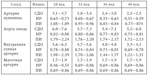
Doppler at normal pregnancy shows the results presented in the table above are possible small deviations, but only a doctor can tell whether they are critical by comparing all the test results obtained. Sometimes Doppler is prescribed again in order to evaluate the indicators over time, for example, after a course of treatment.
If a problem or question arises, of course, you can contact the doctor who supplied you with the device. Outpatient blood pressure measurement is a risk-free test. The recorded measurements will then be transferred from the box to a computer which will analyze the data. Electrophysiological imaging allows one to study the electrical activity of the heart. Abnormalities in this electrical activity can result in discomfort with or without loss of consciousness, severe fainting, or palpitations associated with arrhythmias.
Doppler during pregnancy: where to do it
Having realized the need for this type of research, every mother asks the question: “Where can I do a Doppler ultrasound during pregnancy?” Every city in Russia has a number of free antenatal clinics, as well as paid clinics that provide pregnancy care at a professional level. Therefore, there should be no problems with doing a Doppler test during pregnancy. Usually they do it in the same place where they are registered, and you probably decided on this a long time ago. If your particular clinic does not provide such a service, you can contact any other clinic for a fee. The cost of this procedure is about 1200 rubles.
The principle of the examination is to record the electrical activity of the heart by installing probes located in various predetermined locations in the cardiac cavities. Depending on the results of the examination, you may be offered several procedures: treatment, treatment, creation of a pacemaker, radiofrequency ablation of the intracardiac zone, installation of an implantable defibrillator.
Electrophysiological studies require a short hospital stay. The examination is carried out on post for at least 6 hours, without general anesthesia, a small prescription may be offered. Depending on the purpose of the test, your doctor may ask you to temporarily stop your usual treatment until the test. Subcutaneous anticoagulant therapy is often given before the test to prevent blood clots. The catheter will be inserted most often in the crook of the groin, requiring preliminary shaving of the area.

Let's sum it up
Doppler - required type research that everyone should undergo future mom at least 2 times throughout your pregnancy. It is absolutely safe and at the same time very useful, because some diagnoses can only be confirmed with its help! And a timely diagnosis and correctly taken measures based on it will help the baby to be born absolutely healthy and save his mother nerve cells, which are known to not be restored. But at the same time, there is no need to do Doppler without a doctor’s prescription. Just to make sure the baby is okay. This is simply a whim, unnecessary interventions without important reasons are still undesirable.
The exam is conducted by a qualified physician in a cathedral room equipped with revival material. The area through which the catheter is to be inserted is thoroughly disinfected and isolated with sterile tissues. It is important to avoid moving the pierced limb or placing your hands on the area, which can cause infection. The practitioner conducts local anesthesia region and then punctures a vein, inside which he places an introducer that allows him to place probes in the vessels in part of the heart.
As the probes progress, no pain is felt. Electrical stimulation tests are usually performed in various areas hearts. Probes and all used accessories are discarded with each use and for each patient. Sometimes you can enter certain products by infusion to sensitize the study. An electrophysiological study, like any invasive or surgical procedure, carries the risk of incidents or accidents. Benign complications are complications at the puncture site that can persist for several days, but most often without consequences.
Remember that Doppler ultrasound during pregnancy has norms, but you should not compare your results with them. Moreover, these same standards may vary on different Internet resources! Entrust this matter to your doctor, because you once chose him to accompany such an important period in your life, which means he knows his business!
Video " Pregnancy tests: ultrasound, CTG, fetal movement test, Doppler.”
Each examination method during pregnancy is safe and effective. Particularly accurate and full information You can get information about the health status of the woman and baby using Doppler ultrasound. This technique is simple, but provides an excellent opportunity to listen to the fetal heartbeat and blood flow in the umbilical cord arteries. This procedure is necessary for specific indications and situations. The process is absolutely painless and safe.
Ultrasound and Doppler during pregnancy
Quite often it happens that a doctor prescribes a Dopplerometry procedure for a woman during pregnancy. Behind this mysterious word lies something familiar and familiar to everyone. ultrasound examination, which has advanced capabilities. This method has the main purpose of application - this is the study of blood flow in the arteries and vessels of the umbilical cord, as well as blood flow in the uterine arteries.
Unlike regular ultrasound Doppler allows you to determine the following parameters:
- The state and level of development of the child’s heart health;
- Check the fetal heartbeat, patency of the umbilical cord vessels;
- Determine the level of blood supply to blood vessels;
- Identify any disturbances in the functioning of the placenta or hypoxia at the most early stages.
The device for the procedure allows you to examine the body of the baby and mother without harming health. The principle of operation is simple: ultrasound reacts to blood flow in vessels and arteries and displays the data on the monitor. Thus, you can get the maximum exact information about any symptoms or to make sure there are no various pathologies fetal development.
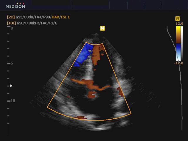
Such a study can be carried out in two ways. The duplex method helps to detect the causes of blood flow disturbances if similar symptom observed, as well as fully assess the patency of each vessel. The Doppler effect gives an idea of the condition and level of development of the fetus. Triplex technology makes it possible to clearly highlight the movement of red blood cells on the monitor, thereby giving this species Ultrasound is more accurate and useful.
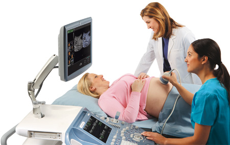
Majority modern devices for ultrasound examinations already have a built-in Doppler function. The procedure is very simple. The patient must be in horizontal position, and no special preliminary preparation is required. Indications for ultrasound may vary, but most often it is included in a routine examination.
Doppler ultrasound
In order to determine the level and norms of a baby’s development, specialists often use Doppler. In this way, disorders can be detected at the earliest stages during pregnancy. Timely treatment will help avoid serious consequences. Main indications for Doppler ultrasound:
- Rh factor, which can lead to serious consequences, for example, miscarriage;
- The presence of any infectious or chronic diseases(diabetes, kidney diseases, hypertension and others);
- If the gestational age is not appropriate for the size of the fetus;
- If there is a suspicion of pathology in the baby’s development;
- If the norm of cardiotography results does not correspond to reality.
Also, an ultrasound of this type can be performed if a woman has abnormal functioning of some organs - gestosis. This symptom occurs quite often during such a period. Doppler can be performed shortly before the expected time of birth. In this case, you can determine the degree of readiness of the woman’s body for the birth of a baby.
In addition to the condition of blood vessels and arteries, the level and patency of blood flow, Doppler ultrasound determines other important medical indicators. Such data include fetal weight, size, condition and development of organs, anatomical features and other. Thus, careful monitoring of the health of mother and baby is carried out.
The Doppler study is carried out quite simply: the woman should be on the couch in a lying position, and the doctor carries out full complex inspection. First of all, in standard mode it is viewed general state fetus, uterus, umbilical cord and other organs. Next, the specialist determines the location of the vessel of interest in order to examine the artery more clearly and carefully. These could be the arteries of the brain or the umbilical cord vessel. Then the monitor displays the movement of blood flow, and an automatic analysis of the obtained data is carried out. If there are any deviations from the norm, the device notifies a specialist, who simultaneously monitors the ongoing analysis.
Process Features
The modern Doppler procedure is extremely informative method to monitor the health of a woman during pregnancy and the development of the baby. Ultrasound allows for timely detection of abnormalities, which will ensure effective treatment or preparing for emergency childbirth.
The main advantages of Doppler are the following features:
- Process safety. Ultrasound does not affect either the woman’s health or the baby’s development;
- The entire process is fairly quick;
- The results obtained are quite accurate and complete, which provides an informative picture of what is happening;
- Doppler makes it much easier to prepare for the birth of your baby.
Based on the inspection results, it is assessed full picture health of women and children. Most often, the total number of ultrasound procedures for the entire period is three to four sessions, Doppler is used once shortly before birth. Identifying any complications allows you to prepare for solving the problem in a timely manner.
Triplex Doppler ultrasound identifies areas of blocked blood flow, thereby providing the opportunity to take action necessary actions. The results also show the cause of any abnormalities, for example, dysfunction of the placenta or the condition of the umbilical cord vessels. Doppler during pregnancy is an extremely effective and necessary examination technique. Each artery and vessel is carefully examined, blood flow speed and lumen are studied. Preliminary preparation The process can be discussed with your doctor depending on your specific situation.
Modern Doppler during pregnancy in the second trimester is sometimes necessary to exclude the presence intrauterine infections, as well as monitor the operation of systems little man. Following all the specialist’s recommendations will help you get pregnant and easy birth, and the baby will delight good health. Research of any type during this period is necessary for women, because pregnancy is an important and responsible time. Every mother strives to give her child maximum attention, and caring for him begins during pregnancy.
