Coronary heart disease: types and clinical picture. Classification, manifestations, outcomes of ischemia
Ischemia- violation peripheral circulation, which is based on restriction or complete cessation of inflow arterial blood.
Clinic: blanching of the organ area, decreased temperature, impaired sensitivity in the form of paresthesia, pain syndrome, decrease in blood flow speed, decrease in organ volume, decrease blood pressure in the area of the artery located below the obstacle, decreased tissue turgor, dysfunction of an organ or tissue, degenerative changes.
Classification of ischemia by etiology:
A) compression ischemia occurs when the adductor artery is compressed by a scar, tumor, etc.;
B) obstructive ischemia occurs when the lumen of the artery is partially or completely closed by a thrombus or embolus. Inflammatory and productive-infiltrative changes in the arterial wall during atherosclerosis, obliterating endarteritis, periarteritis nodosum also lead to limitation of local blood flow as obstructive ischemia;
C) angiospastic ischemia occurs due to irritation of the vasoconstrictor apparatus of blood vessels and their reflex spasm.
Causes of ischemia
Reasons: emotions (fear, pain, anger), physical factors(cold, injury), chemical agents, biological irritants (toxins), etc.
Angiospasm leads to a slowdown in blood flow until it stops completely.
Develops most often according to the type of vascular unconditioned reflexes from the corresponding interoreceptors. It may also have a conditioned reflex character.
Irritation of the vasomotor center by toxins, mechanical irritation of subcortical formations that regulate vascular tone, the presence pathological process in area diencephalon also often lead to severe angiospastic phenomena.
In the development of angiospastic ischemia, not only irritation matters various departments reflex arc, but also the functional state of the muscle fibers of the vascular wall, electrolyte and other types of exchange in it.
For example, sodium ions, accumulating in the muscle fibers of the vessel, increase its sensitivity to pressor substances - catecholamines, vasopressin and angiotensin.
The nature of all types of changes in the ischemic area of tissue or organ is determined by the degree oxygen starvation, the severity of which depends on the rate of development and type of ischemia, its duration, location, nature collateral circulation, functional state organ or tissue.
Ischemia vital important organs has more severe consequences. Brain, heart are characterized high level energy metabolism, but despite this, their collateral vessels are functionally absolutely or relatively unable to compensate for circulatory disorders. Skeletal muscles and especially connective tissue, thanks to low level energy metabolism in them are more stable under ischemic conditions.
Ischemia in conditions of increased functional activity organ or tissue is more dangerous than at rest.
Changes in tissues during ischemia:
The first structural changes occur in mitochondria. Their swelling is observed, cysts disappear, the breakdown of mitochondria and endoplasmic reticulum and cell nuclei can result in the formation of a focus of necrosis - infarction. This occurs in organs with hypersensitivity to oxygen starvation and an insufficient network of collaterals.
In the ischemic area, there is an enhanced biosynthesis of connective tissue components.
Stasis is the slowing down and stopping of blood flow.
Types of stasis:
A) true (arises due to pathological changes in capillaries or disorders rheological properties blood);
B) ischemic (occurs due to complete cessation of blood flow from the arteries);
B) venous.
Venous and ischemic stasis is a consequence of a simple slowdown and cessation of blood flow.
Elimination of the cause of stasis leads to the restoration of normal blood flow. Progression of ischemic and venous stasis contributes to the development of the true.
Causes of true stasis: physical (cold, heat), chemical, biological (bacterial toxins).
Mechanisms for the development of stasis: intracapillary aggregation of erythrocytes and, as a consequence, an increase in peripheral resistance. Important in the pathogenesis of true stasis is attributed to a slowdown in blood flow in capillary vessels due to blood thickening. The leading role here is played by the increased permeability of the capillary wall under the influence of biological active substances to a shift in the reaction of the medium to the acidic side.
Increased permeability vascular wall and vasodilation leads to blood thickening, slowing blood flow, red blood cell aggregation and stasis.
Thrombosis is a process of lifelong education inner surface the walls of blood vessels are blood clots consisting of its elements. Thrombi can be wall or obstructive.
Depending on which components predominate in the structure of the thrombus, they are distinguished:
A) red (red blood cells predominate);
B) white (thrombosis is formed by platelets, leukocytes, plasma proteins);
B) mixed blood clots.
Thrombosis is most often caused by diseases that affect the vascular wall:
Diseases of an inflammatory nature (rheumatism, syphilis, typhus);
Atherosclerosis;
Cardiac ischemia;
Allergic processes.
Inadequate oxygen supply to the heart muscle caused by coronary atherosclerosis, can lead to disruption of the mechanical, biochemical and electrical functions of the myocardium. Sudden development ischemia usually affects the function of the left ventricular myocardium, which leads to disruption of the processes of relaxation and contraction. Due to the fact that the subendocardial parts of the myocardium are less well supplied with blood, ischemia of these areas develops first. Ischemia involving large segments of the left ventricle leads to the development of transient failure of the latter. If ischemia also affects the area of the papillary muscles, then it can be complicated by insufficiency of the left atrioventricular valve. If ischemia is transient, it is manifested by the occurrence of an attack of angina. With prolonged ischemia, myocardial necrosis may occur, which may or may not be accompanied by a clinical picture acute heart attack myocardium. Coronary atherosclerosis is local process, which can cause ischemia varying degrees. Focal disturbances in left ventricular contractility resulting from ischemia cause segmental bulging or dyskinesia and may to a large extent reduce the pumping function of the myocardium.
Based on the above mechanical problems lies wide range changes in cell metabolism, their function and structure. In the presence of oxygen, normal myocardium metabolizes fatty acid and glucose into carbon dioxide and water. Under conditions of oxygen deficiency, fatty acids cannot be oxidized, and glucose is converted into lactate; The pH inside the cell decreases. In the myocardium, the reserves of high-energy phosphates, adenosine triphosphate (ATP) and creatine phosphate are reduced. Dysfunction cell membranes leads to a lack of K ions and absorption of Na ions by myocytes. Whether these changes are reversible or whether they lead to the development of myocardial necrosis depends on the degree and duration of the imbalance between the myocardial oxygen supply and the need for it.
During ischemia, the electrical properties of the heart are also disrupted. The most characteristic early electrocardiographic changes are repolarization disturbances, which represent wave inversion T, a later - segment offset ST(ch. 178). Transient depression of a segment ST often reflects subendocardial ischemia, while transient elevation of the segment ST, is believed to be a consequence of more severe transmural ischemia. In addition, due to myocardial ischemia, electrical instability occurs, which can lead to the development ventricular tachycardia or ventricular fibrillation (Chapter 184).
state of microcirculation. Local manifestations and consequences.
Heart attack as a consequence of ischemia.
Ischemia – a decrease in blood supply to an organ or tissue as a result of obstruction of blood flow through the afferent vessels.
The reasons for the increase in resistance to blood flow in the arteries are: 3 (three) groups of reasons:
Compression (external pressure) afferent vessels (tumor, scar, ligature, foreign body). This type of ischemia is called compression ischemia.
Obstruction of afferent vessels – as a result of complete or partial closure from the inside of the artery lumen by a thrombus or embolus.
Angiospasm of the afferent arteries – as a result of vasoconstriction of vascular smooth muscle. Mechanisms of arterial spasm: a) extracellular – associated with long-term circulation of vasoconstrictor substances in the blood. These are: catecholamines, serotonin; b) membrane – associated with disruption of the process of repolarization of membranes of smooth muscle cells; c) intracellular - the intracellular transport of calcium ions is disrupted, hence the unrelaxing contraction of smooth muscle cells.
Microcirculation during ischemia.
reduced due to a decrease in hydrostatic pressure in the arterial part of the bed.
Resistance to blood flow in the arterial part of the bed it is increased due to obstruction of blood flow in the afferent arteries.
Volumetric blood flow velocity reduced by reducing the arteriovenous pressure difference and increasing resistance to blood flow.
Linear blood flow velocity reduced due to a decrease in the arteriovenous pressure difference and increased resistance to blood flow.
reduced due to the closure of part of the functioning capillaries.
Symptoms of ischemia.
Reducing diameter and quantity visible arterial vessels due to their narrowing and decreased blood supply.
Pallor of tissues or organs due to decreased blood supply and a decrease in the number of functioning capillaries.
Reducing the magnitude of arterial pulsation as a result of filling them with blood.
Decrease in temperature of ischemic tissue or organ a consequence of a decrease in the flow of warm arterial blood, a further decrease in metabolism.
Decreased lymph formation as a result of a decrease in perfusion pressure in tissue microvessels.
Reduction in volume and turgor of tissues and organs due to insufficient blood and lymph supply.
Consequences of ischemia. The main pathogenetic factor of ischemia is hypoxia. In the future: reduction of under-oxidized products, ions, biologically active substances. It follows from this:
Decreased specific functions.
Reduction of non-specific functions and processes: local defensive reactions, lymph formation, plastic processes.
Development of dystrophic processes, hypotrophy and tissue atrophy.
Necrosis and infarction.
The importance of the level of tissue and organ functioning, shunting and collateral circulation in the outcome of ischemia. Heart attack as a consequence of ischemia.
The nature of the consequences of ischemia depends on:
The rate of development of ischemia. The higher it is, the more significant the degree of tissue damage.
The diameter of the damaged artery or arteriole.
"Sensitivity" of the organ to ischemia. This sensitivity is especially high in the brain, kidneys, and heart.
The significance of an ischemic organ or tissue for the body.
The degree of development of collateral vessels and the rate of “switching on” or activation of collateral blood flow in a tissue or organ. Under collateral blood flow understand the circulatory system in the vessels around the ischemic area of tissue and in it itself. Its activation is facilitated by a number of factors, namely: a) the presence of a blood pressure gradient above and below the narrowed area; b) accumulation in the ischemic zone of biologically active substances with a vasodilating effect (adenosine, Ax, prostaglandins, kinins, etc.); c) “emergency” activation of local parasympathetic influences, promoting the expansion of collateral arterioles; d) the degree of development of the vascular network in the affected organ or tissue.
5. Venous hyperemia. Definition, causes and mechanism of development.
Changes in microcirculation and hemodynamic parameters.
Manifestations, significance in pathology and consequences.
Venous hyperemia – this is an increase in blood supply to an organ or tissue due to a mechanical obstruction to outflow venous blood from an organ or tissue. This might be consequence:
Narrowing of the lumen of a venule or vein when it: a) compression (edematous fluid, tumor, scar, tourniquet, etc.); b) obstruction (thrombus, embolus, tumor).
heart failure, when the heart does not pump blood from the systemic circle to the small circle and the central venous pressure in the large veins increases.
In case of pathology of venous vessels, which is accompanied by low elasticity of the venous walls. This pathology is usually accompanied by the formation of dilations (varicose veins) and contractions.
The mechanism of development of venous hyperemia. It consists of creating a mechanical obstacle to the outflow of venous blood from tissues and disrupting the laminarity of blood properties.
Changes in microcirculation.
Arteriovenous pressure difference decreased due to an increase in hydrostatic pressure in the venous part of the bed.
Resistance to blood flow in the venous part of the bed it is increased due to obstruction of blood flow in the drainage vessels.
Volumetric blood flow velocity reduced due to a decrease in the arteriovenous pressure difference and increased resistance to blood flow.
Linear blood flow velocity reduced due to a decrease in the arteriovenous pressure difference and increased resistance to blood flow.
Total cross-sectional area of the capillary bed increased due to the opening of some previously non-functioning capillaries.
Macrosymptoms of venous hyperemia.
Increased number and diameter of visible venous vessels due to an increase in their lumen.
Cyanosis of organs and tissues. The bluish tint is associated with: a) an increase in the amount of venous blood in them; b) an increase in the content of oxygen-free forms of hemoglobin in it (the result of a pronounced transition of oxygen into the tissue due to its slow flow through the capillaries).
Decrease in temperature of organs and tissues due to: a) an increase in the volume of venous blood in them (in comparison with warmer arterial blood); b) reducing the intensity of tissue metabolism.
Swelling of tissues and organs as a result of the increase blood pressure in capillaries, postcapillaries and venules. With prolonged venous hyperemia, edema is potentiated due to the “switching on” of osmotic, oncotic and membranogenic pathogenetic factors.
Hemorrhages into tissue or bleeding (internal and external) as a result of overstretching and micro-tears of the walls of venous vessels.
Microsymptoms of venous hyperemia.
Increased diameter of capillaries, postcapillaries and venules.
Increase in the number of functioning capillaries on initial stage VG and decrease - at later stages, when the blood flow in them stops due to the formation of microthrombi and blood cell aggregates.
Slowing down the flow of venous blood up to complete cessation of outflow.
Significant expansion of the axial cylinder of blood cells(up to the size of the venule lumen) and the disappearance of the “band” of plasma current in them.
“Push-like” and “pendulum-like” movement of blood in the venules. In front of an obstruction in a blocked vein, hydrostatic pressure increases. If its value reaches diastolic blood pressure, then during diastole ………………………….
Consequences and significance of venous hyperemia.
VG has a damaging effect on tissue. The main pathogenetic factor is hypoxia of the corresponding tissue or organ. Venous hyperemia is accompanied by tissue swelling, often with hemorrhages or bleeding. Therefore, against the background of VG:
Specific functions of an organ or tissue are reduced.
Nonspecific functions and processes are suppressed.
Malnutrition and hypoplasia develop structural elements of cells and tissues.
Necrosis of parenchyma and proliferation of connective tissue(sclerosis, scars).
Ischemia- disturbance of peripheral circulation, which is based on restriction or complete cessation of arterial blood flow.
Clinic: pallor of the organ area, decreased temperature, impaired sensitivity in the form of paresthesia, pain syndrome, decreased blood flow speed, decreased organ volume, decreased blood pressure in the area of the artery located below the obstacle, decreased tissue turgor, dysfunction of the organ or tissue, degenerative changes.
1. compression ischemia occurs when the adductor artery is compressed by a scar, tumor, etc.;
2. obstructive ischemia occurs when the arterial lumen is partially or completely closed by a thrombus or embolus. Inflammatory and productive-infiltrative changes in the arterial wall during atherosclerosis, obliterating endarteritis, periarteritis nodosum also lead to limitation of local blood flow as obstructive ischemia;
3. Angiospastic ischemia occurs due to irritation of the vasoconstrictor apparatus of blood vessels and their reflex spasm.
Causes: emotions (fear, pain, anger), physical factors (cold, injury), chemical agents, biological irritants (toxins), etc.
Angiospasm leads to a slowdown in blood flow until it stops completely.
It develops most often according to the type of vascular unconditioned reflexes from the corresponding interoreceptors. It may also have a conditioned reflex character.
Irritation of the vasomotor center by toxins, mechanical irritation of subcortical formations that regulate vascular tone, and the presence of a pathological process in the diencephalon also often lead to pronounced angiospastic phenomena.
In the development of angiospastic ischemia, not only the irritation of various parts of the reflex arc is important, but also the functional state of the muscle fibers of the vascular wall, electrolyte and other types of exchange in it.
For example, sodium ions, accumulating in the muscle fibers of the vessel, increase its sensitivity to pressor substances - catecholamines, vasopressin and angiotensin.
The nature of all types of changes in an ischemic area of tissue or organ is determined by the degree of oxygen starvation, the severity of which depends on the rate of development and type of ischemia, its duration, localization, the nature of collateral circulation, the functional state of the organ or tissue.
Ischemia of vital organs has more severe consequences. The brain and heart are characterized by a high level of energy metabolism, but despite this, their collateral vessels are functionally absolutely or relatively unable to compensate for circulatory disorders. Skeletal muscles and especially connective tissue, due to the low level of energy metabolism in them, are more stable under ischemic conditions.
Ischemia under conditions of increased functional activity of an organ or tissue is more dangerous than at rest.
Stasis, types, etiology, pathogenesis, signs.
Stasis- this is a slowdown, up to a complete stop, of blood flow in the vessels of the microvasculature.
Blood stasis may be preceded by venous congestion (stagnant stasis) or ischemia (ischemic stasis).
Blood stasis is characterized by stopping blood in capillaries and venules with expansion of the lumen and gluing of red blood cells into homogeneous columns - this distinguishes stasis from venous hyperemia. Hemolysis and blood clotting do not occur.
Stasis must be differentiated from the “sludge phenomenon”. Sludge is the phenomenon of red blood cells sticking together not only in capillaries, but also in vessels of various sizes, including veins and arteries.
Stasis- the phenomenon is reversible. Stasis is accompanied by dystrophic changes in the organs where it is observed. Irreversible stasis leads to necrosis.
Causes of stasis
Ischemia and venous hyperemia. They lead to stasis due to a significant slowdown in blood flow (with ischemia due to a decrease in arterial blood inflow, with venous hyperemia as a result of a slowdown or cessation of its outflow) and the creation of conditions for the formation and/or activation of substances that cause adhesion shaped elements blood, the formation of aggregates and blood clots from them.
Proaggregants are factors that cause aggregation and agglutination of blood cells.
Pathogenesis of stasis
At the final stage of stasis There is always a process of aggregation and/or agglutination of blood cells, which leads to thickening of the blood and a decrease in its fluidity. This process is activated by proaggregants, cations and high molecular weight proteins.
All types of stasis are divided into primary and secondary.
Primary (true) stasis. The formation of stasis primarily begins with the activation of blood cells and their release of a large number of proaggregants and/or procoagulants. At the next stage, the formed elements aggregate, agglutinate and attach to the wall of the microvessel. This causes the blood flow in the vessels to slow down or stop.
Secondary stasis (ischemic and congestive).
Ischemic stasis develops as an outcome of severe ischemia due to a decrease in arterial blood flow, a slowdown in the speed of its flow, and its turbulent nature. This leads to aggregation and adhesion of blood cells.
The congestive (venous-congestive) version of stasis is the result of a slowdown in the outflow of venous blood, its thickening, and changes physical and chemical properties, damage to blood cells (in particular due to hypoxia). Subsequently, blood cells adhere to each other and to the wall of microvessels.
Manifestations of stasis
With stasis are happening characteristic changes in the vessels of the microvasculature:
Reduction of the internal diameter of microvessels during ischemic stasis,
Increasing the lumen of microvasculature vessels in the stagnant version of stasis,
A large number of aggregates of blood cells in the lumen of blood vessels and on their walls,
Microbleeds (more often with congestive stasis).
At the same time, manifestations of ischemia or venous hyperemia may overlap manifestations of stasis.
Consequences of stasis
With the rapid elimination of the cause of stasis, blood flow in the vessels of the microvasculature is restored and no significant changes develop in the tissues.
Prolonged stasis leads to the development dystrophic changes in tissues, often to the death of a section of tissue or organ (infarction).
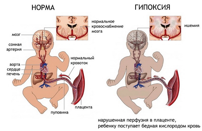
Any brain damage is accompanied by life-threatening consequences for the patient organic changes in his tissues. Oxygen starvation, insufficiency of incoming nutrients, manifested nearby characteristic symptoms determine the dynamics of the functioning of the brain, and the absence of a therapeutic effect can lead to an aggravation of the process and the occurrence of serious side conditions. Cerebral ischemia caused by pathology blood vessels and capillaries in the brain tissues, is accompanied by a constantly ongoing progression of pathological processes in the brain tissues, which are accompanied by a significant deterioration in its blood supply.
Having an idea of what cerebral ischemia is, what are its main manifestations and possible Negative consequences for health, can be diagnosed in a timely manner this state, conduct necessary treatment and maintain brain health. Since the main cause of this condition is organic lesion central (cerebral) vessels in the brain, it is their condition that should be considered the most indicative in the process of identifying the lesion in question. Indeed, it is precisely due to the violation of their integrity, the appearance of excessive permeability of the walls and the tendency to fragility that the risk of possible side effects in the functioning of the brain, which are caused by manifestations of oxygen starvation and lack of required quantity nutrients.
General description of the disease
A disease such as cerebrovascular ischemia has a certain classification depending on the stage of the pathological process, its prevalence and negative influence on brain tissue. Also negative impact The condition of the cerebellar nerve, the anterior lobe of the brain, responsible for the processes of memorization and long-term memory, is affected. All manifestations of this pathology appear in stages, the first stage is considered the most easily treatable therapeutic effects and therefore it is the first stage of the disease that should be detected as early as possible. Thanks to the availability characteristic manifestations Cerebrovascular ischemia can be detected early and treatment can begin.
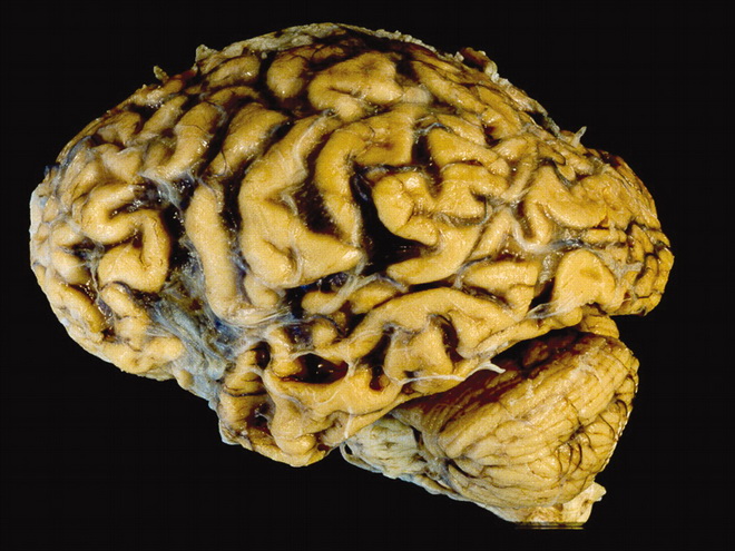
Cerebrovascular ischemia in the photo
Today, according to medical statistics, cerebral ischemia is observed in more cases in older people (about 50-55% of all people over 65 years of age have one of the stages of this lesion) against the background of existing provoking diseases. Hypertension, gradually progressing, can gradually provoke a disruption in the blood supply to the brain, which leads to the appearance of signs of the disease in question. It should be noted that cerebral ischemia cannot be treated complete cure, it is only possible to stop the speed of pathological processes occurring in the vessels of the brain, stabilization general condition the patient and elimination of the most characteristic manifestations.
A chronic disturbance in the process of blood circulation largely determines both the quality of the brain and the general condition of a person, because the brain is one of the target organs that are primarily affected by the most severely manifested pathologies in the body. Therefore, even minor changes in the state of brain function, which have characteristic clinical manifestations(frequent headaches, sudden changes moods and difficulties with remembering) should be considered the first signs, and which need to be paid close attention and adequate therapeutic intervention be initiated.
Development mechanism
The process of disease development looks like in the following way: first, there is some deterioration in the functioning of the brain (anterior lobe of the hypothalamus and cerebellum) due to the development of pathology of the cerebral (central) blood vessels, which are responsible for the process of supplying brain tissue with both oxygen and nutrients. In this case, there is often damage to the nerve connecting the brain (or parts thereof) with the central nervous system, as a result of which there is a pronounced imbalance in his work. Then, as the disease progresses, an ever-increasing degenerative process in the brain is noted against the background of oxygen starvation, which leads to a gradual disruption of organic processes in its tissues.
In this case, pathological processes are first noted transitory nature, and then chronic course in the brain. This condition is fraught with persistent manifestations of the current pathology. Characteristic signs illnesses attract attention and allow you to go to a medical facility in time for examination.
The main complication of this condition is considered high probability stroke, as the disease progresses, its symptoms and manifestations negatively affect daily life sick. Therefore, to prevent the occurrence serious consequences For the health and preservation of the patient’s life, it is recommended to begin treatment immediately after diagnosing this disease.
Symptoms of the disease
External manifestations are quite characteristic: frequent shifts mood, changes in it - a sharp transition from positive attitude to a state of depression, fatigue, frequent depression and a decrease in the quality of intellectual functioning, as well as an increase in the degree of irritability, fatigue during the day. Many patients experience a decrease in intelligence, which initial stages the pathological process is imperceptible, but as it progresses it can seriously change a person’s life - reduce its quality.
In the absence of treatment, the listed manifestations are significantly aggravated: headaches become stronger, worsening night sleep and fatigue during the day are noted, brief fainting spells and loss of performance.
Any deviations from the norm in the condition of the blood vessels provoke a qualitative change in the patient’s daily life, which immediately attracts attention and requires immediate attention. medical care. Treatment at home for this lesion, especially in advanced stages, does not produce significant results. positive result and may result in a health-damaging delay in drug treatment, as a result of which the risk of developing negative complications increases.

Basic principles of disease classification
There are several various types classification of the disease in question. All of them are divided according to the degree of damage to brain tissue, the presence and number of symptoms, and the magnitude of manifestations. The most commonly used classification is based on the nature of the manifestations and the impact on the patient’s health. A classification is also used based on the duration of the disorders caused by the current pathological process.
The classification of the disease according to the nature of its manifestations is as follows:
- transient type - this condition is characterized by an ischemic attack or cerebral crisis, in which there is a sharp change in the general condition with characteristic manifestations in the form of prolonged and acute headache, decreased attention and concentration;
- acute phase of the disease, which should include hemorrhagic stroke, acute form encephalopathy and ischemic stroke and unspecified nature of occurrence;
- the chronic course is characterized by manifestations of occlusion of blood vessels, cerebral thrombosis, subcortical encephalopathy. Chronic cerebral ischemia is accompanied by long-term preservation characteristic manifestations, gradual deterioration of the patient’s general condition with worsening symptoms of the disease.
Cerebral ischemia grade 1 manifests itself in the initial stages of the pathological process. In this case, the manifestations of the disease are not clearly expressed; the patient is characterized by sudden changes in mood, increased fatigue. Cerebral ischemia of the 1st degree is observed in many elderly people; its impact on everyday life is not very pronounced, which significantly interferes with its timely diagnosis.
Cerebral ischemia grade 2- a more in-depth process, clinical picture which is more pronounced. Headaches become constant, and there is a loss of focus and ability to concentrate. The second stage may also be characterized by a deterioration in the quality of night sleep, the appearance of unusual sensations during wakefulness: pain in the frontal lobe of the head, noise in the head for no apparent reason.
Cerebral ischemia grade 3 has maximum strong manifestation symptoms of the disease, there is a sharp decrease in the patient’s performance. At the third stage of development of the pathological process, he requires urgent hospitalization and active drug therapy.
There is also a type of disease called ischemia spinal cord. In this case, there is tissue damage in the adjacent area, insufficient blood supply to the spinal cord with a gradual deterioration in its functioning. There is a similar classification for this disease, which allows the identified lesion to be classified into one of the classes, which makes it possible to quickly select the most appropriate treatment.
Causes
The causes of cerebrovascular ischemia can be different. Basically they are organic in nature, damage to the heart and its accompanying illnesses often become a trigger for the occurrence of this pathology.
Most common reasons The following should be considered cerebrovascular ischemia:
- thromboembolism of blood vessels;
- thrombosis of arteries and veins;
- heart defects, which can be either congenital or acquired;
- vasculitis;
- changes in the condition of the walls of blood vessels (their increased fragility and permeability);
- disturbances in blood clotting processes;
- metabolic disorders.
It is also worth highlighting a number of risk factors that can provoke the disease.
One of the risk factors for the development of the pathology in question is long-term depressive states and stress, which have an extremely negative impact on blood circulation processes in brain tissue. Gout, advanced stages cervical osteochondrosis, smoking and frequent use alcoholic beverages should also be considered serious risk factors in the development of the pathology in question.
Among brain disorders It is by considering the disease that it occupies one of the first places in the number of probable side effects for health, therefore, when identifying it, mandatory professional treatment using appropriate medicines and immediate hospitalization of the patient. Ischemic diseases also have a high degree of risk to the patient’s life: in case of untimely assistance or its insufficiency, the risk fatal outcome increases.
