Displacement and compression of the brain stem. Dislocation syndrome - dislocation of the brain: types, causes, diagnosis and treatment
Acute pathological processes in dislocation syndromes involve the same anatomical structures with a stereotypical clinical manifestation. In other words, the clinical picture of acute dislocation syndrome does not depend on the etiology of the process. The difference is in clinical manifestation in different patients depends on the rate of its development, localization and volume.
The fact is that dislocation syndromes are essentially internal hernias of the brain, that is, the protrusion of its departments into holes and crevices, formed by bones and dura mater. There are 3 degrees of brain dislocation: protrusion, wedging and infringement. There are lateral and axial (along the trunk axis) dislocations of the brain.
Drawing a parallel with hernias, it should be remembered that sharp violation The life of the patient arises not from the fact of the existence of a hernia, but from its infringement. Infringement is a protrusion, which is accompanied by a cessation of blood flow due to compression of the vessels.
Types of dislocation syndromes
Most medical literature describes 4 types of dislocation syndromes (1-4), which have highest value in clinical practice, as they can be accompanied by a significant deterioration in the patient's condition. At the same time, 8 dislocation syndromes are distinguished in the Great Medical Encyclopedia. Some sources also describe an external dislocation (number 9), which is a bulging of brain regions through a traumatic skull defect.
- Displacement of the cerebral hemispheres under the falciform process
- Temporotentorial displacement
- Cerebellar-tentorial displacement
- Displacement of the tonsils of the cerebellum in the foramen magnum of the occipital bone
- Displacement of the pons of the brain through the foramen of the cerebellum
- Filling the middle and side tanks of the bridge
- Bias back section corpus callosum dorsally into the cistern of the same name
- Displacement of the gyri of the frontal lobe into the cistern of the chiasm
- External dislocation of the brain
Displacement of the cerebral hemispheres under the falciform process
With compression of the brain on one side by an acute volumetric process, with an increase in CSF pressure in one lateral ventricle due to acute occlusive hydrocephalus, a difference in CSF pressure develops between the right and left lateral ventricles. This creates conditions for the dislocation of the hemisphere big brain transversely into the gap between the falx cerebrum and the corpus callosum. The wedged area is mainly the cingulate gyrus and the areas adjacent to it. Thus, the cingulate gyrus fills the cistern of the corpus callosum. The cingulate gyrus, which bulges in the opposite direction, displaces the anterior cerebral arteries, pressing them against the falciform process. The lateral and third ventricles are deformed. Perifocal edema can be generalized due to a slowdown in blood circulation, as a result of compression of the arteries and veins. All this leads to insufficient intake in the brain tissue. nutrients and oxygen, which leads to the development of even more edema-swelling of the brain and to even more dislocation. These processes can lead to blockage of the liquor outflow. CSF produced in the lateral ventricles, having no outflow tracts, creates an area of distension in the supratentorial space with the development of a temporotentorial herniation, which leads to compression of the midbrain.
Temporotentorial and cerebellartentorial herniation
These 2 dislocation syndromes can be combined due to the commonality of the affected structures and, accordingly, the developing clinic. They differ in the location (sub- or supratentorially) of the area of distension. Temporotentorial herniation is a protrusion into the tentorial foramen (Bish's fissure) of the area temporal lobe the brain and, above all, the medial sections of the hippocampal gyrus, partly anterior section lingual gyrus and isthmus of the vaulted gyrus. Wedged between the free edge of the cerebellar mantle and the oral sections of the brain stem, the temporal lobe area can occupy various positions in relation to the trunk: anterior - when located in front of the trunk; anterolateral - when located anterior to the trunk and along its anterior quadrant, posterolateral, posterior. Wedging of brain regions into the tentorial foramen can occur not only from the side hemispheres brain, but also in the opposite direction, that is, from the back cranial fossa. In such cases, the area of the cerebellum protrudes between the free edge of the cerebellar mantle and the quadrigemina. This insertion is called cerebellar-tentorial. It occurs during pathological processes in the posterior cranial fossa. The occurrence of temporo-tentorial and cerebellar-tentorial herniations is inevitably associated with a displacement of the trunk in the opposite direction, which depends on the size and characteristics of the herniation.
The ipsilateral oculomotor nerve is compressed - the pupil expands after a short-term miosis.
Lateral displacement of the homolateral parahippocampal gyrus causes midbrain move through the midline towards the opposite edge of the cerebellar tenon. This slight change in the configuration of the midbrain can reduce the level of consciousness of the patient and cause targeted arousal.
Eventually, the globus pallidus, internal capsule, and thalamus on the homolateral side move caudally, and the parahippocampal gyrus protrudes beyond the edge of the cerebellar mantle into the subtentorial space. The mammillary bodies wedged into the narrowed interpeduncular fossa. Under these conditions, the midbrain experiences intense pressure, which causes the development of coma. The midbrain may be pressed against the opposite edge of the cerebellum tenon so strongly that the descending motor fibers in the compressed brain stem are damaged. The hemiplegia due to this type of herniation is not contralateral, but homolateral in relation to the volumetric process - Kernogan's brain stem syndrome . Due to compression, hemorrhages occur in the lower temporal and occipital regions of the brain, the midbrain, which can often lead to irreversible consequences and death of the patient
Intrusion of the tonsils of the cerebellum into the foramen magnum
The tonsils of the cerebellum, shifting into the foramen magnum, sometimes reach the level of the second cervical vertebra. Wedging between the caudal part of the medulla oblongata, the adjacent part of the spinal cord and the occipito-cervical ring, they tightly cover and squeeze this part of the trunk from the dorsal surface.
This creates disturbances cerebral circulation, inevitably leading to hypoxia and to an even greater increase in edema-swelling of the brain. Thus, a vicious circle is closed, which leads to the progression of compression of the brain stem, which leads to a violation of the function of the vital centers of blood circulation and respiration embedded in them and, as a result, to the death of the patient.
Displacement of the pons of the brain through the foramen of the cerebellum
The pons varolii is displaced in the oral direction into the interpeduncular cistern.
Filling the middle and side tanks of the bridge
Occurs as a result of pressing the bridge of the brain to the slope of the base of the skull.
Displacement of the posterior part of the corpus callosum in the dorsal direction into the cistern of the same name
Occurs with closed hydrocephalus of the brain, which can also occur with the above dislocation syndromes. at the expense high blood pressure in the ventricles, the corpus callosum is shifted into the cistern of the same name. The free edge of the sickle forms a notch on the corpus callosum. Since this dislocation syndrome occurs already against the background of deep violations of the body's vital functions, it is rarely described in the literature devoted to this problem.
CSF circulation disorders
Dislocations of the brain are dangerous not only by the development of cerebrovascular accident. Also, there is a displacement and compression of the CSF-containing spaces both as a whole and their individual parts, in particular the cerebral aqueduct, the IV ventricle, etc. The compression leads to a violation of the CSF circulation. The normal state of the CSF pathways is disturbed due to their narrowing at the level of the tentorial foramen or foramen magnum, which leads to the development of hydrocephalus. Naturally, if the outflow of cerebrospinal fluid from the ventricles is disturbed, a pressure gradient occurs between the ventricles and the spinal sac. This pressure difference leads to even greater displacements of the brainstem in the caudal direction, to the development of new dislocation syndromes.
Clinic
At temporo-tentorial and cerebellar-tentorial herniations oculomotor disorders appear (horizontal, rotatory, vertical nystagmus, difference eyeballs, symptom of Hertwig-Magendie, partial or complete paresis of gaze up or to the sides or complete immobility of the eyeballs, weakening or lack of pupillary response to light, etc.). Unilateral pyramidal insufficiency, which has arisen during an acute volumetric process (Kernogan's brain stem syndrome), becomes bilateral. This fact indicates the involvement of the second leg of the brain in the pathological process. The clinic of these types of dislocations depends on the arteries to which areas of the brain are transferred in the tentorial foramen.
Compression of vessels leading to basal ganglia extrapyramidal system, leads to dysregulation muscle tone first in the form of an increase in extensor tone, the appearance of symptoms of a cogwheel. Then hormonia develops, muscle rigidity and as a result, muscle atony is evidence of the separation of the midbrain from the higher lying sections. Diencephalic disorders are detected in the form of tachycardia, which replaces bradycardia. Recorded on the ECG various forms arrhythmias, infarction-like ischemia and other disorders that are functional in nature, secondary to cerebral pathology and disappear without a trace upon elimination acute period. There is also tachypnea, hyperthermia, in some patients up to 39-40 ° C, hyperemia and greasiness skin and others. The consciousness of patients is depressed, up to coma varying degrees expressiveness.
Displacement of the tonsils of the cerebellum into the foramen magnum clinically the most severe. At the same time, vital centers and, first of all, centers of respiration and blood circulation suffer. Breathing problems occur central genesis varying degrees, which depends on the rate of development, degree and duration of herniation. These disorders are detected in tachypnea (at the beginning of the process), which then develops into deeper forms of pathology, up to Cheyne-Stokes, Biot, terminal types and apnea. In parallel with the violation of breathing, the depth of the disturbance of consciousness increases, and anabolic disorders develop. Swallowing is grossly disturbed, the pharyngeal reflex decreases or disappears - develops bulbar syndrome. Decreased vascular tone. Arterial hypotension develops.
Diagnostics
Diagnostic methods:
- Echoencephalography - determines the degree of displacement of the median structures in one direction or another. At the same time, it should be taken into account that according to the Echo-EG data, only lateral (displacement of the cerebral hemispheres under the falciform process) can be assumed, since according to its data it is impossible to diagnose axial dislocation of the brain.
- barbituric anesthesia
- moderate hypothermia
- intermittent deep hyperventilation
- glucocorticoids
- Big medical encyclopedia. Main Ed. B.V. Petrovsky. Ed. 3rd. M., "Owls. Encyclopedia", 1977 p. 347-349
- Duus P. Topical diagnosis in neurology Anatomy. Physiology. Clinic - M. IPC "Vazar-Ferro", 1995 p. 173-179
- Lebedev V.V., Bykovnikov L.D., Kariev M. Emergency Diagnostics and assistance in neurosurgery - T .: Medicine 1988 p. 44-67
- Lebedev VV, Krylov VV Urgent neurosurgery: A guide for physicians. - M.: Medicine, 2000 p.103-141
- Misyuk N. S., Evstigneev V. V., Rogulchenko S. M. Displacements and infringements of the brain stem. Minsk, "Belarus", 1968
- Neurotraumatology. Directory / Edited by: Academician of the Russian Academy of Medical Sciences A, N, Konovalov, Professor L. B. Likhterman, Professor A. A. Potapov. - M.: IPC "Vazar-Ferro", 1993
- Practical Neurosurgery: A Guide for Doctors / Ed. B. V. Gaidar. - St. Petersburg: Hippocrates, 2002
Treatment
The first and indispensable condition for the treatment of dislocation is the elimination of the cause that caused it.
Non-surgical measures used for dislocation syndromes include:
An operation for vital indications in the development of cerebral edema and processes that lead to the dislocation of some parts of the brain in relation to others is decompressive craniotomy. To remove the pathological focus, a wide (at least 5-6 x 6-7 cm) decompressive craniotomy is performed, which does not have to be resectional, but must be decompressive. With temporo-tentorial herniation, trepanation is performed in the temporo-parietal region as low as possible. With bilateral symptoms, decompressive trepanation is performed on both sides. After removal of the pathological focus, the dura mater is not sutured.
Also, to reduce intracranial pressure and, accordingly, reduce the risk of developing life-threatening processes of herniation, drainage of the ventricular system is performed. Puncture the anterior or posterior horn from standard points(Kochera or Dandy). The effect of puncture of the ventricles of the brain is stronger if the drainage is made for more early stages wedging. With a lateral displacement of the ventricles, it is not easy to get into the displaced and compressed ventricle of the brain. Puncture of the hydrocephalic ventricle on the side opposite to the focus ("healthy") with the remaining pathological focus is fraught with an increase in dislocation and an increase in vital disorders.
Literature
The brain, in contrast to, for example, the organs abdominal cavity or chest, is located in the cranial cavity, the volume of which is static and cannot be increased if necessary. The bones of the skull form a rigid "framework", due to which, with an increase in pressure inside the cranium, more soft structures, located inside it, are pushed aside and squeezed in the holes and under the outgrowths of the solid meninges. It is necessary to distinguish between the concepts of "displacement" and "wedging". In the first case we are talking about the dislocation of brain structures under the influence of some volumetric process that creates excess pressure from the opposite side (without infringement), in the second - the displacement of the brain into the natural openings of the skull or under the outgrowths of the shell with possible infringement.
Reasons leading to the occurrence of dislocations of the brain
- Ischemic stroke. Dislocations at ischemic stroke from small to moderate, depending on the volume of the lesion and the severity of cytotoxic cerebral edema. However, even a small dislocation, for example, in a stroke with localization in the cerebellum, can cause respiratory and heart failure due to compression of the medulla oblongata.
Ischemic stroke in the basin of the right middle cerebral artery. CT scan of the brain. When describing, it should be noted that the entire parietal and frontal lobes are in edema on the right, as a result of which the right lateral and 3rd ventricles are completely compressed. There is a slight dislocation of the median structures to the left.
- Hemorrhagic stroke. Intracerebral hematomas lead to more pronounced displacements of brain structures, which are clearly visible on computed tomography and MRI. The degree of displacement depends on the size of the hematoma and the severity of the perifocal cerebral edema. 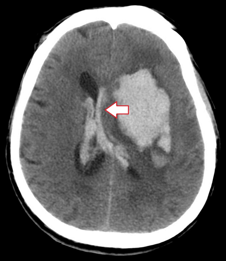
CT signs of lateral dislocation syndrome in hemorrhagic stroke(left). The left lateral ventricle and the 3rd ventricle of the brain are compressed, the right lateral ventricle in both cases looks enlarged. Pronounced dislocation of median structures to the right side.
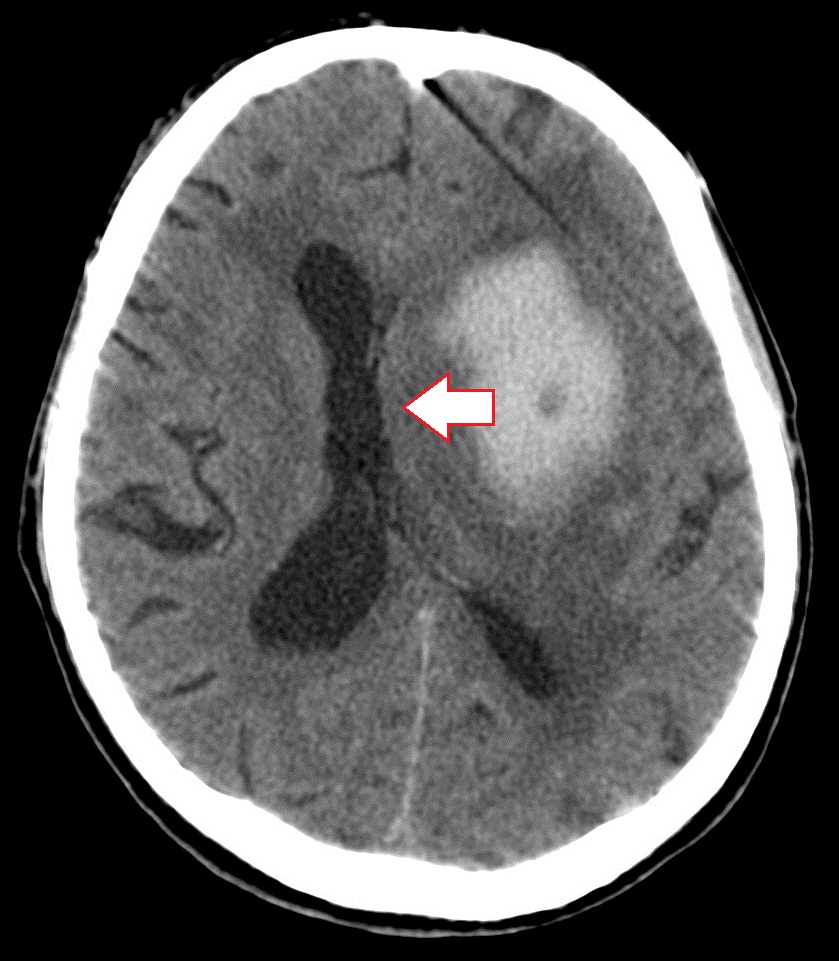
- Subdural hematoma. It causes significant dislocations of brain structures, since the volume of blood in subdural hematomas is large and can reach several hundred ml. Both acute and chronic subdural hematoma lead to displacement of midline structures and to other types of dislocations.
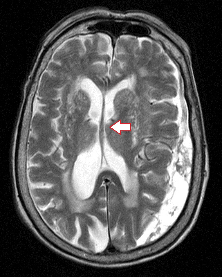
Dislocation syndrome (direction of displacement marked with an arrow) in a left-sided acute subdural hematoma (far left image). Pronounced displacement of the ventricles of the brain to the right. The image in the middle shows the dislocation of brain structures caused by chronic subdural hematoma, the far right image shows MRI of the brain.
- Epidural hematoma. It usually occurs with fractures of the skull bones, leads to the appearance of local displacements, almost never causes significant dislocations, since the volume of blood pouring into the epidural space is small - rarely reaches 100 ml.
- Contusion of the brain. By its nature, the contusion focus is an intracerebral hematoma, in conjunction with areas of crushing of the brain substance, and areas of ischemia resulting from damage to the arteries. The severity of dislocations of brain structures in CT and MRI studies depends on the volume of the hematoma, as well as the size of the zone of perifocal edema.
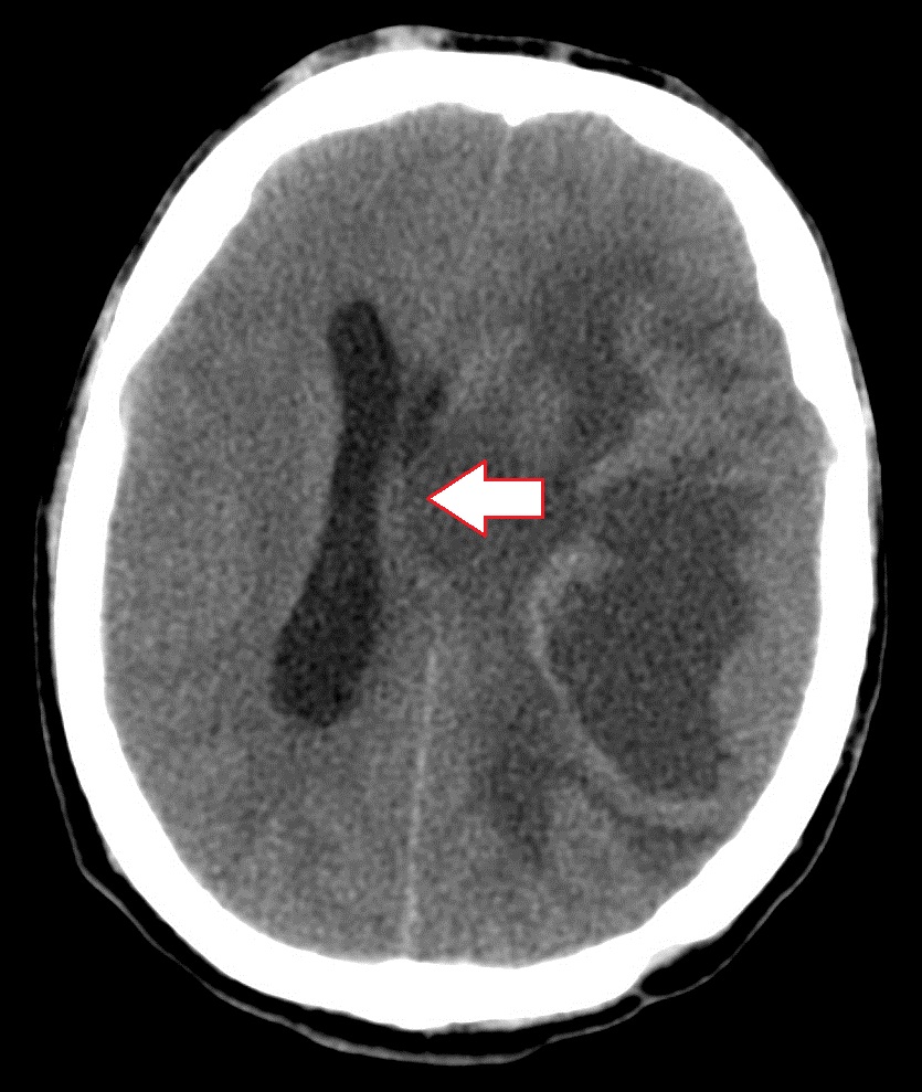
CT scan of the brain. When deciphering, a tumor of the left parietal lobe, "mass effect", displacement of the ventricles of the brain to the left
- Brain tumor. Dislocations are due to volume pathological tissue, as well as cerebral edema. The larger the tumor and stronger swelling the greater the displacement. Tumors of the meninges (meningiomas) behave similar to epidural hematomas - they cause minimal displacement of the brain in a limited area.
- Monoventricular hydrocephalus (due to occlusion of the CSF pathways). Expansion of the right or left lateral ventricle leads to displacement of the septum pellucidum and other median structures of the brain in the opposite direction.
Classification of displacements and herniations of brain structures in CT and MRI
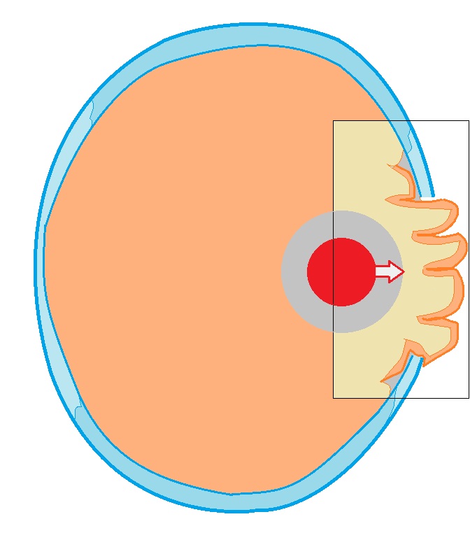
Lateral dislocation of the median structures of the brain (lateral dislocation syndrome). The most common type of displacement found on CT and MRI of the head. Measurements are usually made relative to the transparent septum and falxcerebri (or conditional midline of the head). Dislocations of the median structures are very often accompanied by compression of the lateral ventricle on the one hand, and expansion on the other. 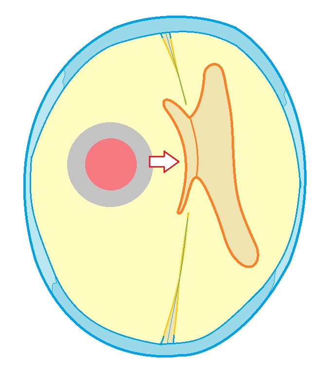 Schematic representation of changes in lateral dislocation syndrome. The pathological focus is marked in red, the direction of pressure is marked with an arrow. Falxcerebri and lateral ventricles shifted to the side, there is multiventricular hydrocephalus against the background of compression of the ventricle adjacent to the pathological process.
Wedging of the frontal lobe under the crescent of the brain occurs during volumetric processes localized in the frontal lobe of the brain. As a result, one of the convolutions (or several) of the frontal lobe is wedged from the medial side and infringed under the “sickle” of the brain.
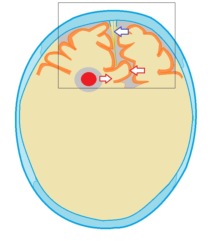 Wedging of the medial gyrus of the frontal lobe under the "sickle" of the brain. The volumetric pathological process that caused the wedging is marked in red, the direction of displacement is indicated by the arrow from it, the “sickle” of the brain is indicated by the blue arrow.
Temporo-tentorial herniation. With volumetric processes in the middle cranial fossa, a displacement of the temporal lobe and a wedging of the hippocampus under the cerebellum can occur.
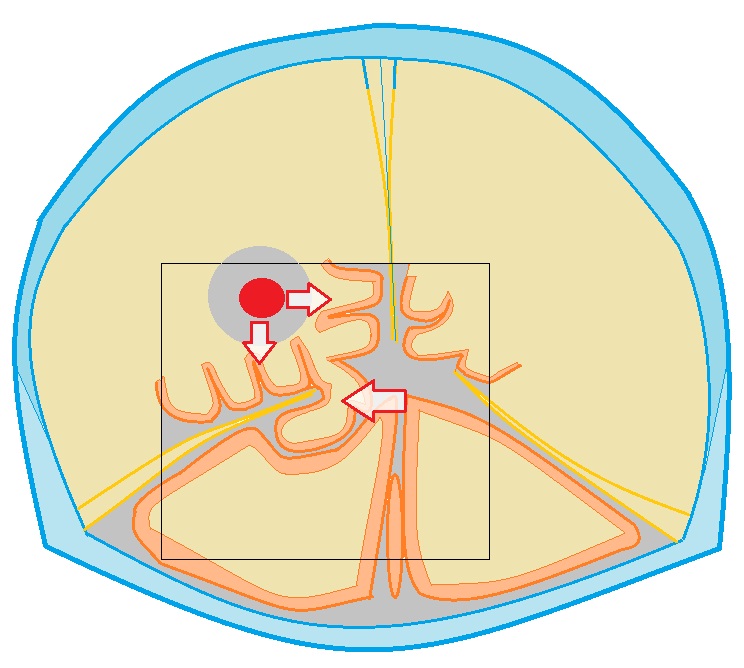
Temporo-tentorial herniation (diagram). The volumetric pathological process is marked in red, the direction of displacement is marked by arrows. The gyrus of the temporal lobe, displaced under the tentorium, was also noted.
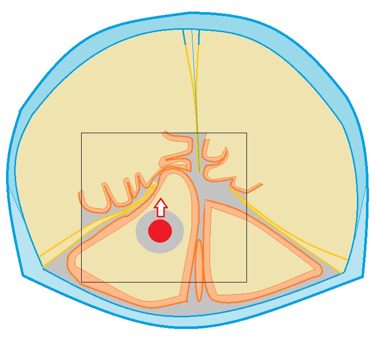
Cerebellar-tentorial herniation: one of the hemispheres of the cerebellum is displaced upward and is located above the tenon, closely adjacent to temporal lobe brain
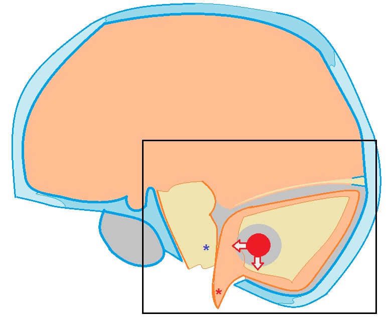
Intrusion of the tonsils of the cerebellum into the foramen magnum occipital bone can occur with volumetric processes (tumors) of the cerebellum, with strokes with localization in the cerebellum, cerebellar hematomas. The tonsils (marked with a red asterisk) bulge into the dural funnel and lie below the plane of the foramen magnum. The fourth ventricle is compressed and deformed. This condition is also dangerous by the possibility of compression of the brain stem (marked with an asterisk of blue color) with the development of respiratory and cardiac disorders).
External dislocation of the brain (scheme). Occurs after resection trepanation of the skull. A section of the brain bulges into the burr hole and can be squeezed by its edges
(Late Latin dislocatio displacement, movement)
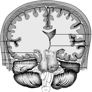
displacement of the brain due to changes in intracranial topography in various pathological processes in the central nervous system. The causes of D. m. are an increase in intracranial pressure, an increase in brain volume or deformation of its various departments. With an intracranial tumor or hematoma, brain abscess, craniocerebral injury, accompanied by cerebral edema, the syntopy of brain structures changes, redistributes intracranial pressure in the spaces separated by the cerebellum and the greater falciform process of the dura mater (the falx cerebrum). Dislocation of the brain may be accompanied by descent and infringement of the tonsils of the cerebellum in the foramen magnum and upper part spinal canal with the development of life-threatening occipital herniation syndrome ( rice. one
). It is characteristic of tumors of the posterior cranial fossa, however, it can also develop with tumors located above the cerebellum. Clinical picture in this syndrome is primarily due to compression of the medulla oblongata and the associated violation of vital important functions. With occipital herniation, it sharply increases headache, especially in the occipital region; vomiting, dizziness, which increases with a change in the position of the head and body, a forced position of the head, which helps to reduce headaches, are noted, meningeal syndrome, choking when taking liquid food, hiccups, increased sweating, increasing disturbances in the rhythm of breathing (arrhythmic, intermittent, such as Cheyne-Stokes) and activity of cardio-vascular system. Significant symptoms of intracranial hypertension (see Intracranial Hypertension) -
progressive lethargy, congested discs optic nerves, diplopia, decreased corneal reflexes, smell, hearing, etc. The rapid increase in the phenomena of herniation leads to the development of coma and respiratory arrest. With tumors of the temporal lobe of the brain, less often tumors of the frontal or occipital lobes, the brain can be pushed back from front to back and medially. At the same time, the medial parts of the hemispheres wedged into the opening of the cerebellar tenon, squeezing the brain stem passing through this opening ( rice. 2
). Such a tentorial herniation, depending on the degree and symmetry of the compression of the trunk, may initially be clinically manifested by the syndrome of intracranial hypertension and symptoms of a unilateral lesion of the brain stem. However, soon the stem symptoms become bilateral, the headache and vomiting intensify, and the impairment of consciousness deepens. The stem syndrome of tentorial herniation is characterized by paresis of the upward gaze, the absence of a pupillary light reflex, weakness of convergence, the presence of bilateral pathological pyramidal reflexes, etc. Compression at the base of the brain of the oculomotor nerve causes ptosis, strabismus, pupil dilation on the side of the lesion. The progression of the phenomena of tentorial herniation leads to the development of paralysis, decerebrate rigidity (Decerebrate rigidity) ,
coma. Dislocation of the brain caused by swelling around the focus of the brain contusion, intracranial hematoma, a tumor of the parietal, frontal lobe, etc., may consist in the lateral displacement of the median structures (around the third ventricle, etc.) of the brain and the medial parts of the cerebral hemispheres. In this case, a wedging occurs under the large falciform process of the dura mater. The displacement of the median structures of the brain exacerbates the severity of clinical symptoms due to the focal process, enhances vegetative-vascular, endocrine-metabolic and metabolic disorders. The development of D. g. m should be suspected in cases of acute deterioration in the condition of patients with intracranial processes, primarily of a tumor nature, accompanied by intracranial hypertension, hydrocephalus (Hydrocephalus) ,
occlusion syndrome (occlusion syndrome) .
Echoencephalography helps to clarify the diagnosis, CT scan heads, craniography. Diagnostic spinal tap contraindicated due to the possibility of increasing the dislocation of the brain. Suspicion of occipital or tentorial herniation requires immediate administration of dehydrating agents and an urgent solution to the issue of neurosurgical operation, which is the only radical medical event. Question about character surgical intervention should be addressed before the development of violations of vital functions. Lateral dislocation of the brain can sometimes be eliminated by symptomatic means (corticosteroid and dehydrating drugs), ventricular puncture of the brain, followed by neurosurgery or radiation therapy. The prognosis is always very serious, depending on the location and nature of the underlying process that caused the dislocation of the brain. Bibliography: Blinkov S.M. and Smirnov N.A. Displacements and deformations of the brain, L., 1967; Diseases nervous system, ed. P.V. Melnichuk, vol. 1-2, M., 1982; Multivolume guide to surgery, ed. B V. Petronsky, v. 3, book. 1. p. 87, M., 1968.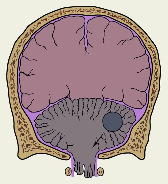
- - displacement of the brain associated with deformation and displacement medulla under the influence of increased intracranial pressure, cerebral edema, hydrocephalus, hemorrhage, tumors and ...
- - An abscess that forms in the brain tissues as a result of pathogens entering them purulent infection from other foci or with traumatic brain injury ...
Medical Encyclopedia
- - metastatic brain abscess, in which the source of metastasis is an inflammatory endocardium; occurs in children with birth defects blue-type hearts, mainly in tetralogy of Fallot ...
Medical Encyclopedia
- - Brain abscess that develops as a result of the spread of infectious agents from a closely located focus of purulent inflammation ...
Medical Encyclopedia
- - see Hydrocephalus ...
Medical Encyclopedia
- - common name cavities located in the brain, lined with ependyma, communicating with each other and with the subarachnoid space and containing cerebrospinal fluid ...
Medical Encyclopedia
- - see Compression of the brain ...
Medical Encyclopedia
- - closed damage of the brain, characterized by the occurrence of a focus of destruction of its tissue and manifested by neurological and psychopathological symptoms, respectively, the localization of the focus ...
Medical Encyclopedia
- - a significant increase in brain volume, accompanied by an increase in intracranial pressure and dysfunction of the nerve centers ...
Medical Encyclopedia
- is a pathological process characterized by excessive accumulation fluid in brain tissue. The resulting increase in brain volume leads in most cases to an increase in intracranial pressure ...
Medical Encyclopedia
- - cm....
Medical Encyclopedia
- - see the cerebral hemisphere ...
Medical Encyclopedia
- - a combination of signs of increased intracranial pressure with focal neurological symptoms due to the presence of a volumetric formation in the cranial cavity ...
Medical Encyclopedia
- - clinical form mild closed craniocerebral injury, characterized by a predominance of reversible functional changes brain, - see Traumatic brain injury ...
Medical Encyclopedia
- part of the base of the brain that contains nuclei cranial nerves and vital centers...
Medical Encyclopedia
- - see Contusion of the brain ...
Medical Encyclopedia
"Dislocation of the brain" in books
Grandfather Matvey
From the book the Lord will rule author Alexander AvdyuginGrandfather Matvey Grandfather Matvey is old. He himself says: "So many now do not live." He speaks correctly, probably because there are no peers left in the district, especially those who went through the war: with trenches, attacks, injuries and other fears, which we can now judge only from books
5. Levi Matvey
From the book Woland and Margarita author Pozdnyaeva Tatiana5. Levi Matthew At the foot of the Mount of Olives, in Bethphage, Yeshua Ha-Nozri met his only disciple Levi Matthew. Yeshua tells about this during interrogation, and although the Yershalaim topography does not indicate the proximity of this small village to the Olivet
Matvey
From the book The Secret of the Name the author Zima DmitryMatvey The meaning and origin of the name: from the Hebrew name Matthew - God's gift granted by the Lord. Energetics and Karma of the name: today the name Matvey is quite rare, although it is possible that it may soon become fashionable. At least today they are
MATVEY
From the book of 100 happiest Russian names author Ivanov Nikolai NikolaevichMATVEY Origin of the name: "granted by God" (Jewish). Name day (according to the new style): July 13; August 22; October 11, 18; November 29. Positive character traits: calmness, responsibility, harmony, absence of contradictions, complexes. Matthew is reliable
Matthew of Paris
From the book Russian-Livonian War of 1240-1242 author Shkrabo DMatthew of Paris Matthew of Paris, French author of the 1st half. 13th century, wrote that the Danish king sent princes Knut and Abel with an army and settlers to settle the Novgorod possessions devastated by the Tatars. He mixed two events: the German-Danish campaign of 1240 on
Cat Matvey
From the book Knitted Toys author Kaminskaya Elena AnatolievnaCat Matvey Such a knitted cat can not only become your baby's favorite toy, but also a wonderful gift for a friend for the birth of a child. Cat Matvey You will need 50 g of yarn in the main color, 20 g of white yarn for the muzzle and paws,
Matvey
From the book All Monarchs of the World. Western Europe author Ryzhov Konstantin Vladislavovich author From the book Orthodox names. Name choice. Heavenly patrons. Saints author Pecherskaya Anna IvanovnaMatvey Name meaning: from ancient Hebrew. Mattityahu - “gift of Yahweh” (“granted by the Lord”). Main features: honesty, modesty, morality. Character features. In his family, Matvey is usually a long-awaited child, parents have high hopes for him. He
Dislocation of the brain (dislocation syndrome) is a pathology that occurs as a result of displacement of brain tissue in relation to hard formations. This causes the space inside the skull to be limited and divided.
What causes brain dislocation?
The development of the disease leads to processes that increase pressure inside the skull:
- abscesses;
- tumors;
- swelling of the brain;
- hematomas.
In some cases, craniocerebral hernias are observed, the nature of which is congenital. There are three stages of dislocation syndrome:
- protrusion;
- wedging;
- infringement.
 When a certain area of the brain tissue protrudes into a large hole located in the back of the head, or into a gap, the limiter of which is the hard shell of the brain, it develops venous congestion. Also among possible consequences swelling of a local nature and small outpourings of blood.
In the wedged area, the growth of the phenomena of local edema does not stop, as a result of which its size increases, the form becomes herniated.
The pathology under consideration exists in two forms:
- Lateral dislocation. Under the conditions of a literal supratentorial process, a part of the brain is displaced under the falciform process.
- Axial dislocation (displacement of the brain is carried out along the axis into the opening of the cerebellum and into a large opening in the back of the head).
Forms of dislocation of the brain
Let us consider the forms of the dislocation syndrome, the significance of which is decisive.
Displacement of the cerebellum into the cerebellar cistern
As a rule, this process indicates neoplasms of the posterior cranial fossa. There are cases of its development when there is swelling of the brain. There are signs of increased intracranial pressure:
- severe headache;
- vomit;
- nausea.
Bulbar disturbances are possible.
Dislocation of the parts of the temporal lobe (basal) in the foramen of the cerebellum
The result of this type of dislocation is flattening and compression of the adjacent parts of the midbrain.
Wedging of this kind can be both bilateral and unilateral. With this form of dislocation syndrome, intracranial pressure increases sharply. Occlusal crises are possible. In addition, to negative consequences this includes ptosis, mydriasis.
Symptoms of brain dislocation
 With this pathology, a person in most cases is in a coma, but loss of consciousness does not always occur - for example, if the cause of the disorder was a sudden process, infections of the central nervous system and swelling of the brain.
Dislocation syndrome can also develop for other reasons, when the displacement of structures occurs at a slower pace. This process is accompanied by the following symptoms:
- convulsions;
- severe headaches;
- transient or permanent loss of vision;
- repeated vomiting and nausea.
Diagnostic methods
Brain dislocation is diagnosed using the following methods:
- echoencephalography. Sets how much the median structures are shifted to either side. Echo-EG data reveal only a lateral dislocation, in which the cerebral hemispheres are displaced under the falciform process.
- Angiography is a method of X-ray contrast study of blood vessels.
- Computed tomography is a method of layer-by-layer non-destructive examination of an object, more precisely, its internal structure.
- Magnetic resonance imaging - method radiodiagnosis, which in a non-invasive way allows you to get an image internal structures human body.
Treatment
 In order to eliminate the pathological focus, a wide trepanation of the skull is carried out, which, in without fail is decompressive. At the same time, it may not be resectional. Temporotentorial herniation is an indication for trepanation in the lower region of the temporo-parietal region. If the symptoms are bilateral, decompressive trepanation is performed on both sides. The hard shell of the brain is not sutured after the elimination of the focus of pathology.
To bring in normal condition intracranial pressure and minimize the likelihood of herniation processes, life threatening, the ventricular system is drained.
To enhance the effectiveness of puncture of the cerebral ventricles allows drainage carried out on early stages wedging. If the displacement of the ventricles is lateral, getting into the ventricle of the brain (compressed and displaced) is difficult.
In addition, a number of non-surgical measures can be used for dislocation syndrome: hyperventilation, barbituric anesthesia, glucocorticoids, moderate hypothermia. Lateral dislocation of the brain in some cases can be eliminated with the help of symptomatic remedies(drugs).
Finally, we bring to your attention a video about a brain tumor - the most common cause development of dislocation syndrome:
According to classical concepts, the symptoms of herniation are caused by the pressure of the medulla displaced into the hard openings of the skull (wedging) (as a result of the action of a volumetric formation or increased ICP) on other structures of the central nervous system. These ideas are criticized on the basis of the hypothesis that the herniation may be an epiphenomenon that develops later in pathological process and is not the cause of the observed changes. Be that as it may, shedding models are used as a useful approximation.
There are several herniation syndromes, 5 of which are the most common:
1. central (transtentorial) herniation
2. temporal herniation (hook of the temporal lobe)
3. herniation of the cingulate gyrus: the cingulate gyrus can wedged under the falx (the so-called wedging under the falx). Usually asymptomatic until ACA kinks and occludes, leading to bilateral infarction frontal lobes. Usually a sign of impending transtentorial herniation
4. superior herniation of the cerebellum
5. herniation of the tonsils of the cerebellum
(1, 2 - supratentorial herniation, 4, 5 - infratentorial herniation)
Coma with supratentorial volumetric education
Central and temporal herniation cause different forms rostral-caudal deterioration. Central herniation results in sequential damage to the diencephalon, midbrain, pons, and medulla oblongata. The "classic" signs of increased ICP (high blood pressure, bradycardia, changes in the nature of breathing) are usually observed with PCF lesions, but may be absent in slowly developing supratentorial formations.
The distinction between the central and temporal herniations is difficult to make when the lesions reach the midbrain and lower regions. Determining the localization of the lesion based on the dislocation syndrome is unreliable.
Clinical signs to distinguish between temporal and central herniation
Disturbances in the level of consciousness are observed in early period with central herniation and late with temporal herniation
. temporal herniation rarely results in decortication rigidity
Differential diagnosis of causes of supratentorial herniation
1. vascular: NMK, VMK, SAH
2. inflammatory: brain abscess, subdural empyema, herpetic encephalitis
4. traumatic: EDH, SDH, depressed skull fracture
Coma with infratentorial volumetric education
NB: important to identify patients with primary lesions ZCHYA, because they may require urgent surgery.
Differential diagnosis of causes of infratentorial herniation
1. Vascular: brainstem infarction (including OA occlusion), cerebellar infarction, or hematoma
2. inflammatory: cerebellar abscess, central pontine myelinolysis, brainstem encephalitis
3. tumor: primary or mts
4. traumatic: EDG or SDG
Superior herniation of the cerebellum
Sometimes seen with bulk formations PCOS, may be exacerbated by ventriculostomy. The cerebellar vermis rises above the tenon, compresses the midbrain, can compress the PCA → infarction. Can compress plumbing → GCF.
Herniation of the tonsils of the cerebellum
The cerebellar tonsils are wedged into the BZO, squeezing medulla→ stop breathing. Usually, death occurs quickly.
It is observed both with supra- and with infratentorial space-occupying formations or with ICP. May be caused by LP. In many cases, there may simply be pressure on the brainstem without true herniation. There are cases of significant wedging of the tonsils into the BZO with the patient's consciousness preserved.
Greenberg. Neurosurgery
