Loose type of structure of the bronchi. What are "bronchi" and where are they located
The bronchi are an important part of the respiratory system. Studying the human anatomy from the photo, you can understand what exactly they deliver to the air saturated with oxygen and remove the exhaust with a high content of carbon dioxide. With their help, small particles that have entered the lungs, such as dust particles or pieces of soot, are removed from the respiratory system. Here, the incoming air acquires a temperature and humidity favorable for humans.
Bronchial hierarchy
Features of the anatomy of the bronchi are in the strict sequence of their division and location. For any person, they are divided into:
- Main bronchi with a diameter of 14-18 mm, which depart directly from the trachea. They are not the same size: the right one is wider and shorter, while the left one is longer and narrower. This is due to the fact that the volume of the right lung is larger than the left;
- Lobar bronchi of the 1st order, which provide oxygen to the lobar zones of the lung. There are 2 on the left side, and 3 on the right;
- Zonal, or large 2nd order;
- Segmental and subsegmental, which belong to the 3rd-5th order. There are 11 of them on the right side, and 10 on the left;
- Small bronchi related to the 6-15th order;
- Terminal, or terminal bronchioles, which are considered the smallest parts of the system. They are directly adjacent to the lung tissue and alveoli.
Such anatomy of the human bronchi provides air flow to each lobe of the lung, which allows gas exchange throughout the entire lung tissue. Due to the structural features of the bronchi, they resemble the crown of a tree, and they are often called that - the bronchial tree.

The structure of the bronchi
The wall of the bronchus consists of several layers, which vary depending on the hierarchy of the bronchus. Wall anatomy includes three basic layers:

- Fibromuscular-cartilaginous layer located on the outside of the body. This layer has the greatest thickness in the main bronchi, and with their further division it becomes smaller, up to its complete absence in the bronchioles. If outside the lung this layer is completely covered with cartilaginous semirings, then deepening inward, the semirings are replaced by separate plates with a lattice structure. The main components of the fibrous-muscular-cartilaginous layer are:
- Cartilage tissue;
- Collagen fibers;
- elastic fibers;
- Smooth muscles collected in bundles.
The fibrocartilaginous layer plays the role of a framework, thanks to which the bronchi do not lose their shape and allow the lungs to increase and decrease in size.

muscle layer, which changes the lumen of the tube, is part of the fibromuscular-cartilaginous. With its contraction, the diameter of the bronchus decreases. This happens, for example. The contraction contributes to a slower flow of air within the respiratory system, which is necessary for its warming. Relaxation of the muscles provokes the opening of the lumen, which occurs during active exercises and is necessary to prevent the occurrence of shortness of breath. The muscle layer includes smooth muscle tissues, collected in the form of bundles of oblique and circular types.
- Slime layer located in the inner part of the bronchus, its structure includes connective tissue, muscle fibers and cylindrical epithelium.
The anatomy of the columnar epithelium includes several different types of cells:

- Ciliated, designed for bronchial drainage and cleansing of the epithelium from foreign particles. They make wave-like movements with a frequency of 17 times per minute. Relaxing and straightening, the cilia push out foreign elements from the lungs. They create the movement of mucus, the speed of which can reach 6 mm / s;
- Goblet secrete mucus designed to protect the epithelium from damage. Getting on the mucous membrane, foreign bodies cause irritation, provoking increased secretion of mucus. In this case, a person develops a cough, with the help of which the cilia move the foreign object outward. The secreted mucus is necessary to protect the lungs from drying out, as it moisturizes the air mixture entering them;
- Basal, necessary to restore the inner layer;
- Serous, synthesize a special secret necessary for cleansing and drainage;
- Clara cells, which are located to a greater extent in the bronchioles and are intended for the synthesis of phospholipids. Inflammation can transform into goblet cells;
- Cells of Kulchitsky. They produce hormones and belong to the APUD system (neuroendocrine system).
- Adventitia or outer layer, which consists of fibrous connective tissue and ensures contact of the bronchus with its external environment.
Find out what to do with such a diagnosis.
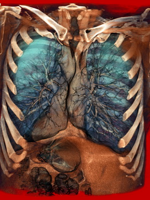
It is important to know what the lungs are, where they are in a person, what functions they perform. The respiratory organ is located in humans in the chest. The chest is one of the most interesting anatomical systems. There are also bronchi, heart, some other organs and large vessels. This system is formed by the ribs, spine, sternum and muscles. It reliably protects all important internal organs and, due to the pectoral muscles, ensures the smooth operation of the respiratory organ, which almost completely occupies the chest cavity. The respiratory organ expands and contracts several thousand times a day.
Where are the human lungs located?
The lungs are a paired organ. The right and left lungs play a major role in the respiratory system. It is they who distribute oxygen throughout the circulatory system, where it is absorbed by red blood cells. The work of the respiratory organ leads to the release of carbon dioxide from the blood, which breaks down into water and carbon dioxide.
Where are the lungs located? The lungs are located in a person's chest and have a very complex connecting structure with air, circulatory systems and lymphatic vessels and nerves. All these systems are intertwined in the area, which is called the "gate". Here is the pulmonary artery, main bronchus, branches of nerves, bronchial artery. In the so-called "root" lymphatic vessels and pulmonary veins are concentrated.

The lungs look like a vertically dissected cone. They have:
- one convex surface (costal, adjacent to the ribs);
- two convex surfaces (diaphragmatic, medial or median, separate the respiratory organ from the heart);
- interstitial surfaces.
The lungs are separated from the liver, spleen, colon, stomach, and kidney. Separation is carried out using a diaphragm. These internal organs border on large vessels and the heart. Behind them is limited by the back.
The shape of the respiratory organ in humans depends on the anatomical features of the body. They can be narrow and elongated or short and wide. The shape and size of the organ also depend on the phase of respiration.
To better understand where and how the lungs are located in the chest and how they border on other organs and blood vessels, you need to pay attention to the photos that are located in the medical literature.
The respiratory organ is covered with a serous membrane: smooth, shiny, moist. In medicine, it is called the pleura. The pleura in the region of the pulmonary root passes to the surface of the chest cavity and forms the so-called pleural sac.
Anatomy of the lungs
It is important to remember that the right and left lungs have their own anatomical features and differ from each other. First of all, they have a different number of lobes (separation occurs due to the presence of so-called gaps located on the surface of the organ).
In the right - there are three lobes: lower; average; upper (in the upper lobe there is an oblique fissure, a horizontal fissure, lobar right bronchi: upper, lower, middle).
In the left there are two lobes: the upper one (here is the reed bronchus, tracheal keel, intermediate bronchus, main bronchus, left lobar bronchi - lower and upper, oblique fissure, cardiac notch, uvula of the left lung) and lower. The left one differs from the right one in larger size and the presence of a tongue. Although, according to such an indicator as the volume of the right lung, it is larger than the left.
The base of the lungs rests on the diaphragm. The upper part of the respiratory organ is located in the region of the collarbone.

The lungs and bronchi should be in close relationship. The work of some is impossible without the work of others. In each lung are the so-called bronchial segments. There are 10 of them in the right, and 8 in the left. There are several bronchial lobules in each segment. It is believed that there are only 1600 bronchial lobules in the human lungs (800 each in the right and left).
The bronchi branch out (bronchioles form alveolar ducts and small alveoli, which form a breathing tissue) and form an intricately woven network or bronchial tree, which provides oxygen to the circulatory systems. Alveoli contribute to the fact that during exhalation the human body releases carbon dioxide, and when inhaling, it is from them that oxygen enters the blood.
Interestingly, when inhaling, not all alveoli are filled with oxygen, but only a small part of them. The other part is a kind of reserve that comes into action during physical exertion or stressful situations. The maximum amount of air that a person can inhale characterizes the vital capacity of the respiratory organ. It can range from 3.5 liters to 5 liters. In one breath, a person absorbs about 500 ml of air. This is called tidal volume. Vital capacity and tidal volume are different for women and men.
The blood supply to this organ occurs through the pulmonary and bronchial vessels. Some perform the function of a gas outlet and gas exchange, others provide nutrition to the organ, these are the vessels of the small and large circles. The physiology of respiration will necessarily be disturbed if the ventilation of the respiratory organ is knocked down or the speed of blood flow decreases or increases.
Lung functions
- normalization of blood pH;
- protection of the heart, for example, from mechanical impact (it is the lungs that suffer when hit in the chest);
- protecting the body from various respiratory infections (parts of the lung secrete immunoglobulins and antimicrobial compounds);
- storage of blood (this is a kind of blood reservoir of the human body, about 9% of the volume of all blood is located here);
- creating voice sounds;
- thermoregulation.
The lungs are a very vulnerable organ. Its diseases are very common throughout the world and there are a lot of them:
- COPD;
- asthma;
- bronchitis of different types and types;
- emphysema;
- cystic fibrosis;
- tuberculosis;
- pneumonia;
- sarcoidosis;
- pulmonary hypertension;
- pulmonary embolism, etc.
They can be provoked by various pathologies, gene diseases, and an unhealthy lifestyle. The lungs are very closely related to other organs found in the human body. It often happens that they suffer even if the main problem is related to the disease of another organ.
Initially, the trachea divides into two main bronchi (left and right), going to both lungs. Then each main bronchus is divided into lobar bronchus: the right one into 3 lobar bronchi, and the left one into two lobar bronchi. The main and lobar bronchi are bronchi of the first order, and extrapulmonary in location. Then come the zonal (4 in each lung) and segmental (10 in each lung) bronchi. These are the interlobar bronchi. The main, lobar, zonal and segmental bronchi have a diameter of 5-15 mm and are called large-caliber bronchi. Subsegmental bronchi are interlobular and belong to the bronchi of medium caliber (d 2 - 5 mm). Finally, small bronchi include bronchioles and terminal bronchioles (d 1 - 2 mm), which are intralobular in location.
Main bronchi (2) extrapulmonary
Equity (2 and 3) I order large
Zonal (4) II order interlobar bronchi
Segmental (10) III order 5 - 15
Subsegmental IV and V order interlobular media
Small intralobular bronchioles
terminal bronchioles bronchi
The segmental structure of the lungs allows the clinician to easily establish the exact localization of the pathological process, especially radiologically and during surgical operations on the lungs.
In the upper lobe of the right lung there are 3 segments (1, 2, 3), in the middle - 2 (4, 5), in the lower - 5 (6, 7, 8, 9, 10).
There are 3 segments in the upper lobe of the left lung (1, 2, 3), in the lower lobe - 5 (6, 7, 8, 9, 10), in the uvula - 2 (4, 5).
The structure of the bronchial wall
The mucous membrane of the bronchi of large caliber is lined with ciliated epithelium, the thickness of which gradually decreases, and in the terminal bronchioles the epithelium is single-row ciliated, but cubic. Among the ciliated cells there are goblet, endocrine, basal, as well as secretory cells (Clara cells), border, non-ciliated cells. Clara cells contain numerous secretory granules in the cytoplasm and are characterized by high metabolic activity. They produce enzymes that break down the surfactant that covers the respiratory compartments. In addition, Clara cells secrete some surfactant components (phospholipids). The function of non-ciliated cells has not been established.
Border cells have numerous microvilli on their surface. It is believed that these cells perform the function of chemoreceptors. An imbalance of hormone-like compounds of the local endocrine system significantly disrupts morphofunctional changes and can be the cause of immunogenic asthma.
As the caliber of the bronchi decreases, the number of goblet cells decreases. The epithelium covering the lymphoid tissue contains special M-cells with a folded apical surface. Here they are assigned an antigen presenting function.
The lamina propria is characterized by a high content of longitudinally located elastic fibers, which provide stretching of the bronchi during inhalation and their return to their original position during exhalation. The muscle layer is represented by oblique bundles of smooth muscle cells. As the caliber of the bronchus decreases, the thickness of the muscle layer increases. The contraction of the muscle layer causes the formation of longitudinal folds. Prolonged contraction of muscle bundles in bronchial asthma leads to difficulty in breathing.
In the submucosa are numerous glands located in groups. Their secret moisturizes the mucous membrane and promotes adhesion and enveloping of dust and other particles. In addition, mucus has bacteriostatic and bactericidal properties. As the caliber of the bronchus decreases, the number of glands decreases, and they are completely absent in the bronchi of small caliber. The fibrocartilaginous membrane is represented by large plates of hyaline cartilage. As the caliber of the bronchi decreases, the cartilage plates become thinner. In the bronchi of medium caliber, cartilaginous tissue in the form of small islands. In these bronchi, there is a replacement of hyaline cartilage with elastic. In the small bronchi, the cartilaginous sheath is absent. Because of this, small bronchi have a stellate lumen.
Thus, as the caliber of the airways decreases, there is a thinning of the epithelium, a decrease in the number of goblet cells and an increase in the number of endocrine cells and cells in the epithelial layer; the number of elastic fibers in its own layer, a decrease and complete disappearance of the number of mucous glands in the submucosa, thinning and complete disappearance of the fibrocartilaginous membrane. The air in the airways is warmed, cleaned, moistened.
Gas exchange between blood and air takes place in respiratory department lungs, the structural unit of which is acinus. The acinus begins with a respiratory bronchiole of the 1st order, in the wall of which single alveoli are located.
Then, as a result of dichotomous branching, respiratory bronchioles of the 2nd and 3rd orders are formed, which in turn are divided into alveolar passages containing numerous alveoli and ending in alveolar sacs. In each pulmonary lobule, which has a triangular shape, with a diameter of 10-15 mm. and 20-25 mm high, contains 12-18 acini. At the mouth of each alveoli there are small bundles of smooth muscle cells. Between the alveoli there are messages in the form of openings-alveolar pores. Between the alveoli are thin layers of connective tissue containing a large number of elastic fibers and numerous blood vessels. The alveoli have the form of vesicles, the inner surface of which is covered with a single-layer alveolar epithelium, consisting of several types of cells.
Alveolocytes of the 1st order(small alveolar cells) (8.3%) have an irregular elongated shape and a non-nuclear part thinned in the form of a plate. Their free surface, facing the alveolar cavity, contains numerous microvilli, which significantly increases the area of air contact with the alveolar epithelium.
In their cytoplasm there are mitochondria and pinocytic vesicles. These cells are located on the basement membrane, which merges with the basement membrane of the capillary endothelium, due to which the barrier between blood and air is extremely small (0.5 microns.). This is an air-blood barrier. In some areas, thin layers of connective tissue appear between the basement membranes. Another numerous type (14.1%) are type 2 alveolocytes(large alveolar cells), located between type 1 alveolocytes and having a large rounded shape. There are also numerous microvilli on the surface. The cytoplasm of these cells contains numerous mitochondria, a lamellar complex, osmiophilic bodies (granules with a large amount of phospholipids) and a well-developed endoplasmic reticulum, as well as acid and alkaline phosphatase, nonspecific esterase, redox enzymes. It is assumed that these cells can be a source of education type 1 alveolocytes. However, the main function of these cells is the secretion of merocrine-type lipoprotein substances, collectively called surfactant. In addition, the composition of the surfactant includes proteins, carbohydrates, water, electrolytes. However, its main components are phospholipids and lipoproteins. The surfactant coats the alveolar lining in the form of a surfactant film. The surfactant is very important. So it lowers the surface tension, which prevents the alveoli from sticking together when exhaling, and when inhaling, it protects against overstretching. In addition, the surfactant prevents the perspiration of tissue fluid and thereby prevents the development of pulmonary edema. Surfactant is involved in immune reactions: it contains immunoglobilins. Surfactant performs a protective function by activating the bactericidal activity of pulmonary macrophages. The surfactant is involved in the absorption of oxygen and its transport through the air-blood barrier.
The synthesis and secretion of surfactant begins at the 24th week of intrauterine development of the human fetus, and by the time the child is born, the alveoli are covered with a sufficient amount and complete surfactant, which is very important. When a newborn baby takes his first deep breath, the alveoli expand, filling with air, and thanks to the surfactant, they no longer collapse. In premature babies, as a rule, there is still an insufficient amount of surfactant, and the alveoli can again subside, which causes a violation of the act of breathing. There is shortness of breath, cyanosis, and the child dies in the first two days.
It is important to note that even in a healthy full-term baby, part of the alveoli remains in a collapsed state and straightens out a little later. This explains the predisposition of infants to pneumonia. The degree of maturity of the lungs of the fetus is characterized by the content of surfactant in the amniotic fluid, which gets there from the lungs of the fetus.
However, the bulk of the alveoli of newborns is filled with air at birth, straightens out, and such a lung does not sink when lowered into water. This is used in jurisprudence to decide whether a child was born alive or dead.
Surfactant is constantly updated due to the presence of an antisurfactant system: (Klara cells secrete phospholipids; basal and secretory cells of bronchioles, alveolar macrophages).
In addition to these cellular elements, the composition of the alveolar lining includes another type of cell - alveolar macrophages. These are large, rounded cells that spread both inside the wall of the alveolus and as part of the surfactant. Their thin processes spread out on the surface of alveolocytes. Two adjacent alveoli account for 48 macrophages. The source of macrophage development is monocytes. The cytoplasm contains many lysosomes and inclusions. Alveolar macrophages are characterized by 3 features: active movement, high phagocytic activity and a high level of metabolic processes. Overall, alveolar macrophages represent the most important cellular defense mechanism of the lung. Lung macrophages are involved in phagocytosis and removal of organic and mineral dust. They perform a protective function, phagocytize various microorganisms. Macrophages have a bactericidal effect due to the secretion of lysozyme. They participate in immune responses by primary processing of various antigens.
Chemotaxis stimulates the migration of alveolar macrophages to the area of inflammation. Chemotactic factors include microorganisms penetrating into the alveoli and bronchi, their metabolic products, as well as dying of the body's own cells.
Alveolar macrophages synthesize more than 50 components: hydrolytic and proteolytic enzymes, complement components and their inactivators, arachidonic acid oxidation products, reactive oxygen species, monokines, fibronectins. Alveolar macrophages express more than 30 receptors. The most important functional receptors are Fc receptors, which determine the selective recognition, binding and recognition antigens, microorganisms, receptors for the C3 component of complement necessary for effective phagocytosis.
Contractile protein filaments (active and myosin) were found in the cytoplasm of lung macrophages. Alveolar macrophages are very sensitive to tobacco smoke. So, in smokers, they are characterized by an increase in oxygen uptake, a decrease in their ability to migrate, adhere, phagocytosis, as well as inhibition of bactericidal activity. The cytoplasm of the alveolar macrophages of smokers contains numerous electron-dense kaolinite crystals formed from tobacco smoke condensate.
Viruses have a negative effect on pulmonary macrophages. Thus, the toxic products of the influenza virus inhibit their activity and lead them (90%) to death. This explains the predisposition to a bacterial infection when infected with a virus. The functional activity of macrophages is significantly reduced during hypoxia, cooling, under the influence of drugs and corticosteroids (even at a therapeutic dose), as well as with excessive air pollution. The total number of alveoli in an adult is 300 million with a total area of 80 sq.m.
Thus, alveolar macrophages perform 3 main functions: 1) clearance, aimed at protecting the alveolar surface from pollution. 2) modulation of the immune system, i.e. participation in immune reactions due to phagocytosis of antigenic material and its presentation to lymphocytes, as well as due to enhancement (due to interleukins) or suppression (due to prostaglandins) of proliferation, differentiation and functional activity of lymphocytes. 3) modulation of the surrounding tissue, i.e. influence on the surrounding tissue: cytotoxic damage to tumor cells, influence on the production of elastin and fibroblast collagen, and therefore on the elasticity of the lung tissue; produces a growth factor that stimulates the proliferation of fibroblasts; stimulates the proliferation of type 2 alveocytes. Emphysema develops under the action of elastase produced by macrophages.
The alveoli are quite closely spaced relative to each other, which is why the capillaries braiding them border on one alveolus with one of their surfaces, and on the neighboring one with the other. This creates optimal conditions for gas exchange.
Thus, aerohematic barer includes the following components: a surfactant, a lamellar part of type 1 alveocytes, a basement membrane that can merge with the basement membrane of the endothelium, and the cytoplasm of endotheliocytes.
Blood supply in the lung carried out through two vascular systems. On the one hand, the lungs receive blood from the systemic circulation through the bronchial arteries, which extend directly from the aorta and form arterial plexuses in the wall of the bronchi, and nourish them.
On the other hand, venous blood enters the lungs for gas exchange from the pulmonary arteries, i.e. from the pulmonary circulation. The branches of the pulmonary artery intertwine the alveoli, forming a narrow capillary network through which red blood cells pass in one row, which creates optimal conditions for gas exchange.
In the wall of the trachea and main bronchi, the mucous, fibrocartilaginous membrane and adventitia are distinguished
The mucous membrane is lined from the inside with a multi-row ciliated prismatic epithelium, in which there are 4 main types of cells: ciliated, goblet, intermediate and basal (Fig. 4). In addition to them, Clara cells and Kulchitsky cells and the so-called brush cells are described under electron microscopy.
Ciliated cells perform the function of cleansing the airways. Each of them carries on the free surface about 200 ciliated cilia with a thickness of 0.3 microns and a length of about 6 microns, which move in concert 16-17 times per second. Thus, the secret is promoted, moisturizing the surface of the mucosa, and the removal of various dust particles, free cellular elements and microbes that enter the respiratory tract. Between the cilia on the free surface of the cells there are microvilli.
The ciliated cells are irregularly prismatic and attach at their narrow end to the basement membrane. They are richly supplied with mitochondria, the endoplasmic reticulum, which is associated with energy costs. In the upper part of the cell there is a row of basal bodies, to which cilia are attached.
Rice. 4. Schematic representation of the human tracheal epithelium (according to Rhodin, 1966).
Four types of cells: 1 - ciliated; 2 - goblet; 3 - intermediate and 4 - basal.
Electron-optical density of the cytoplasm is low. The nucleus is oval, vesicular, usually located in the middle part of the cell.
Goblet cells are present in varying numbers, averaging one per 5 ciliated cells, being denser in the area of bronchial ramifications. They are unicellular glands that function according to the merocrine type and secrete a mucous secretion. The shape of the cell and the level of location of the nucleus depend on the phase of secretion and the filling of the supranuclear part with mucus granules that can merge. The wide end of the cell on the free surface is provided with microvilli, the narrow end reaches the basement membrane. The cytoplasm is electron-dense, the nucleus is irregular in shape.
Basal and intermediate cells are located deep in the epithelial layer and do not reach its free surface. They are less differentiated cellular forms, due to which the physiological regeneration of the epithelium is mainly carried out. The shape of the intermediate cells is elongated, the basal cells are irregularly cubic. Both are characterized by a round, DNA-rich nucleus and a meager amount of electron-dense cytoplasm (especially in basal cells), in which tonofibrils are found.
Clara cells are found at all levels of the respiratory tract, but are most typical of small ramifications that lack goblet cells. They perform integumentary and secretory functions, contain secretion granules and, when irritated by the mucous membrane, can turn into goblet cells.
The function of Kulchitsky's cells is unclear. They are found at the base of the epithelial layer and differ from basal cells in the low electron density of the cytoplasm. They are compared with similar cells of the intestinal epithelium and are presumably referred to as neurosecretory elements.
Brush cells are considered as modified ciliated cells adapted to perform a resorptive function. They also have a prismatic shape, carry microvilli on the free surface, but are devoid of cilia.
In the integumentary epithelium, non-fleshy nerves are found, most of which terminate at the level of basal cells.
Under the epithelium there is a basal membrane about 60-80 mm thick, indistinctly delimited from the own layer following it. It consists of the smallest network of reticular fibers immersed in a homogeneous amorphous substance.
The proper layer is formed by loose connective tissue containing argyrophilic, delicate collagen and elastic fibers. The latter form longitudinal bundles in the subepithelial zone and are loosely located in a meager amount in the deep zone of the mucosa. Cellular elements are represented by fibroblasts and free cells (lymphocytes and histiocytes, more rarely mast cells, eosinophilic and neutrophilic leukocytes). There are also blood and lymphatic vessels and non-fleshy nerve fibers. Blood capillaries reach the basement membrane and are adjacent to it or separated from it by a thin layer of collagen fibers.
The number of lymphocytes and plasma cells in the own layer of the mucous membrane is often
significant that Policard and Galy (1972) associate with recurrent respiratory tract infections. There are also lymphocytic follicles. In embryos and newborns, cellular infiltrates are not observed.
In the depths of the mucous membrane there are tubular-acinic mixed (protein-mucous) glands, which include 4 sections: mucous and serous tubules, collecting and ciliary canals. The serous tubules are much shorter than the mucous ones and connect with them. Both are formed by epithelial cells that secrete, respectively, a mucous or protein secret.
Mucous tubules drain into a wider collecting duct, whose epithelial cells may play a role in the regulation of water and ion balance in the mucus. The collecting duct, in turn, passes into the ciliary duct, which opens into the lumen of the bronchus. The epithelial lining of the ciliary canal is similar to that of the bronchus. In all departments of the glands, the epithelium is located on the basement membrane. In addition, near the mucous, serous and collecting ducts, myoepithelial cells are found, the contraction of which contributes to the excretion of secretions. Motor nerve endings are found between the secretory cells and the basement membrane. The stroma of the glands is formed by loose connective tissue.
The fibrocartilaginous membrane consists of cartilaginous plates and dense collagenous connective tissue. At the same time, in the trachea and the parts of the main bronchi closest to it, the cartilages look like arcs or rings, open in the back of the wall, which is called the membranous part. The connective tissue connects the cartilaginous arches and their open ends to each other and forms the perichondrium, in which there are elastic fibers.
cartilaginous skeleton. In the trachea, there are from 17 to 22 cartilaginous rings, which have median and side connections in the bifurcation area. In the distal parts of the main bronchi, the cartilaginous rings are often divided into 2-3 plates, which are arranged arcuately in one row. Occasionally, in humans, as an anomaly, there are supernumerary cartilaginous plates in the second row, which, however, in animals (dogs, rabbits) is a common occurrence.

Rice. 5. Scheme of the structure of the walls of the bronchi of various calibers.
In the main bronchi, K. D. Filatova (1952) distinguished 4 types of cartilaginous skeleton: 1) the trellis cartilaginous skeleton (found in 60% of cases) is formed from transverse cartilaginous arches fastened with longitudinal joints; 2) a fragmentary skeleton (20%) is characterized by separation of the cartilaginous lattice into 2-3 parts: proximal, middle and distal; 3) the fenestrated framework (12%), the most powerful, is represented by one massive cartilaginous plate, in the body of which there are holes of various sizes and shapes; 4) a sparse framework (8%) is formed by thin arcuate, interconnected cartilages. In all types, the cartilaginous skeleton reaches its greatest capacity in the distal section of the main bronchus. The fibrocartilaginous membrane outwardly passes into a loose adventitia, rich in vessels and nerves, which provides the possibility of some displacement of the bronchi in relation to the surrounding parts of the lungs.
In the membranous part of the trachea, between the ends of the cartilaginous arches, there are smooth muscles arranged in bundles in the transverse direction. In the main bronchi, muscles are contained not only in the membranous part, but in the form of rare groups they are found throughout the entire circumference.
In the lobar and segmental bronchi, the number of muscle bundles increases, and therefore it becomes possible to isolate the muscle and submucosal layers (Fig. 5). The latter is formed by loose connective tissue with small vessels and nerves. It contains most of the bronchial glands. According to A. G. Yakhnitsa (1968), the number of glands in the main and lobar bronchi is 12-18 per 1 sq. mm of the mucosal surface. At the same time, part of the glands lies in the fibrocartilaginous membrane, and some penetrate into the adventitia.
As the bronchi branch out and the caliber decreases, the wall becomes thinner. The height of the epithelial layer and the number of cell rows in it decrease, and in the bronchioles the integumentary epithelium becomes single-row (see below).
The cartilaginous plates of the lobar and segmental bronchi are smaller than in the main bronchi, they number from 2 to 7 around the circumference. Toward the periphery, the number and size of the cartilaginous plates decrease, and there are no cartilages in the small generations of the bronchi (membrane bronchi). In this case, the submucosal layer passes into the adventitia. The mucous membrane of the membranous bronchi forms longitudinal folds. Typically, cartilage plates are found in the bronchi up to the 10th generation, although, according to Bucher and Reid (1961), the number of generations of bronchi containing cartilaginous plates^ varies from 7 to 21, or, in other words, the number
distal generations, devoid of cartilage, ranges from 3 to 14 (usually 5-6).
The number of bronchial glands and goblet cells decreases towards the periphery. At the same time, some of their thickening in the area of branching of the bronchi is noted.
A. G. Yakhnitsa (1968) found glands throughout the bronchi containing cartilaginous plates. According to Bucher and Reid (1961), bronchial glands do not extend as far to the periphery as cartilage does, and are found only in the proximal third of the bronchial tree. Goblet cells are found in all cartilaginous bronchi, but are absent in membranous bronchi.
Smooth muscle bundles in small, but still containing cartilage, bronchi are located densely in the form of intersecting spirals. With their reduction, a decrease in diameter and shortening of the bronchus occurs. In membranous bronchi, muscle fibers form a continuous layer and are circular, which makes it possible to narrow the lumen by x/4. The hypothesis of peristaltic movements of the bronchi was not confirmed. Lambert (1955) described communications between the lumen of the smallest bronchi and bronchioles, on the one hand, and the peribronchial alveoli, on the other. They are narrow canals lined with low prismatic or flattened epithelium and are involved in collateral respiration.
In the structure of the human body, such an “anatomical structure” as the chest, where the bronchi and lungs, the heart and large vessels, as well as some other organs are located, is quite interesting. This part of the body, formed by the ribs, sternum, spine and muscles, is designed to reliably protect the organ structures located inside it from external influences. Also, due to the respiratory muscles, the chest provides breathing, in which one of the most important roles is played by the lungs.
The human lungs, the anatomy of which will be discussed in this article, are very important organs, because it is thanks to them that the breathing process is carried out. They fill the entire chest cavity, with the exception of the mediastinum, and are the main ones in the entire respiratory system.
In these organs, oxygen contained in the air is absorbed by special blood cells (erythrocytes), and carbon dioxide is also released from the blood, which then breaks down into two components - carbon dioxide and water.
Where are the human lungs located (with photo)
Approaching the question of where the lungs are located, you should first pay attention to one very interesting fact regarding these organs: the location of the human lungs and their structure are presented in such a way that the airways, blood and lymphatic vessels and nerves are very organically combined in them. .
Externally, the anatomical structures considered are quite interesting. In their shape, each of them looks like a vertically dissected cone, in which one can distinguish one convex and two concave surfaces. The convex is called costal, due to its direct fit to the ribs. One of the concave surfaces is diaphragmatic (adjacent to the diaphragm), the other is medial, or, in other words, median (that is, located closer to the median longitudinal plane of the body). In addition, interlobar surfaces are also distinguished in these organs.
With the help of the diaphragm, the right part of the anatomical structure we are considering is separated from the liver, and the left part from the spleen, stomach, left kidney and transverse colon. The median surfaces of the organ border on large vessels and the heart.
It is worth noting that the place where the human lungs are located also affects their shape. If a person has a narrow and long chest, then the lungs are correspondingly elongated and vice versa, these organs have a short and wide appearance with a similar shape of the chest.
Also in the structure of the described organ there is a base that lies on the dome of the diaphragm (this is the diaphragmatic surface) and an apex protruding into the neck about 3-4 cm above the collarbone.
To form a clearer idea of what these anatomical formations look like, as well as to understand where the lungs are, the photo below will probably be the best visual aid:

Anatomy of the right and left lung
Do not forget that the anatomy of the right lung is different from the anatomy of the left lung. These differences are primarily in the number of shares. The right one has three (the bottom one is the largest, the top one is slightly smaller, and the smallest of the three is the middle one), while the left one has only two (top and bottom). In addition, in the left lung there is a tongue located on its front edge, as well as this organ, due to the lower position of the left dome of the diaphragm, is slightly longer than the right one.
Before entering the lungs, the air first passes through other equally important sections of the respiratory tract, in particular the bronchi.
The anatomy of the lungs and bronchi overlaps, so much so that it is difficult to imagine the existence of these organs separately from each other. In particular, each lobe is divided into bronchopulmonary segments, which are sections of the organ, to some extent isolated from the same neighboring ones. In each of these areas there is a segmental bronchus. In total, there are 18 such segments: 10 on the right and 8 on the left side of the organ.
The structure of each segment is represented by several lobules - areas within which the lobular bronchus branches. It is believed that a person has about 1600 lobules in his main respiratory organ: about 800 on the right and left.
However, the relationship between the location of the bronchi and the lungs does not end there. The bronchi continue to branch out, forming bronchioles of several orders, and already they, in turn, give rise to alveolar passages, dividing from 1 to 4 times and ending, in the end, with alveolar sacs, into the lumen of which the alveoli open.


A similar branching of the bronchi forms the so-called bronchial tree, otherwise called the airways. In addition to them, there is also an alveolar tree.
Anatomy of the blood supply to the lungs in humans

The anatomy connects the blood supply to the lungs with the pulmonary and bronchial vessels. The former, entering the pulmonary circulation, are mainly responsible for the function of gas exchange. The second, belonging to a large circle, nourish the lungs.
It should be noted that the provision of the body largely depends on the extent to which various pulmonary areas are ventilated. It is also affected by the relationship between blood flow velocity and ventilation. A significant role is given to the degree of blood saturation with hemoglobin, as well as the rate of passage of gases through the membrane located between the alveoli and capillaries, and some other factors. With a change in even one indicator, the physiology of respiration is disturbed, which negatively affects the entire body.
The article has been read 97,894 times.
