Regeneration as a property of the living: the ability to self-renewal and restoration. Types of regeneration
REGENERATION
restoration by the body of lost parts at one stage or another of the life cycle. Regeneration usually occurs when an organ or part of the body is damaged or lost. However, in addition to this, in every organism throughout its life, processes of restoration and renewal are constantly going on. In humans, for example, the outer layer of the skin is constantly updated. Birds periodically shed their feathers and grow new ones, while mammals change their coat. In deciduous trees, the leaves fall annually and are replaced by fresh ones. Such regeneration, usually not associated with damage or loss, is called physiological. Regeneration that occurs after damage or loss of any part of the body is called reparative. Here we will consider only reparative regeneration. Reparative regeneration may be typical or atypical. In typical regeneration, the lost part is replaced by the development of exactly the same part. The cause of the loss may be an external influence (for example, amputation), or the animal deliberately tears off part of its body (autotomy), like a lizard breaking off part of its tail to escape from the enemy. In atypical regeneration, the lost part is replaced by a structure that differs quantitatively or qualitatively from the original. In a regenerated tadpole limb, the number of fingers may be less than the original, and in a shrimp, instead of an amputated eye, an antenna may grow.
REGENERATION IN ANIMALS
The ability to regenerate is widespread among animals. Generally speaking, lower animals are more often capable of regeneration than more complex, highly organized forms. Thus, among invertebrates there are many more species capable of restoring lost organs than among vertebrates, but only in some of them is it possible to regenerate an entire individual from its small fragment. Nevertheless, the general rule about a decrease in the ability to regenerate with an increase in the complexity of the organism cannot be considered absolute. Such primitive animals as ctenophores and rotifers are practically incapable of regeneration, while this ability is well expressed in much more complex crustaceans and amphibians; other exceptions are known. Some closely related animals differ greatly in this respect. So, in an earthworm, a new individual can completely regenerate from a small piece of the body, while leeches are unable to restore one lost organ. In tailed amphibians, a new limb is formed in place of the amputated limb, while in the frog, the stump simply heals and no new growth occurs. Many invertebrates are capable of regenerating a significant portion of their body. In sponges, hydroid polyps, flat, tape and annelids, bryozoans, echinoderms and tunicates, a whole organism can regenerate from a small fragment of the body. Especially remarkable is the ability of sponges to regenerate. If the body of an adult sponge is pressed through a mesh tissue, then all the cells will separate from each other, as if sifted through a sieve. If you then place all these individual cells in water and carefully, thoroughly mix, completely destroying all the bonds between them, then after a while they begin to gradually approach each other and reunite, forming a whole sponge, similar to the previous one. This involves a kind of "recognition" at the cellular level, as evidenced by the following experiment. Sponges of three different species were divided into individual cells in the manner described and mixed well. At the same time, it was found that cells of each species are able to "recognize" cells of their own species in the total mass and reunite only with them, so that as a result, not one, but three new sponges, similar to the three original ones, were formed.
The tapeworm, which is many times longer than its width, is able to recreate a whole individual from any part of its body. It is theoretically possible, by cutting one worm into 200,000 pieces, to obtain 200,000 new worms from it as a result of regeneration. A single starfish beam can regenerate an entire star.

Mollusks, arthropods, and vertebrates are not able to regenerate a whole individual from a single fragment, but many of them recover the lost organ. Some, if necessary, resort to autotomy. Birds and mammals, as evolutionarily the most advanced animals, are less capable of regeneration than others. In birds, the replacement of feathers and some parts of the beak is possible. Mammals can regenerate integument, claws, and partially liver; they are also capable of healing wounds, and deer are capable of growing new antlers to replace those shed.
regeneration processes. Two processes are involved in regeneration in animals: epimorphosis and morphallaxis. During epimorphic regeneration, the lost part of the body is restored due to the activity of undifferentiated cells. These embryonic-like cells accumulate under the injured epidermis at the surface of the incision, where they form the primordium, or blastema. Blastema cells gradually multiply and turn into tissues of a new organ or body part. In morphallaxis, other tissues of the body or organ are directly transformed into the structures of the missing part. In hydroid polyps, regeneration occurs mainly by morphallaxis, while in planarians, both epimorphosis and morphallaxis are involved in it simultaneously. Regeneration by blastema formation is widespread in invertebrates and plays a particularly important role in amphibian organ regeneration. There are two theories of the origin of blastema cells: 1) blastema cells originate from "reserve cells", i.e. cells left unused in the process of embryonic development and distributed to different organs of the body; 2) tissues, the integrity of which was violated during amputation, "dedifferentiate" in the area of the incision, i.e. disintegrate and transform into individual blastema cells. Thus, according to the theory of "reserve cells", the blastema is formed from cells that remained embryonic, which migrate from different parts of the body and accumulate at the surface of the cut, and according to the theory of "dedifferentiated tissue", blastema cells originate from cells of damaged tissues. In support of both one and the other theory, there is enough data. For example, in planarians, reserve cells are more sensitive to x-rays than cells in differentiated tissue; therefore, they can be destroyed by strictly dosing radiation so as not to damage the normal tissues of the planarian. Individuals irradiated in this way survive, but lose the ability to regenerate. However, if only the front half of the body of a planarian is exposed to radiation and then cut, then regeneration occurs, albeit with some delay. The delay indicates that the blastema is formed from reserve cells migrating to the cut surface from the unirradiated half of the body. The migration of these reserve cells along the irradiated part of the body can be observed under a microscope. Similar experiments have shown that in the newt limb regeneration occurs due to blastema cells of local origin; due to dedifferentiation of damaged stump tissues. If, for example, the entire newt larva is irradiated, with the exception of, say, the right forelimb, and then this limb is amputated at the level of the forearm, then the animal grows a new forelimb. Obviously, the blastema cells necessary for this come from the stump of the forelimb, since the rest of the body has been irradiated. Moreover, regeneration occurs even if the entire larva is irradiated, except for a 1 mm wide area on the right forepaw, and then the latter is amputated by making an incision through this unirradiated area. In this case, it is quite obvious that the blastema cells come from the cut surface, since the entire body, including the right forepaw, was deprived of the ability to regenerate. The described processes were analyzed using modern methods. An electron microscope makes it possible to observe changes in damaged and regenerating tissues in all details. Dyes have been created that reveal certain chemicals contained in cells and tissues. Histochemical methods (using dyes) make it possible to judge the biochemical processes that occur during the regeneration of organs and tissues.
Polarity. One of the most puzzling problems in biology is the origin of polarity in organisms. A tadpole develops from a globular frog egg, which from the very beginning has a head with a brain, eyes and mouth at one end of the body, and a tail at the other. Similarly, if you cut the body of a planarian into separate fragments, a head develops at one end of each fragment, and a tail at the other. In this case, the head is always formed at the front end of the fragment. Experiments clearly show that the planaria has a gradient of metabolic (biochemical) activity running along the anterior-posterior axis of its body; at the same time, the most anterior end of the body has the highest activity, and activity gradually decreases towards the posterior end. In any animal, the head is always formed at the end of the fragment, where the metabolic activity is higher. If the direction of the gradient of metabolic activity in an isolated planarian fragment is reversed, then the formation of the head will also occur at the opposite end of the fragment. The gradient of metabolic activity in the body of planarians reflects the existence of some more important physicochemical gradient, the nature of which is still unknown. In the regenerating limb of the newt, the polarity of the newly formed structure is apparently determined by the preserved stump. For reasons that still remain unclear, only structures located distal to the wound surface are formed in the regenerating organ, and those that are located proximal (closer to the body) never regenerate. So, if the triton's hand is amputated, and the remaining part of the forelimb is inserted with the cut end into the body wall and this distal (distant from the body) end is allowed to take root in a new, unusual place for it, then the subsequent transection of this upper limb near the shoulder (freeing it from the connection shoulder) leads to the regeneration of the limb with a complete set of distal structures. Such a limb has the following parts at the time of transection (starting from the wrist, which has merged with the body wall): wrist, forearm, elbow and distal half of the shoulder; then, as a result of regeneration, appear: another distal half of the shoulder, elbow, forearm, wrist and hand. Thus, the inverted (inverted) limb regenerated all parts distal to the wound surface. This striking phenomenon indicates that the tissues of the stump (in this case, the stump of the limb) control the regeneration of the organ. The task of further research is to find out exactly what factors control this process, what stimulates regeneration, and what causes cells that provide regeneration to accumulate on the wound surface. Some scientists believe that damaged tissue releases some kind of chemical "wound factor". However, it has not yet been possible to isolate a chemical specific for wounds.
REGENERATION IN PLANTS
The widespread use of regeneration in the plant kingdom is due to the preservation of meristems (tissues consisting of dividing cells) and undifferentiated tissues. In most cases, regeneration in plants is, in essence, one of the forms of vegetative propagation. So, at the tip of a normal stem there is an apical bud, which ensures the continuous formation of new leaves and the growth of the stem in length throughout the life of this plant. If this bud is cut off and kept moist, then new roots often develop from the parenchymal cells present in it or from the callus formed on the cut surface; while the bud continues to grow and gives rise to a new plant. The same thing happens in nature when a branch breaks off. Scourges and stolons are separated as a result of the death of old sections (internodes). In the same way, the rhizomes of iris, wolf's foot or ferns are divided, forming new plants. Usually tubers, such as potato tubers, continue to live after the death of the underground stem on which they grew; with the onset of a new growing season, they can give rise to their own roots and shoots. In bulbous plants, such as hyacinths or tulips, shoots form at the base of the scales of the bulb and can in turn form new bulbs that eventually give rise to roots and flowering stems, i.e. become independent plants. In some lilies, air bulbs form in the axils of the leaves, and in a number of ferns, brood buds grow on the leaves; at some point they fall to the ground and resume growth. Roots are less capable of forming new parts than stems. For this, a dahlia tuber needs a bud that forms at the base of the stem; however, the sweet potato can give rise to a new plant from the bud formed by the root cone. Leaves are also capable of regeneration. In some species of ferns, for example, the krivokuchnik (Camptosorus), the leaves are very elongated and look like long hair-like formations ending in a meristem. From this meristem develops an embryo with a rudimentary stem, roots and leaves; if the tip of the leaf of the parent plant leans down and touches the ground or moss, the primordium begins to grow. The new plant is separated from the parent after the depletion of this hairy formation. The leaves of the succulent houseplant Kalanchoe bear well-developed plants along the edges, which easily fall off. New shoots and roots form on the surface of begonia leaves. Special little bodies, called germinal buds, develop on the leaves of some club mosses (Lycopodium) and liverworts (Marchantia); falling to the ground, they take root and form new mature plants. Many algae reproduce successfully, dismembering into fragments under the impact of waves.
see also SYSTEMATICS OF PLANTS. LITERATURE Mattson P. Regeneration - present and future. M., 1982 Gilbert S. Developmental biology, vols. 1-3. M., 1993-1995
Collier Encyclopedia. - Open society. 2000 .
Synonyms:See what "REGENERATION" is in other dictionaries:
REGENERATION- REGENERATION, the process of formation of a new organ or tissue at the site of a part of the body removed in one way or another. Very often, R. is defined as the process of restoring the lost, i.e., the formation of an organ similar to the removed one. Such… … Big Medical Encyclopedia
- (late lat., from lat. re again, again, and genus, eris genus, generation). Revival, renewal, restoration of what was destroyed. In a figurative sense: a change for the better. Dictionary of foreign words included in the Russian language. ... ... Dictionary of foreign words of the Russian language
REGENERATION, in biology, the body's ability to replace one of the lost parts. The term regeneration also refers to a form of asexual reproduction in which a new individual arises from a separated part of the mother organism... Scientific and technical encyclopedic dictionary
Recovery, recovery; compensation, regeneration, renewal, heteromorphosis, pettenkoffering, rebirth, morphallaxis Dictionary of Russian synonyms. regeneration n., number of synonyms: 11 compensation (20) ... Synonym dictionary
1) recovery with the help of certain physicochemical processes of the original composition and properties of waste products for their reuse. In military affairs, air regeneration has become widespread (especially on submarines ... ... Marine Dictionary
Regeneration- - return to the used product of its original properties. [Terminological dictionary for concrete and reinforced concrete. Federal State Unitary Enterprise "Research Center" Construction "NIIZHB them. A. A. Gvozdeva, Moscow, 2007, 110 pages] Regeneration - recovery of waste ... ... Encyclopedia of terms, definitions and explanations of building materials
REGENERATION- (1) restoration of the original properties and composition of spent materials (water, air, oils, rubber, etc.) for their reuse. It is carried out with the help of certain physical. chem. processes in special devices regenerators. Wide... ... Great Polytechnic Encyclopedia
- (from late Latin regeneratio rebirth, renewal), in biology, the restoration of lost or damaged organs and tissues by the body, as well as the restoration of the whole organism from its part. To a greater extent inherent in plants and invertebrates ... ...
In technology, 1) the return of the used product to its original qualities, for example. restoring the properties of spent sand in foundries, cleaning used lubricating oil, turning worn rubber products into plastic ... ... Big Encyclopedic Dictionary
REGENERATION, regeneration, pl. no, female (lat. regeneratio restoration, return). 1. Heating of gas and air entering the furnace with waste products of combustion (tech.). 2. Reproduction of lost organs by animals (zool.). 3. Radiation ... ... Explanatory Dictionary of Ushakov
Regeneration(from lat. regeneratio- rebirth) - the process of restoring biological structures in the course of the life of the organism. Regeneration maintains the structure and functions of the body, its integrity. Regeneration processes are implemented at different levels of organization - molecular genetic, subcellular, cellular, tissue, organ, organism. DNA replication, its repair, synthesis of new enzymes, ATP molecules are carried out at the molecular genetic level. etc. All these processes are included in the metabolism of the cell. At the subcellular level, the cell structures are restored due to the formation of new structural units and the assembly of organelles or the division of the remaining organelles. For example, mobile structures of the cell membrane - receptors, ion channels and pumps - can move, concentrate or be distributed within the membrane. In addition, they leave the membrane, are destroyed and replaced by new ones. So, in myoblasts, about 1 µm2 of the surface degrades every minute and is replaced by new molecules. In photoreceptor cells - rods (Fig. 8.73) there is an outer segment consisting of about a thousand so-called photoreceptor discs - densely packed sections of the cell membrane in which light-sensitive proteins associated with visual pigment are immersed. These discs are continuously updated - they degrade at the outer end and reappear at the inner end at a rate of 3-4 discs per hour. Similarly, the processes of recovery after damage are carried out. Exposure to mitochondrial poisons causes loss of mitochondrial cristae. After the cessation of the action of the poison in the liver cell, the mitochondria restore their structure in 2-3 days. The cellular level of regeneration implies the restoration of the structure and, in some cases, the functions of the cell. Examples of this kind include the restoration of the outgrowth of a nerve cell of a neuron. In mammals, this process occurs at a rate of 1 mm per day. Restoration of cell functions can be carried out by hyperplasia- an increase in the number of intracellular organelles (intracellular regeneration). At the next level - tissue or cell-population - the lost cells of a certain direction of differentiation are replenished. Reorganizations occur within cell populations, and their result is the restoration of tissue functions. So, in humans, the lifetime of intestinal epithelial cells is 4-5 days, platelets - 5-7 days, erythrocytes - 120-125 days. Every second, about 1 million erythrocytes are destroyed and the same number is formed again in the red bone marrow. The ability to restore lost cells is ensured by the fact that there are two cell compartments in tissues. One is differentiated working cells, and the other is cambial cells capable of division and subsequent differentiation. These latter are currently called regional stem cells (see paragraphs 3.1.2, 3.2). They are committed, i.e. their fate is predetermined (see section 8.3.1), so they are able to give rise to one or more specific cell types. Their further differentiation is determined by signals coming from outside: from the environment (intercellular interactions) and distant ones (for example, hormones), depending on which specific genes are selectively activated in cells. So, in the epithelium of the small intestine, cambial cells are located in the near-bottom zones of the crypts (Fig. 8.74). Under certain influences, they are able to give rise to the cells of the "border" suction epithelium and some unicellular glands. The organ level of regeneration involves the restoration of the function or structure of an organ. At this level, not only transformations of cell populations are observed, but also morphogenetic processes. In this case, the same mechanisms are realized as in the formation of organs in embryogenesis. Ta- Rice. 8.73. Schematic representation of the retinal photoreceptor - rods: 1 - synaptic body adjacent to the neural layer of the retina, 2 - nucleus, 3 - Golgi apparatus, 4 - inner segment with mitochondria, 5 - connecting cilium, 6 - outer segment with photoreceptor discs What kind of regeneration can be carried out byepimorphosis, morpholaxis, regenerative hypertrophy.Thesemethods and mechanisms of regeneration are discussed below. At the organismic level, it is possible in some cases to recreate a whole organism from one or a group of cells. There are two types of regeneration:physiologicalAndreparative.Physiological (homeostatic) regeneration is a process of restoring structures that wear out in the course of normal life. Thanks to it, structural homeostasis is maintained and it is possible for the organs to constantly perform their functions. From a general biological point of view, physiological regeneration, like metabolism, is a manifestation of such an important property of life as self-renewal. Self-renewal ensures the existence of the organism in time and space. It is based on the biogenic migration of atoms. At the intracellular level, the significance of physiological regeneration is especially great for the so-called "eternal" tissues that have lost the ability to regenerate through cell division. First of all, this applies to the nervous tissue, the retina of the eye. At the cellular and tissue levels, physiological regeneration is carried out in "labile" tissues, where Rice. 8.74. Localization of regional stem cells in the epithelium of the small intestine: 1 - non-dividing cells; 2 - dividing stem cells; 3 - rapidly dividing cells; 4 - non-dividing differentiated cells; 5 — direction of cell movement; 6 - cells desquamated from the surface of the intestinal villus, the intensity of cell renewal is very high, and in "growing" tissues, the cells of which are renewed much more slowly. The first group includes, for example, the cornea of the eye, the epithelium of the intestinal mucosa, peripheral blood cells, the epidermis of the skin and its derivatives - hair and nails. Cells of organs such as the liver, kidney, adrenal gland constitute the second of these groups. The intensity of proliferation is judged by the number of mitoses per 1000 counted cells. Considering that mitosis itself lasts about 1 hour on average, and the entire mitotic cycle in somatic cells takes 22-24 hours on average, it becomes clear that in order to determine the intensity of renewal of the cellular composition of tissues, it is necessary to count the number of mitoses within one or several days. It turned out that the number of dividing cells is not the same at different hours of the day. Thus, the daily rhythm of cell divisions was discovered, an example of which is shown in Fig. 8.75. The daily rhythm of the number of mitoses was found not only in normal, but also in tumor tissues. It reflects a more general pattern, Rice. 8.75. Daily changes in the mitotic index (MI) in the epithelium of the esophagus (1) and cornea (2) of mice. The mitotic index is expressed in ppm (0/00), reflecting the number of mitoses in a thousand cells counted. namely, the rhythm of all body functions. One of the modern areas of biology ischronobiology- studies, in particular, the mechanisms of regulation of circadian rhythms of mitotic activity, which is of great importance for medicine. The existence of a daily periodicity in the number of mitoses indicates that physiological regeneration is regulated by the organism. In addition to daily, there are lunar and annual cycles of renewal of tissues and organs. Physiological regeneration is inherent in organisms of all species, but it proceeds especially intensively in warm-blooded vertebrates, since they generally have a very high intensity of functioning of all organs in comparison with other animals. Reparative regeneration(from lat.repair - recovery) - restoration of biological structures after injuries and other damaging factors. Such factors may include toxic substances, pathogens, high and low temperatures (burns and frostbite), radiation exposure, starvation, etc. The ability to regenerate does not have an unambiguous dependence on the level of organization, although it has long been noted that lower organized animals have a better ability to regenerate external organs. This is confirmed by amazing examples of the regeneration of hydra, planarians, annelids, arthropods, echinoderms, lower chordates, such as sea squirts. Of the vertebrates, caudate amphibians have the best regenerative capacity. It is known that different species of the same class can differ greatly in their ability to regenerate. In addition, when studying the ability to regenerate internal organs, it turned out that it is much higher in warm-blooded animals, for example, in mammals, compared with amphibians. Regeneration in mammals is unique. For the regeneration of some external organs, special conditions are needed. The tongue, ear, for example, do not regenerate in case of marginal damage (in fact, we are talking about amputation of the marginal part of the structure). If a through defect is applied through the entire thickness of the organ, the recovery goes well. Regeneration of internal organs can go very actively. A whole organ is restored from a small fragment of the ovary. There is an assumption that the impossibility of regeneration of limbs and other external organs in mammals is adaptive in nature and is due to selection, since with an active lifestyle, morphogenetic processes that require complex regulation would make life difficult. A number of researchers believe that organisms originally had two ways of healing from wounds - the action of the immune system and regeneration. But in the course of evolution they became incompatible with each other. While regeneration may seem like the best choice, what matters most to us is the immune system's T cells, the main weapon against tumors. Regeneration of a limb becomes meaningless if cancer cells are rapidly developing in the body. It turns out that the immune system, while protecting us from infections and cancer, simultaneously suppresses our ability to recover. The amount of reparative regeneration can be very different. The extreme option is to restore the whole organism from a separate small part of it, actually from a group of somatic cells. Among animals, such a restoration is possible in sponges and coelenterates. Hydra can be regenerated from a group of cells obtained by forcing it through a sieve. Among plants, it is possible to develop a whole new plant even from a single somatic cell, as is the case with carrots and tobacco. This type of recovery processes is accompanied by the emergence of a new morphogenetic axis of the organism and is named by B.P. Tokin "somatic embryogenesis", as in many respects it resembles embryonic development. Experimental cloning of a whole organism from a single somatic cell in mammals can be considered as such a variant of regeneration. An example is the regeneration of hydra, ciliary worm (planaria), starfish (Fig. 8.76). When a part of the animal is removed from the remaining fragment, even a very small one, it is possible to restore a full-fledged organism. For example, the restoration of a starfish from a preserved ray. Next in this series is the regeneration of individual organs, which is widespread in the animal kingdom, for example, the tail of a lizard, the eyes of arthropods, the eyes, limbs, tail of a newt. Healing of the skin, wounds, injuries bones and other internal organs is the least voluminous process, but no less important for restoring the structural and functional integrity of the body. There are several ways of reparative regeneration. These include epimorphosis, morphallaxis, regenerative hypertrophy, compensatory hypertrophy, epithelial wound healing, and tissue regeneration. Rice. 8.76. Regeneration of the organ complex in some species of invertebrates: a — hydra; b - flatworm; c - starfish; d - restoration of a starfish from a beam Epimorphosis is the most obvious way of regeneration, which consists in the growth of a new organ from the amputation surface. An illustration is the regeneration of the lens or limb in caudate amphibians (Fig. 8.77). Let us consider the process of regeneration in more detail using the epimorphosis of a newt's limb as an example. In the process of recovery, regressive and progressive phases of regeneration are distinguished. The regressive phase begins with wound healing, during which the following major events occur: Rice. 8.77. Regeneration of the lens (1) from the dorsal iris (2) in a bleeding newt, contraction of the soft tissues of the limb stump, formation of a fibrin clot over the wound surface, and migration of the epidermis covering the amputation surface. Then tissue destruction begins immediately proximal to the amputation site. At the same time, cells involved in the inflammatory process penetrate into the destroyed soft tissues, phagocytosis and local edema are observed. Following this, in the area under the wound epidermis, the dedifferentiation of specialized cells begins: muscle, bone, cartilage, etc. Cells acquire the features of mesenchymal, form an accumulation and form regeneration blastema(Fig. 8.78). At the same time, the wound epidermis rapidly thickens and forms apical ectodermal cap. At this stage, vessels and nerve fibers grow into the regeneration blastema and the ectodermal cap. Then the progressive phase begins, for which the processes of growth and morphogenesis are most characteristic. The length and mass of the regeneration blastema rapidly increase. It takes on a conical shape. The mesenchymal cells of the blastema dedifferentiate, giving rise to all the specialized cell types that are necessary for the formation of limb structures. The growth of the limb and its morphogenesis (shaping) is carried out. When the shape of the limb has already taken shape in general terms, the regenerate is still smaller than the normal limb. The larger the animal, the greater this difference in size. The completion of morphogenesis requires time, after which the regenerate reaches the size of a normal limb. 8.79. Rice. 8.78. Limb regeneration in a newt: a — normal limb, b — amputation; c — formation of the apical cap and blastema; d — redifferentiation of cells; e — newly formed limb. 1 - blastema; 2 - apical ectodermal cap; 3 - redifferentiation of blastema cells (explanations in the text) In young axolotl larvae, the limb can regenerate in 3 weeks, in adult newts and axolotls - in 1-2 months, and in terrestrial ambistomas this takes about 1 year. Morphallaxis- regeneration by restructuring the regenerating area. An example is the regeneration of a hydra from a ring cut from the middle of its body, or the restoration of a planaria from one tenth or twentieth of its part. In this case, there are no significant shaping processes on the wound surface. The cut piece shrinks, the cells inside it are rearranged, and a whole individual of reduced size appears, which then grows. This method of regeneration was first described by T. Morgan in 1900. In accordance with his description, morphallaxis occurs without mitoses. Often there is a combination of epimorphic growth at the site of amputation with reorganization by morphallaxis in adjacent parts of the body. Regenerative hypertrophy (endomorphosis) refers to internal organs. This method of regeneration consists in increasing the size of the remnant of the organ without restoring the original shape. An illustration is the regeneration of the liver of vertebrates, including mammals. With a marginal injury to the liver, the removed part of the organ is never restored. The wound surface heals. At the same time, inside Rice. 8.79. Regeneration of the forelimb in a newt in the experiment Rice. 8.80. Influence of age on the increase in the number of glomeruli of nephrons after removal of one kidney in rats shortly after birth: 1 — curve of increase in the number of glomeruli in normal postnatal development in one kidney; 2 - curves of an increase in the number of newly formed glomeruli after removal of a kidney at different periods of ontogeny, but the remaining part increases cell reproduction (hyperplasia) and even after removal of 2/3 of the liver, the original mass and volume are restored, but not the shape. The internal structure of the liver is normal, the lobules have a typical size for them. Liver function also returns to normal. Compensatory (vicar) hypertrophy consists in changes in one of the organs with a violation in another, related to the same organ system. An example is hypertrophy in one of the kidneys when the other is removed or an increase in lymph nodes when the spleen is removed. Changes in the ability for this type of regeneration depending on age are shown in Fig. 8.80. The last two methods differ in the place of regeneration, but their mechanisms are the same: hyperplasia and hypertrophy (Fig. 8.81)1. 1 Hypertrophy(gr. hyper-+ trophy— food, meals)- an increase in the volume and mass of an organ of the body or a separate part of it. Hyperplasia (gr. hyper-+ plasis- education, formation) - an increase in the number of structural elements of tissues through their excessive neoplasm. This is not only cell reproduction, but also an increase in cytoplasmic ultrastructures (first of all, mitochondria, myofilaments, endoplasmic reticulum, ribosomes change). Rice. 8.81. Scheme illustrating the mechanisms of hypertrophy and hyperplasia: a — normal; b - hyperplasia; c - hypertrophy; d - combined change epithelialization during the healing of wounds with a disturbed epithelial cover, the process is approximately the same, regardless of whether the organ regenerates further by epimorphosis or not. Epidermal wound healing in mammals, when the wound surface dries out with the formation of a crust, proceeds as follows (Fig. 8.82). The epithelium at the edge of the wound thickens due to an increase in cell volume and expansion of intercellular spaces. The fibrin clot plays the role of a substrate for the migration of the epidermis into the depth of the wound. There are no mitoses in migrating epithelial cells, only Rice. 8.82. Scheme of some events that occur during the epithelialization of a skin wound in mammals: a - the beginning of the ingrowth of the epidermis under the necrotic tissue, b - the fusion of the epidermis and the separation of the scab; 1 - connective tissue; 2 - epidermis; 3 - scab; 4 - necrotic tissue, they have phagocytic activity. Cells from opposite edges come into contact. Then comes the keratinization of the wound epidermis and the separation of the crust covering the wound. By the time the epidermis of opposite edges meets in the cells located directly around the edge of the wound, an outbreak of mitoses is observed, which then gradually fades away. The restoration of individual mesodermal tissues, such as muscle and skeletal, is called tissue regeneration. For muscle regeneration, it is important to preserve at least its small stumps at both ends, and for bone regeneration, periosteum is necessary. Thus, there are many different methods or types of morphogenetic phenomena in the restoration of lost and damaged parts of the body. The differences between them are not always obvious, and a deeper understanding of these processes is required. Regeneration does not always produce an exact copy of the removed structure. When typical regeneration restores the lost part of the correct structure (homomorphosis), what doesn't happen when atypical regeneration. An example of the latter is the appearance of a different structure in place of the lost one - heteromorphosis. It may appear in the form homeotic regeneration, which consists in the appearance of an antenna or limb in place of the eye in arthropods. Another option is hypomorphosis, regeneration with partial replacement of the amputated structure. For example, in a lizard, an awl-shaped structure appears instead of a limb (Fig. 8.83). Cases can be attributed to atypical regeneration polarity reversal structures. Thus, a bipolar planaria can be obtained stably from a short planarian fragment. There is the formation of additional structures, or excessive regeneration. After an incision in the stump during amputation of the head section of a planaria, regeneration of two or more heads occurs (Fig. 8.84). The study of regeneration concerns not only external manifestations. There are a number of aspects that are problematic and theoretical in nature. These include issues of regulation and conditions in which recovery processes take place, issues of the origin of cells involved in regeneration, the ability to regenerate in various groups of animals, and the features of recovery processes in mammals. It has been established that during regeneration processes such as determination, differentiation and differentiation, growth, morpho- Rice. 8.83. Examples of atypical regeneration: a — normal cancer head; b - formation of an antenna instead of an eye; c - the formation of an awl-shaped structure instead of a limb in a salamander. 1 - eye; 2 - antenna; 3 - place of amputation; 4 - nerve ganglion Rice. 8.84. Examples of atypical regeneration: a - bipolar planaria; b — a multi-headed planarian obtained after amputation of the head and incisions on the stump, similar to the processes taking place in embryonic development. The data obtained so far indicate that the restoration of lost structures, in fact, is carried out on the basis of the same development programs, which directs their formation in the embryo, and on the basis of cellular and systemic mechanisms of development. However, during regeneration, all development processes are already secondary, i.e. in the formed organism, therefore, the restoration of structures has a number of differences and specific features. Undoubtedly, in the course of regeneration, great importance belongs to systemic mechanisms - intercellular and inter-germural interactions, nervous and humoral regulation. Thus, during the epimorphosis of a newt's limb, the epidermis formed during epithelization stimulates the lysis of the underlying mesodermal tissues. In its absence or with the formation of a scar, regeneration does not occur. The cells under the formed epidermis dedifferentiate and form the blastema. At this stage, reciprocal inductive influences are observed between the epidermis, which forms the apical ectodermal cap, and the mesodermal blastema. In the course of embryonic development, during the formation of a limb, similar interactions occurred between the mesodermal bud of the limb and the apical ectodermal ridge. During dedifferentiation in cells, the activity of type-specific genes that determine the specialization of the cell, for example, genes MRFAndMif5in muscle fibers. Then the genes necessary for cell proliferation are activated. One of themmsx1. At this stage, the nerve processes and epidermis that grow into the blastema produce trophic and growth factors necessary for the proliferation and survival of blastema cells. Among them, fibroblast growth factor FGF-10. The same factor is necessary for the proliferation of the epidermis itself. The blastema, in turn, synthesizes in response neurotrophic factors that stimulate nerve ingrowth. Nerves are needed to form the apical ectodermal cap. In addition, the blastema, like the apical epidermal cap, produces FGF-8,which stimulates capillary ingrowth. The differences observed at this stage between regeneration and embryonic development should be noted. Innervation is necessary for the implementation of regeneration. Without it, cell dedifferentiation can take place, but there is no subsequent development. During the period of embryonic morphogenesis of the limb (during cellular differentiation), the nerves are not yet formed. In addition to innervation, the action of metalloproteinase enzymes is required at an early stage of regeneration. They destroy matrix components, which allows cells to divide (dissociate) and actively proliferate. Cells in contact with each other cannot continue regeneration and respond to the action of growth factors. Thus, during regeneration, all variants of intercellular interactions are observed: through the release of paracrine factors diffusing from one cell to another, interactions through the matrix, and through direct contact of cell surfaces. In the stage of dedifferentiation, homeotic genes are expressed in stump cellsHoxD8AndHoxDlo,and with the onset of differentiation, genesHoxD9AndHoxD13.As was shown in Section 8.3.4, these same genes are also actively transcribed in embryonic limb morphogenesis. It is important to note that in the course of regeneration, cell differentiation is lost, while their determination is preserved. Already at the stage of undifferentiated blastema, the main features of the regenerating limb are laid. This does not require the activation of genes that provide limb specification. (Tbx-5for front andTbx-4 for the back). The limb is formed depending on the localization of the blastema. Its development occurs in the same way as in embryogenesis: first, the proximal sections, and then the distal ones. The proximal-distal gradient, which determines which parts of the growing primordium will become the shoulder, which - the forearm, and which - the hand, is set by the protein gradient Prod 1. It is localized on the surface of blastema cells and its concentration is higher at the base of the limb. This protein plays the role of a receptor, and the signal molecule (ligand) for it is the protein nag. It is synthesized by Schwann cells surrounding the regenerating nerve. In the absence of this protein, which through the ligand-receptor interaction triggers the activation of the gene cascade necessary for the development, regeneration does not occur. This explains the phenomenon of the lack of recovery of the limb when the nerve is transected, as well as when an insufficient number of nerve fibers grow into the blastema. Interestingly, if the nerve of the newt's limb is taken under the skin of the base of the limb, then an additional limb is formed. If it is taken to the base of the tail, the formation of an additional tail is stimulated. Retraction of the nerve to the lateral region does not cause any additional structures. All this led to the creation of the concept regeneration fields. Rice. 8.85. Experiment with rotation of the limb blastema (explanations in the text) Similar to the process of embryogenesis, the anterior-posterior axis is also formed in the field of the developing limb. A zone of polarizing activity appears in the developing rudiment, which determines the asymmetry of the limb. By turning the end of the limb stump by 180°, one can obtain a limb with a mirror doubling of the fingers (Fig. 8.85). Thus, the statement is true that the formation of the limb occurs in the field of the organ, and the blastema is a self-regulating system. Along with the above, this is evidenced by the results obtained in a series of experiments on the transplantation of the blastema of the forelimb to the blastema of the middle thigh (Fig. 8.86). When transplanted into the regeneration field of another limb, the graft is positioned in accordance with the received positional information (substance gradients): the shoulder blastema is displaced to the middle of the thigh, the forearm to the lower leg, and the wrist to the foot. The development of the transplanted blastema in the corresponding part of the forelimb occurs in accordance with its determination, which is determined by the level of amputation. In addition to intercellular and induction interactions, which are less diverse than in the course of embryonic morphogenesis, regeneration is significantly affected by nervous and humoral regulation. This is quite understandable by the fact that regeneration is carried out in an already formed organism, where the latter are the main regulatory mechanisms. Among the humoral influences, one should dwell on the action of hormones. Aldosterone, thyroid and pituitary hormones have a stimulating effect on the restoration of lost Rice. 8.86. Experiments on transplantation of the blastema of the forelimb in the field of the posterior (explanations in the text) structures. Metabolites secreted by damaged tissue and transported by blood plasma or transmitted through intercellular fluid have a similar effect. That is why additional damage in some cases accelerates the regeneration process. In addition to the above, regeneration is also influenced by other factors, including the temperature at which recovery occurs, the age of the animal, the functioning of the organ that stimulates regeneration, and, in certain situations, a change in the electrical charge in the regenerate. It has been established that real changes in electrical activity occur in the limbs of amphibians after amputation and in the process of regeneration. When conducting an electric current through an amputated limb in adult clawed frogs, an increase in the regeneration of the forelimbs is observed. In the regenerates, the amount of nervous tissue increases, from which it is concluded that the electric current stimulates the growth of nerves into the edges of the limbs, which do not normally regenerate. Attempts to stimulate limb repair in mammals in this way have been unsuccessful. Under the action of an electric current or by combining the action of an electric current with a nerve growth factor, it was possible to obtain in the rat only the growth of skeletal tissue in the form of cartilage and bone calluses, which did not resemble normal elements of the skeleton of the extremities. One of the most intriguing in the theory of regeneration is the question of its cellular sources. Where do undifferentiated blastema cells, morphologically similar to mesenchymal ones, come from or how do they arise? There are currently three possiblesources of regeneration.The first one isdedifferentiated cells,second -regional stem cellsand third -stem cells from other structures,migrated to the place of regeneration. Most researchers recognize dedifferentiation and metaplasia during lens regeneration in amphibians. The theoretical significance of this problem lies in the assumption that it is possible or impossible for a cell to change its program to such an extent that it returns to a state where it is again able to divide and reprogram its synthetic apparatus. The presence of regional stem cells has been established to date in many tissues: in muscles, bones, skin epidermis, liver, retina and others. Such cells are found even in the nervous tissue - in certain areas of the brain. In many cases, it is believed that they are the source from which differentiated cells are formed during regeneration (regenerative medicine, regenerative veterinary medicine). It is assumed that as the age of the individual increases, the number of populations of regional stem cells decreases. If an organ lacks its own regional stem cells, then cells from others can migrate into it and give rise to the desired tissue. It has recently been shown that stem cells isolated from one adult tissue can give rise to mature cells of other cell lines, regardless of the purpose of the classical germ layer. Thus, the endothelium of large main arteries does not have its own stocks of stem cells. Its renewal occurs due to bone marrow stem cells entering the bloodstream. However, the comparative inefficiency of such transformations in vivo(in the body), even in the presence of tissue damage, raises the question of whether this mechanism is of physiological significance. Interestingly, among adult stem cells, the ability to change lines is greatest in stem cells that can be cultured in a medium for a long time .If it is possible to solve the problem of transformation of cell lines, then it will be quite possible to use these technologies in reparative medicine for the treatment of a wide range of diseases. However, despite the achievements of biology in recent years, there are still a lot of unresolved issues in the problem of regeneration.
Regeneration (in pathology) is the restoration of the integrity of tissues, disturbed by some painful process or external traumatic influence. Recovery occurs due to neighboring cells, filling the defect with young cells and their subsequent transformation into mature tissue. This form is called reparative (reimbursing) regeneration. In this case, two options for regeneration are possible: 1) the loss is compensated for by tissue of the same type as the deceased (complete regeneration); 2) the loss is replaced by young connective (granulation) tissue, which turns into cicatricial (incomplete regeneration), which is not regeneration in the proper sense, but the healing of a tissue defect.
Regeneration precedes the release of this site from dead cells by their enzymatic melting and absorption into the lymph or blood or by (see). The products of melting are one of the stimulators of reproduction of neighboring cells. In many organs and systems, there are areas whose cells are a source of cell reproduction during regeneration. For example, in the skeletal system, such a source is the periosteum, whose cells, multiplying, first form osteoid tissue, which later turns into bone; in mucous membranes - cells of deep-lying glands (crypts). Regeneration of blood cells occurs in the bone marrow and outside it in the system and its derivatives (lymph nodes, spleen).
Not all tissues have the ability to regenerate, and not to the same extent. Thus, the muscle cells of the heart are not capable of reproduction, culminating in the formation of mature muscle fibers, therefore, any defect in the muscles of the myocardium is replaced by a scar (in particular, after a heart attack). With the death of brain tissue (after hemorrhage, arteriosclerotic softening), the defect is not replaced by nervous tissue, but an icon case is formed.
Sometimes the tissue that occurs during regeneration differs in structure from the original (atypical regeneration) or its volume exceeds the volume of the dead tissue (hyperregeneration). Such a course of the regeneration process can lead to the occurrence of tumor growth.
Regeneration (lat. regenerate - rebirth, restoration) - restoration of the anatomical integrity of an organ or tissue after the death of structural elements.
Under physiological conditions, regeneration processes occur continuously with varying intensity in different organs and tissues, corresponding to the intensity of the obsolescence of the cellular elements of a given organ or tissue and their replacement by newly formed ones. Formed elements of blood, cells of the integumentary epithelium of the skin, mucous membranes of the gastrointestinal tract, and respiratory tract are continuously replaced. Cyclic processes in the female genital area lead to rhythmic rejection and renewal of the endometrium through its regeneration.
All these processes are the physiological prototype of pathological regeneration (it is also called reparative). Features of the development, course and outcome of reparative regeneration are determined by the size of tissue death and the nature of pathogenic effects. The latter circumstance should be especially taken into account, since the conditions and causes of tissue death are essential for the regeneration process and its outcomes. So, for example, scars after skin burns, which differ from scars of other origin, have a special character; syphilitic scars are rough, lead to deep retractions and disfigurement of the organ, etc. Unlike physiological regeneration, reparative regeneration covers a wide range of processes leading to the replacement of a defect caused by tissue loss due to tissue damage. There are complete reparative regeneration - restitution (replacement of a defect with a tissue of the same type and the same structure as the deceased) and incomplete reparative regeneration (filling the defect with tissue that has greater plastic properties than the deceased, i.e., ordinary granulation tissue and connective tissue with further turning it into cicatricial). Thus, in pathology, regeneration is often understood as healing.
The concept of organization is also associated with the concept of regeneration, since both processes are based on the general patterns of tissue neoformation and the concept of substitution, i.e., displacement and replacement of a pre-existing tissue with a newly formed tissue (for example, substitution of a thrombus with fibrous tissue).
The degree of completeness of regeneration is determined by two main factors: 1) the regenerative potential of a given tissue; 2) the volume of the defect and the homogeneity or heterogeneity of the species of the dead tissues.
The first factor is often associated with the degree of differentiation of a given tissue. However, the very concept of differentiation and the content of this concept are very relative, and it is impossible to compare tissues on this basis with the establishment of a quantitative gradation of differentiation in functional and morphological respects. Along with tissues with a high regenerative potential (for example, liver tissue, mucous membranes of the gastrointestinal tract, hematopoietic organs, etc.), there are organs with an insignificant potential for regeneration, in which regeneration never ends with a complete restoration of the lost tissue (for example, the myocardium). , CNS). Connective tissue, wall elements of the smallest blood and lymphatic vessels, peripheral nerves, reticular tissue and its derivatives have extremely high plasticity. Therefore, plastic irritation, which is trauma in the broad sense of the word (ie, all its forms), first of all and most fully stimulates the growth of these tissues.
The volume of dead tissue is essential for the completeness of regeneration, and the quantitative boundaries of tissue loss for each organ, which determine the degree of recovery, are more or less empirically known. It is believed that for the completeness of regeneration, not only volume as a purely quantitative category, but also the complex diversity of dead tissues is important (this is especially true for tissue death caused by toxic-infectious effects). To explain this fact, one should, apparently, turn to the general patterns of stimulation of plastic processes in pathological conditions: the stimulants are the products of tissue death themselves (hypothetical "necrohormones", "mitogenetic rays", "trephons", etc.). Some of them are specific stimulants for cells of a certain type, others are nonspecific, stimulating the most plastic tissues. Nonspecific stimulants include decay products and vital activity of leukocytes. Their presence in reactive inflammation, which always develops with the death of not only parenchymal elements, but also the vascular stroma, contributes to the reproduction of the most plastic elements - connective tissue, i.e., the development of a scar in the end.
There is a general scheme for the sequence of regeneration processes, regardless of the area where it occurs. Under conditions of pathology, regeneration processes in the narrow sense of the word and healing processes have a different character. This difference is determined by the nature of tissue death and the selective direction of the action of the pathogenic factor. Pure forms of regeneration, i.e., the restoration of tissue identical to the lost one, are observed in those cases when, under the influence of pathogenic influence, only specific parenchymal elements of the organ die, provided that they have a high regenerating potency. An example of this is the regeneration of the epithelium of the tubules of the kidney, selectively damaged by toxic exposure; regeneration of the epithelium of the mucous membranes during its desquamation; regeneration of lung alveolocytes in desquamative catarrh; skin epithelium regeneration; regeneration of the endothelium of blood vessels and endocardium, etc. In these cases, the source of regeneration is the remaining cellular elements, the reproduction, maturation and differentiation of which leads to the complete replacement of the lost parenchymal elements. With the death of complex structural complexes, the restoration of the lost tissue comes from special parts of the organ, which are original centers of regeneration. In the intestinal mucosa, in the endometrium, such centers are glandular crypts. Their proliferating cells first cover the defect with one layer of undifferentiated cells, from which the glands then differentiate and the mucosal structure is restored. In the skeletal system, such a center of regeneration is the periosteum, in the integumentary squamous epithelium - the Malpighian layer, in the blood system - the bone marrow and extramedullary derivatives of the reticular tissue.
The general law of regeneration is the law of development, according to which, in the process of neoplasm, young undifferentiated cellular derivatives arise, which subsequently go through the stages of morphological and functional differentiation up to the formation of a mature tissue.
The death of parts of the body, consisting of a complex of different tissues, causes reactive inflammation (see) on the periphery. This is an adaptive act, since the inflammatory reaction is accompanied by hyperemia and an increase in tissue metabolism, which contributes to the growth of newly formed cells. In addition, cellular elements of inflammation from the group of histophagocytes are a plastic material for neoplasms of connective tissue.
In pathology, anatomical healing is often achieved with the help of granulation tissue (see) - the stage of neoplasm of a fibrous scar. Granulation tissue develops with almost any reparative regeneration, but the degree of its development and the final outcomes vary over a very wide range. Sometimes these are tender areas of fibrous tissue, hardly distinguishable by microscopic examination, sometimes rough dense strands of hyalinized bradytrophic scar tissue, often subject to calcification (see) and ossification.
In addition to the regenerative potency of this tissue, the nature of its damage, its volume, common factors are important in the regenerative process. These include the age of the subject, the nature and characteristics of nutrition, the general reactivity of the body. With disorders of innervation, beriberi, the usual course of reparative regeneration is perverted, which is most often expressed in a slowdown in the regeneration process, lethargy of cellular reactions. There is also the concept of fibroplastic diathesis as a constitutional feature of the body to respond to various pathogenic stimuli with increased formation of fibrous tissue, which is manifested by the formation of keloid (see), adhesive disease. In clinical practice, it is important to take into account general factors to create optimal conditions for the completeness of the regeneration process and healing.
Regeneration is one of the most important adaptive processes that ensure the restoration of health and the continuation of life under emergency conditions created by the disease. However, like any adaptive process, regeneration at a certain stage and under certain paths of development can lose its adaptive significance and itself create new forms of pathology. Disfiguring scars, deforming the organ, sharply disrupting its function (for example, cicatricial transformation of the heart valves in the outcome of endocarditis), often create a severe chronic pathology that requires special therapeutic measures. Sometimes the newly formed tissue quantitatively exceeds the volume of the deceased (superregeneration). In addition, in any regenerate there are elements of atypism, the sharp severity of which is a stage in the development of the tumor (see). Regeneration of individual organs and tissues - see the relevant articles on organs and tissues.
General information
Regeneration(from lat. regeneratio- revival) - restoration (reimbursement) of the structural elements of the tissue in exchange for the dead. In a biological sense, regeneration is adaptive process, developed in the course of evolution and inherent in all living things. In the life of an organism, each functional function requires the expenditure of a material substrate and its restoration. Therefore, during regeneration, self-reproduction of living matter, moreover, this self-reproduction of the living reflects principle of autoregulation And automation of vital functions(Davydovsky I.V., 1969).
The regenerative restoration of the structure can occur at different levels - molecular, subcellular, cellular, tissue and organ, however, it is always about the replacement of a structure that is capable of performing a specialized function. Regeneration is restoration of both structure and function. The value of the regenerative process is in the material support of homeostasis.
Restoration of structure and function can be carried out using cellular or intracellular hyperplastic processes. On this basis, cellular and intracellular forms of regeneration are distinguished (Sarkisov D.S., 1977). For cellular form regeneration is characterized by cell reproduction in the mitotic and amitotic way, for intracellular form, which can be organoid and intraorganoid, - an increase in the number (hyperplasia) and size (hypertrophy) of ultrastructures (nucleus, nucleoli, mitochondria, ribosomes, lamellar complex, etc.) and their components (see Fig. 5, 11, 15) . intracellular form regeneration is universal, since it is characteristic of all organs and tissues. However, the structural and functional specialization of organs and tissues in phylo- and ontogenesis "selected" for some the predominantly cellular form, for others - predominantly or exclusively intracellular, for the third - equally both forms of regeneration (Table 5). The predominance of one or another form of regeneration in certain organs and tissues is determined by their functional purpose, structural and functional specialization. The need to preserve the integrity of the integument of the body explains, for example, the predominance of the cellular form of regeneration of the epithelium of both the skin and mucous membranes. Specialized function of the pyramidal cell of the brain
of the brain, as well as the muscle cells of the heart, excludes the possibility of division of these cells and makes it possible to understand the need for selection in the phylo- and ontogenesis of intracellular regeneration as the only form of restoration of this substrate.
Table 5 Forms of regeneration in organs and tissues of mammals (according to Sarkisov D.S., 1988)
These data refute the ideas that existed until recently about the loss of the ability of some mammalian organs and tissues to regenerate, about the “bad” and “good” regenerating human tissues, that there is an “inverse relationship law” between the degree of tissue differentiation and their ability to regenerate. . It has now been established that in the course of evolution the ability to regenerate in some tissues and organs did not disappear, but took on forms (cellular or intracellular) corresponding to their structural and functional originality (Sarkisov D.S., 1977). Thus, all tissues and organs have the ability to regenerate, only its forms are different depending on the structural and functional specialization of the tissue or organ.
Morphogenesis regenerative process consists of two phases - proliferation and differentiation. These phases are especially well expressed in the cellular form of regeneration. IN proliferation phase young, undifferentiated cells multiply. These cells are called cambial(from lat. cambium- exchange, change) stem cells And progenitor cells.
Each tissue is characterized by its own cambial cells, which differ in the degree of proliferative activity and specialization, however, one stem cell can be the ancestor of several types.
cells (for example, a stem cell of the hematopoietic system, lymphoid tissue, some cellular representatives of the connective tissue).
IN differentiation phase young cells mature, their structural and functional specialization occurs. The same change of hyperplasia of ultrastructures by their differentiation (maturation) underlies the mechanism of intracellular regeneration.
Regulation of the regenerative process. Among the regulatory mechanisms of regeneration, humoral, immunological, nervous, and functional ones are distinguished.
Humoral mechanisms are implemented both in the cells of damaged organs and tissues (interstitial and intracellular regulators) and beyond (hormones, poetins, mediators, growth factors, etc.). The humoral regulators are keylons (from Greek. chalainino- weaken) - substances that can suppress cell division and DNA synthesis; they are tissue specific. Immunological mechanisms regulation is associated with "regenerative information" carried by lymphocytes. In this regard, it should be noted that the mechanisms of immunological homeostasis also determine structural homeostasis. Nervous mechanisms regenerative processes are associated primarily with the trophic function of the nervous system, and functional mechanisms- with a functional "request" of an organ, tissue, which is considered as a stimulus for regeneration.
The development of the regenerative process largely depends on a number of general and local conditions or factors. TO general should include age, constitution, nutritional status, metabolic and hematopoietic status, local - the state of innervation, blood and lymph circulation of the tissue, the proliferative activity of its cells, the nature of the pathological process.
Classification. There are three types of regeneration: physiological, reparative and pathological.
Physiological regeneration occurs throughout life and is characterized by constant renewal of cells, fibrous structures, the main substance of connective tissue. There are no structures that would not undergo physiological regeneration. Where the cellular form of regeneration dominates, cell renewal takes place. So there is a constant change of the integumentary epithelium of the skin and mucous membranes, the secretory epithelium of the exocrine glands, the cells lining the serous and synovial membranes, the cellular elements of the connective tissue, erythrocytes, leukocytes and blood platelets, etc. In tissues and organs where the cellular form of regeneration is lost, for example, in the heart, brain, intracellular structures are renewed. Along with the renewal of cells and subcellular structures, biochemical regeneration, those. renewal of the molecular composition of all body components.
Reparative or restorative regeneration observed in various pathological processes leading to damage to cells and tissues
her. The mechanisms of reparative and physiological regeneration are the same, reparative regeneration is enhanced physiological regeneration. However, due to the fact that reparative regeneration is induced by pathological processes, it has qualitative morphological differences from the physiological one. Reparative regeneration can be complete or incomplete.
complete regeneration, or restitution, characterized by the compensation of the defect with tissue that is identical to the deceased. It develops predominantly in tissues where cellular regeneration predominates. Thus, in the connective tissue, bones, skin, and mucous membranes, even relatively large defects in an organ can be replaced by a tissue identical to the deceased by cell division. At incomplete regeneration, or substitutions, the defect is replaced by connective tissue, a scar. Substitution is characteristic of organs and tissues in which the intracellular form of regeneration predominates, or it is combined with cellular regeneration. Since during regeneration there is a restoration of a structure capable of performing a specialized function, the meaning of incomplete regeneration is not in replacing the defect with a scar, but in compensatory hyperplasia elements of the remaining specialized tissue, the mass of which increases, i.e. going on hypertrophy fabrics.
At incomplete regeneration, those. tissue healing by a scar, hypertrophy occurs as an expression of the regenerative process, therefore it is called regeneration, it contains the biological meaning of reparative regeneration. Regenerative hypertrophy can be carried out in two ways - with the help of cell hyperplasia or hyperplasia and hypertrophy of cellular ultrastructures, i.e. cell hypertrophy.
Restoration of the initial mass of the organ and its function due mainly to cell hyperplasia occurs with regenerative hypertrophy of the liver, kidneys, pancreas, adrenal glands, lungs, spleen, etc. Regenerative hypertrophy due to hyperplasia of cellular ultrastructures characteristic of the myocardium, brain, i.e. those organs where the intracellular form of regeneration predominates. In the myocardium, for example, along the periphery of the scar that replaced the infarction, the size of the muscle fibers increases significantly; they hypertrophy due to hyperplasia of their subcellular elements (Fig. 81). Both ways of regenerative hypertrophy do not exclude each other, but, on the contrary, often are combined. So, with regenerative hypertrophy of the liver, not only an increase in the number of cells in the part of the organ preserved after damage occurs, but also their hypertrophy, due to hyperplasia of ultrastructures. It cannot be ruled out that regenerative hypertrophy in the heart muscle can proceed not only in the form of fiber hypertrophy, but also by increasing the number of their constituent muscle cells.
The recovery period is usually not limited only to the fact that reparative regeneration unfolds in the damaged organ. If
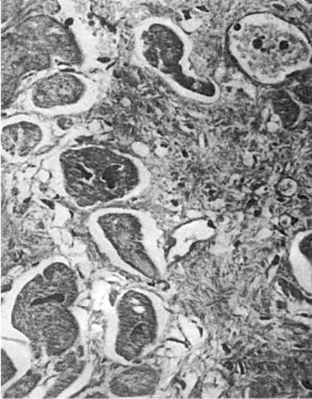 Rice. 81. Regeneration myocardial hypertrophy. Hypertrophied muscle fibers are located along the periphery of the scar
the effect of the pathogenic factor stops before the death of the cell, there is a gradual restoration of damaged organelles. Consequently, the manifestations of the reparative reaction should be expanded by including restorative intracellular processes in dystrophically altered organs. The generally accepted opinion about regeneration only as the final stage of the pathological process is hardly justified. Reparative regeneration is not local, A general reaction organism, covering various organs, but fully realized only in one or another of them.
ABOUT pathological regeneration they say in those cases when, as a result of various reasons, there is perversion of the regenerative process, violation of phase change proliferation
and differentiation. Pathological regeneration is manifested in excessive or insufficient formation of regenerating tissue (hyper- or hyporegeneration), as well as in the transformation during the regeneration of one type of tissue into another [metaplasia - see. Processes of adaptation (adaptation) and compensation]. Examples are hyperproduction of connective tissue with the formation keloid, excessive regeneration of peripheral nerves and excessive callus formation during fracture healing, sluggish wound healing and epithelial metaplasia in the focus of chronic inflammation. Pathological regeneration usually develops with violations of general And local regeneration conditions(violation of innervation, protein and vitamin starvation, chronic inflammation, etc.).
Regeneration of individual tissues and organs
Reparative regeneration of blood differs from physiological regeneration primarily in its greater intensity. In this case, active red bone marrow appears in the long bones in place of fatty bone marrow (myeloid transformation of fatty bone marrow). Fat cells are replaced by growing islands of hematopoietic tissue, which fills the medullary canal and looks juicy, dark red. In addition, hematopoiesis begins to occur outside the bone marrow - extramedullary, or extramedullary, hematopoiesis. Ocha-
GI extramedullary (heterotopic) hematopoiesis as a result of eviction from the bone marrow of stem cells appear in many organs and tissues - the spleen, liver, lymph nodes, mucous membranes, adipose tissue, etc.
Blood regeneration can be sharply oppressed (eg, radiation sickness, aplastic anemia, aleukia, agranulocytosis) or perverted (eg, pernicious anemia, polycythemia, leukemia). At the same time, immature, functionally defective and rapidly collapsing formed elements enter the blood. In such cases, one speaks of pathological regeneration of blood.
The reparative capabilities of the organs of the hematopoietic and immunocompetent systems are ambiguous. Bone marrow has very high plastic properties and can be restored even with significant damage. The lymph nodes they regenerate well only in those cases when the connections of the afferent and efferent lymphatic vessels with the surrounding connective tissue are preserved. Tissue regeneration spleen when damaged, it is usually incomplete, the dead tissue is replaced by a scar.
Regeneration of blood and lymph vessels proceeds ambiguously depending on their caliber.
microvessels have a greater ability to regenerate than large vessels. New formation of microvessels can occur by budding or autogenously. During vascular regeneration by budding (Fig. 82) lateral protrusions appear in their wall due to intensively dividing endothelial cells (angioblasts). Strands are formed from the endothelium, in which gaps appear and blood or lymph from the "mother" vessel enters them. Other elements: the vascular wall is formed due to the differentiation of the endothelium and connective tissue cells surrounding the vessel. Nerve fibers from preexisting nerves grow into the vascular wall. Autogenic neoplasm vessels consists in the fact that foci of undifferentiated cells appear in the connective tissue. In these foci, gaps appear, into which pre-existing capillaries open and blood flows out. Young connective tissue cells differentiate and form the endothelial lining and other elements of the vessel wall.
 Rice. 82. Vessel regeneration by budding
Large vessels do not have sufficient plastic properties. Therefore, if their walls are damaged, only the structures of the inner shell, its endothelial lining, are restored; elements of the middle and outer shells are usually replaced by connective tissue, which often leads to narrowing or obliteration of the vessel lumen.
Connective tissue regeneration begins with the proliferation of young mesenchymal elements and neoplasms of microvessels. A young connective tissue rich in cells and thin-walled vessels is formed, which has a characteristic appearance. This is a juicy dark red fabric with a granular surface, as if strewn with large granules, which was the basis for calling it granulation tissue. Granules are loops of newly formed thin-walled vessels protruding above the surface, which form the basis of granulation tissue. Between the vessels there are many undifferentiated lymphocyte-like cells of the connective tissue, leukocytes, plasma cells and labrocytes (Fig. 83). Later on, it happens maturation granulation tissue, which is based on the differentiation of cellular elements, fibrous structures, and also vessels. The number of hematogenous elements decreases, and fibroblasts - increases. In connection with the synthesis of collagen fibroblasts in the intercellular spaces are formed argyrophilic(see Fig. 83), and then collagen fibers. The synthesis of glycosaminoglycans by fibroblasts serves to form
basic substance connective tissue. As fibroblasts mature, the number of collagen fibers increases, they are grouped into bundles; at the same time, the number of vessels decreases, they differentiate into arteries and veins. The maturation of granulation tissue ends with the formation coarse fibrous scar tissue.
New formation of connective tissue occurs not only when it is damaged, but also when other tissues are incompletely regenerated, as well as during organization (encapsulation), wound healing, and productive inflammation.
The maturation of granulation tissue may have certain deviations. Inflammation that develops in the granulation tissue leads to a delay in its maturation,
 Rice. 83. granulation tissue. There are many undifferentiated connective tissue cells and argyrophilic fibers between the thin-walled vessels. Silver impregnation
and excessive synthetic activity of fibroblasts - to excessive formation of collagen fibers with their subsequent pronounced hyalinosis. In such cases, scar tissue appears in the form of a tumor-like formation of a bluish-red color, which rises above the surface of the skin in the form keloid. Keloid scars are formed after various traumatic skin lesions, especially after burns.
Regeneration of adipose tissue occurs due to the neoplasm of connective tissue cells, which turn into fat (adiposocytes) by accumulating lipids in the cytoplasm. Fat cells are folded into lobules, between which there are connective tissue layers with vessels and nerves. Regeneration of adipose tissue can also occur from the nucleated remnants of the cytoplasm of fat cells.
Bone regeneration in case of bone fracture, it largely depends on the degree of bone destruction, the correct reposition of bone fragments, local conditions (circulatory status, inflammation, etc.). At uncomplicated bone fracture, when bone fragments are motionless, may occur primary bone union(Fig. 84). It begins with growing into the area of the defect and hematoma between bone fragments of young mesenchymal elements and vessels. There is a so-called preliminary connective tissue callus, in which bone formation begins immediately. It is associated with the activation and proliferation osteoblasts in the area of damage, but primarily in the periostat and endostat. In the osteogenic fibroreticular tissue, low-calcified bone trabeculae appear, the number of which increases.
Formed preliminary callus. In the future, it matures and turns into a mature lamellar bone - this is how
 Rice. 84. Primary bone fusion. Intermediary callus (shown by an arrow), soldering bone fragments (according to G.I. Lavrishcheva)
definitive callus, which in its structure differs from bone tissue only in the disorderly arrangement of the bone crossbars. After the bone begins to perform its function and a static load appears, the newly formed tissue undergoes restructuring with the help of osteoclasts and osteoblasts, bone marrow appears, vascularization and innervation are restored. In case of violation of local conditions of bone regeneration (circulatory disorder), mobility of fragments, extensive diaphyseal fractures, secondary bone union(Fig. 85). This type of bone fusion is characterized by the formation between bone fragments, first of cartilage tissue, on the basis of which bone tissue is built. Therefore, with secondary bone fusion they speak of preliminary osteochondral callus, which develops into mature bone over time. Secondary bone fusion compared with the primary is much more common and takes longer.
At adverse conditions bone regeneration may be impaired. Thus, when a wound becomes infected, bone regeneration is delayed. Bone fragments, which, during the normal course of the regenerative process, act as a framework for the newly formed bone tissue, support inflammation under conditions of wound suppuration, which inhibits regeneration. Sometimes primary bone-cartilaginous callus is not differentiated into bone callus. In these cases, the ends of the broken bone remain movable, forming false joint. Excess production of bone tissue during regeneration leads to the appearance of bone outgrowths - exostoses.
Cartilage regeneration in contrast to the bone occurs usually incomplete. Only small defects can be replaced by newly formed tissue due to the cambial elements of the perichondrium - chondroblasts. These cells create the basic substance of cartilage, then turn into mature cartilage cells. Large cartilage defects are replaced by scar tissue.
regeneration of muscle tissue, its possibilities and forms are different depending on the type of this fabric. Smooth mice, whose cells are capable of mitosis and amitosis, with minor defects can regenerate quite completely. Significant areas of damage to smooth muscles are replaced by a scar, while the remaining muscle fibers undergo hypertrophy. New formation of smooth muscle fibers can occur by transformation (metaplasia) of connective tissue elements. This is how bundles of smooth muscle fibers are formed in pleural adhesions, in thrombi undergoing organization, in vessels during their differentiation.
striated muscles regenerate only when the sarcolemma is preserved. Inside the tubes from the sarcolemma, its organelles are regenerated, resulting in the appearance of cells called myoblasts. They stretch, the number of nuclei in them increases, in the sarcoplasm
 Rice. 85. Secondary bone fusion (according to G.I. Lavrishcheva):
a - osteocartilaginous periosteal callus; a piece of bone tissue among the cartilage (microscopic picture); b - periosteal bone and cartilage callus (histotopogram 2 months after surgery): 1 - bone part; 2 - cartilaginous part; 3 - bone fragments; c - periosteal callus soldering displaced bone fragments
myofibrils differentiate, and the sarcolemma tubes turn into striated muscle fibers. Skeletal muscle regeneration may also be associated with satellite cells, which are located under the sarcolemma, i.e. inside the muscle fiber, and are cambial. In the event of an injury, satellite cells begin to divide intensively, then undergo differentiation and ensure the restoration of muscle fibers. If, when the muscle is damaged, the integrity of the fibers is violated, then at the ends of their ruptures, flask-shaped bulges appear, which contain a large number of nuclei and are called muscle kidneys. In this case, the restoration of the continuity of the fibers does not occur. The rupture site is filled with granulation tissue, which turns into a scar (muscle callus). Regeneration heart muscles when it is damaged, as with damage to the striated muscles, it ends with scarring of the defect. However, in the remaining muscle fibers, intense hyperplasia of ultrastructures occurs, which leads to fiber hypertrophy and restoration of organ function (see Fig. 81).
Epithelial regeneration in most cases, it is carried out quite completely, since it has a high regenerative capacity. Regenerates especially well cover epithelium. Recovery keratinized stratified squamous epithelium possible even with fairly large skin defects. During the regeneration of the epidermis at the edges of the defect, there is an increased reproduction of cells of the germinal (cambial), germ (Malpighian) layer. The resulting epithelial cells first cover the defect in one layer. In the future, the layer of the epithelium becomes multi-layered, its cells differentiate, and it acquires all the signs of the epidermis, which includes the growth, granular shiny (on the soles and palmar surface of the hands) and the stratum corneum. In violation of the regeneration of the skin epithelium, non-healing ulcers are formed, often with the growth of atypical epithelium in their edges, which can serve as the basis for the development of skin cancer.
Integumentary epithelium of mucous membranes (stratified squamous non-keratinizing, transitional, single-layer prismatic and multinuclear ciliated) regenerates in the same way as multi-layered squamous keratinizing. The defect of the mucous membrane is restored due to the proliferation of cells lining the crypts and excretory ducts of the glands. Undifferentiated flattened epithelial cells first cover the defect with a thin layer (Fig. 86), then the cells take the form characteristic of the cellular structures of the corresponding epithelial lining. In parallel, the glands of the mucous membrane are partially or completely restored (for example, tubular glands of the intestine, endometrial glands).
Mesothelial regeneration the peritoneum, pleura and pericardial sac is carried out by dividing the remaining cells. Comparatively large cubic cells appear on the surface of the defect, which then flatten. With small defects, the mesothelial lining is restored quickly and completely.
The state of the underlying connective tissue is important for the restoration of the integumentary epithelium and mesothelium, since the epithelialization of any defect is possible only after it has been filled with granulation tissue.
Regeneration of specialized organ epithelium(liver, pancreas, kidneys, endocrine glands, pulmonary alveoli) is carried out according to the type regenerative hypertrophy: in areas of damage, the tissue is replaced by a scar, and along its periphery, hyperplasia and hypertrophy of parenchyma cells occur. IN liver the site of necrosis is always subject to scarring, however, in the rest of the organ, intensive neoplasm of cells occurs, as well as hyperplasia of intracellular structures, which is accompanied by their hypertrophy. As a result, the initial mass and function of the organ are quickly restored. The regenerative possibilities of the liver are almost limitless. In the pancreas, regenerative processes are well expressed both in the exocrine sections and in the pancreatic islets, and the epithelium of the exocrine glands becomes the source of restoration of the islets. IN kidneys with necrosis of the epithelium of the tubules, the surviving nephrocytes reproduce and restore the tubules, but only with the preservation of the tubular basement membrane. When it is destroyed (tubulorhexis), the epithelium is not restored and the tubule is replaced by connective tissue. The dead tubular epithelium is not restored even in the case when the vascular glomerulus dies along with the tubule. At the same time, scar connective tissue grows in place of the dead nephron, and the surrounding nephrons undergo regenerative hypertrophy. in the glands internal secretion recovery processes are also represented by incomplete regeneration. IN lung after the removal of individual lobes, hypertrophy and hyperplasia of tissue elements occur in the remaining part. Regeneration of the specialized epithelium of organs can proceed atypically, which leads to the growth of connective tissue, structural reorganization and deformation of organs; in such cases one speaks of cirrhosis (liver cirrhosis, nephrocyrrhosis, pneumocirrhosis).
Regeneration of different parts of the nervous system happens ambiguously. IN head And spinal cord neoplasms of ganglion cells do not
 Rice. 86. Regeneration of the epithelium in the bottom of a chronic stomach ulcer
even when they are destroyed, the restoration of function is possible only due to the intracellular regeneration of the remaining cells. Neuroglia, especially microglia, are characterized by a cellular form of regeneration; therefore, defects in the tissue of the brain and spinal cord are usually filled with proliferating neuroglia cells - so-called glial (glial) scarring. When damaged vegetative nodes along with hyperplasia of cell ultrastructures, their neoplasm also occurs. In case of violation of integrity peripheral nerve regeneration occurs due to the central segment, which has retained its connection with the cell, while the peripheral segment dies. The multiplying cells of the Schwann sheath of the dead peripheral segment of the nerve are located along it and form a case - the so-called Byungner cord, into which regenerating axial cylinders from the proximal segment grow. The regeneration of nerve fibers ends with their myelination and restoration of nerve endings. Regenerative hyperplasia receptors pericellular synaptic devices and effectors is sometimes accompanied by hypertrophy of their terminal apparatuses. If the regeneration of the nerve is disturbed for one reason or another (significant divergence of parts of the nerve, the development of an inflammatory process), then a scar is formed at the site of its break, in which the regenerated axial cylinders of the proximal segment of the nerve are randomly located. Similar growths occur at the ends of the cut nerves in the stump of the limb after its amputation. Such growths formed by nerve fibers and fibrous tissue are called amputation neuromas.
Wound healing
Wound healing proceeds according to the laws of reparative regeneration. The rate of wound healing, its outcomes depend on the degree and depth of wound damage, the structural features of the organ, the general condition of the body, and the methods of treatment used. According to I.V. Davydovsky, the following types of wound healing are distinguished: 1) direct closure of an epithelial cover defect; 2) healing under the scab; 3) wound healing by primary intention; 4) wound healing by secondary intention, or wound healing through suppuration.
Direct closure of an epithelial defect- this is the simplest healing, which consists in the creeping of the epithelium on the superficial defect and closing it with an epithelial layer. Observed on the cornea, mucous membranes healing under the scab concerns small defects, on the surface of which a drying crust (scab) quickly appears from coagulated blood and lymph; the epidermis is restored under the crust, which disappears 3-5 days after the injury.
Healing by primary intention (per rimamm intentionem) observed in wounds with damage not only to the skin, but also to the underlying tissue,
and the edges of the wound are even. The wound is filled with clots of spilled blood, which protects the edges of the wound from dehydration and infection. Under the influence of proteolytic enzymes of neutrophils, a partial lysis of blood coagulation, tissue detritus occurs. Neutrophils die, they are replaced by macrophages that phagocytize red blood cells, the remnants of damaged tissue; hemosiderin is found in the edges of the wound. Part of the contents of the wound is removed on the first day of injury along with exudate on its own or when treating the wound - primary cleansing. On the 2-3rd day, fibroblasts and newly formed capillaries growing towards each other appear at the edges of the wound, granulation tissue, the layer of which at primary tension does not reach large sizes. By the 10-15th day, it fully matures, the wound defect epithelizes and the wound heals with a delicate scar. In a surgical wound, healing by primary intention is accelerated due to the fact that its edges are pulled together with silk or catgut threads, around which giant cells of foreign bodies that absorb them accumulate and do not interfere with healing.
Healing by secondary intention (per secundam intentionem), or healing through suppuration (or healing by granulation - per granulationem), It is usually observed with extensive wounds, accompanied by crushing and necrosis of tissues, penetration of foreign bodies and microbes into the wound. At the site of the wound, hemorrhages occur, traumatic swelling of the edges of the wound, signs of demarcation quickly appear. purulent inflammation on the border with dead tissue, melting of necrotic masses. During the first 5-6 days, rejection of necrotic masses occurs - secondary cleansing of the wound, and granulation tissue begins to develop at the edges of the wound. granulation tissue, performing the wound, consists of 6 layers passing into each other (Anichkov N.N., 1951): superficial leukocyte-necrotic layer; superficial layer of vascular loops, layer of vertical vessels, maturing layer, layer of horizontally located fibroblasts, fibrous layer. The maturation of granulation tissue during wound healing by secondary intention is accompanied by regeneration of the epithelium. However, with this type of wound healing, a scar is always formed in its place.
Regeneration- restoration by the body of lost or damaged organs and tissues, as well as the restoration of the whole organism from its part. In more
degree inherent in plants and invertebrates, to a lesser extent - vertebrates. Regeneration can be triggered
experimentally.
Regeneration is aimed at restoring damaged structural elements and regeneration processes can
carried out at different levels:
a) molecular
b) subcellular
c) cellular - cell reproduction by mitosis and amitotic way
d) tissue
e) organ.
Types of regeneration:
7. Physiological - ensures the functioning of organs and systems under normal conditions. Physiological regeneration occurs in all organs, but in some more, in others less.
2. Reparative(recovery) - occurs in connection with pathological processes that lead to tissue damage (this is enhanced physiological regeneration)
a) complete regeneration (restitution) - exactly the same tissue appears at the site of tissue damage
b) incomplete regeneration (substitution) - connective tissue appears in place of the dead tissue. For example, in the heart with myocardial infarction, necrosis occurs, which is replaced by connective tissue.
The meaning of incomplete regeneration: regenerative hypertrophy occurs around the connective tissue, which
ensures the preservation of the function of the damaged organ.
Regenerative hypertrophy carried out through:
a) cell hyperplasia (excess formation)
b) cell hypertrophy (an increase in the body in volume and mass).
Regeneration hypertrophy in the myocardium is carried out due to hyperplasia of intracellular structures.
forms of regeneration.
1. Cellular - cell reproduction occurs in a mitotic and amitotic way. It exists in bone tissue, epidermis, gastrointestinal mucosa, respiratory mucosa, urogenital mucosa, endothelium, mesothelium, loose connective tissue, hematopoietic system. In these organs and tissues, complete regeneration occurs (exactly the same tissue).
2. Intracellular - hyperplasia of intracellular structures occurs. Myocardium, skeletal muscles (mainly), ganglion cells of the central nervous system (exclusively).
3. Cellular and intracellular forms. Liver, kidneys, lungs, smooth muscles, autonomic nervous system, pancreas, endocrine system. Usually there is incomplete regeneration.
Connective tissue regeneration.
Stages:
1. Formation of granulation tissue. Gradually there is a displacement of vessels and cells with the formation of fibers. Fibroblasts are fibrocytes that produce fibers.
2. Formation of mature connective tissue. Blood regeneration
1. Physiological regeneration. In the bone marrow.
2. Reparative regeneration. Occurs with anemia, leukopenia, thrombocytopenia. Extramedullary foci of hematopoiesis appear (in the liver, spleen, lymph nodes, yellow bone marrow is involved in hematopoiesis).
3. Pathological regeneration. With radiation sickness, leukemia. In the hematopoietic organs, immature
hematopoietic elements (power cells).
Question 16
HOMEOSTASIS.
homeostasis - maintaining the constancy of the internal environment of the body in continuously changing environmental conditions. Because an organism is a multi-level self-regulating object, it can be considered from the point of view of cybernetics. Then, the body is a complex multi-level self-regulating system with many variables.
Input variables:
Cause;
Irritation.
Output variables:
Reaction;
Consequence.
The reason is a deviation from the norm of the reaction in the body. Feedback plays a decisive role. There is positive and negative feedback.
negative feedback reduces the effect of the input signal on the output. positive feedback increases the effect of the input signal on the output effect of the action.
A living organism is an ultrastable system that searches for the most optimal stable state, which is provided by adaptations.
Question 18:
TRANSPLANTATION PROBLEMS.
Transplantation is the transplantation of tissues and organs.
Transplantation in animals and humans is the engraftment of organs or sections of individual tissues to replace defects, stimulate regeneration, during cosmetic surgeries, as well as for the purposes of experiment and tissue therapy.
Autotransplantation - tissue transplantation within the same organism Allotransplantation - transplantation between organisms of the same species. Xenotransplantation is a transplant between different species.
Question 19
Chronobiology- a branch of biology that studies biological rhythms, the course of various biological processes
(mostly cyclic) in time.
biological rhythms- (biorhythms), cyclic fluctuations in the intensity and nature of biological processes and phenomena. Some biological rhythms are relatively independent (for example, heart rate, respiration), others are associated with the adaptation of organisms to geophysical cycles - daily (for example, fluctuations in the intensity of cell division, metabolism, animal motor activity), tidal (for example, biological processes in organisms associated with the level of sea tides), annual (changes in the number and activity of animals, growth and development of plants, etc.). The science of biological rhythms is chronobiology.
Question 20
PHYLOGENESIS OF THE SKELETON
The skeleton of fish consists of a skull, spine, skeleton of unpaired, paired fins and their belts. In the trunk region, ribs are attached to the transverse processes of the body. The vertebrae articulate with each other with the help of articular processes, providing bending mainly in the horizontal plane.
The skeleton of amphibians, like all vertebrates, consists of a skull, spine, limb skeleton and their belts. The skull is almost entirely cartilaginous (Fig. 11.20). It is movably articulated with the spine. The spine contains nine vertebrae, united into three sections: cervical (1 vertebra), trunk (7 vertebrae), sacral (1 vertebrae), and all caudal vertebrae are fused to form a single bone - the urostyle. Ribs are missing. The shoulder girdle includes bones typical of terrestrial vertebrates: paired shoulder blades, crow bones (coracoids), clavicles, and an unpaired sternum. It has the form of a semicircle lying in the thickness of the trunk muscles, that is, it is not connected to the spine. The pelvic girdle is formed by two pelvic bones, formed by three pairs of iliac, ischial and pubic bones, fused together. The long iliac bones are attached to the transverse processes of the sacral vertebrae. The skeleton of free limbs is built according to the type of a system of multi-membered levers, movably connected by spherical joints. As part of the forelimb. allocate the shoulder, forearm and hand.
The body of the lizard is subdivided into the head, trunk and tail. The neck is well defined in the trunk region. The whole body is covered with horny scales, and the head and belly are covered with large shields. The limbs of the lizard are well developed and armed with five fingers with claws. The shoulder and thigh bones are parallel to the ground, causing the body to sag and touch the ground (hence the class name). The cervical spine consists of eight vertebrae, the first of which is movably connected to both the skull and the second vertebra, which provides the head region with greater freedom of movement. The vertebrae of the lumbothoracic region bear ribs, part of which is connected to the sternum, resulting in the formation of the chest. The sacral vertebrae provide a stronger connection to the pelvic bones than in amphibians.
The skeleton of mammals is basically similar in structure to the skeleton of terrestrial vertebrates, but there are some differences: the number of cervical vertebrae is constant and equal to seven, the skull is more voluminous, which is associated with the large size of the brain. The bones of the skull fuse rather late, allowing the brain to expand as the animal grows. The limbs of mammals are built according to the five-fingered type characteristic of terrestrial vertebrates.
Question 21
PHYLOGENESIS OF THE CIRCULATION SYSTEM
The circulatory system of fish is closed. The heart is two-chambered, consisting of an atrium and a ventricle. Venous blood from the ventricle of the heart enters the abdominal aorta, which carries it to the gills, where it is enriched with oxygen and released from carbon dioxide. Arterial blood flowing from the gills is collected in the dorsal aorta, which is located along the body under the spine. Numerous arteries depart from the dorsal aorta to various organs of the fish. In them, the arteries break up into a network of the thinnest, capillaries, through the walls of which the blood gives off oxygen and is enriched with carbon dioxide. Venous blood is collected in the veins and through them enters the atrium, and from it the ventricle. Therefore, fish have one circle of blood circulation.
The circulatory system of amphibians is represented by a three-chambered heart, consisting of two atria and a ventricle, and two circles of blood circulation - large (trunk) and small (pulmonary). The pulmonary circulation begins in the ventricle, includes the vessels of the lungs and ends in the left atrium. A large circle also begins in the ventricle. Blood, having passed through the vessels of the whole body, returns to the right atrium. Thus, arterial blood from the lungs enters the left atrium, and venous blood from the whole body enters the right atrium. Arterial blood flowing from the skin also enters the right atrium. So, thanks to the appearance of the pulmonary circulation, arterial blood also enters the heart of amphibians. Despite the fact that arterial and venous blood enters the ventricle, complete mixing of the blood does not occur due to the presence of pockets and incomplete septa. Thanks to them, when leaving the ventricle, arterial blood flows through the carotid arteries to the head section, venous blood to the lungs and skin, and mixed blood to all other organs of the body. Thus, in amphibians there is no complete division of blood in the ventricle, therefore the intensity of life processes is low, and the body temperature is unstable.
The heart of reptiles is three-chambered, however, complete mixing of arterial and venous blood does not occur due to the presence of an incomplete longitudinal septum in it. Three vessels departing from different parts of the ventricle - the pulmonary artery, the left and right aortic arches - carry venous blood to the lungs, arterial - to the head and forelimbs, and to the rest of the parts - mixed with a predominance of arterial. Such blood supply, as well as a low ability to thermoregulate, lead to the fact that
The body temperature of reptiles depends on the temperature conditions of the environment.
The high level of vital activity of birds is due to a more advanced circulatory system compared to animals of previous classes. They had a complete separation of arterial and venous blood flow. This is due to the fact that the heart of birds is four-chambered and is completely divided into the left - arterial, and right - venous parts. The aortic arch is only one (right) and departs from the left ventricle. Pure arterial blood flows in it, supplying all the tissues and organs of the body. The pulmonary artery departs from the right ventricle, carrying venous blood to the lungs. Blood moves rapidly through the vessels, gas exchange occurs intensively, a lot of heat is released. The circulatory system of mammals has no fundamental differences from that of birds. Unlike birds, in mammals the left aortic arch departs from the left ventricle.
Question 22
DEVELOPMENT OF ARTERIAL ARCHES
Arterial arches, aortic arches, blood vessels that are laid down in vertebrate embryos in the form of 6-7 (up to 15 in cyclostomes) paired lateral trunks extending from the abdominal aorta. AD pass through the interbranchial septa to the dorsal side of the pharynx and, merging, form the dorsal aorta. The first 2 pairs of arterial arches are usually reduced early; in fish and amphibian larvae, they are preserved in the form of small vessels. The remaining 4-5 pairs of arterial arches become gill vessels. In terrestrial vertebrates, carotid arteries are formed from the third pair of arterial arches, and pulmonary arteries are formed from the sixth. In caudate amphibians, usually the 4th and 5th pairs of arterial arches form the trunks or roots of the aorta, which merge into the dorsal aorta. In tailless amphibians and reptiles, the aortic arches arise only from the 4th pair of arterial arches, and the 5th is reduced. In birds and mammals, the 5th and half of the 4th arterial arches are reduced, in birds the aorta becomes its right half, in mammals - the left. Sometimes, in adults, germinal vessels remain, connecting the aortic arches with the carotid (carotid ducts) or pulmonary (botallian ducts) arteries.
Question 23
Respiratory system.
Most animals are aerobes. Diffusion of gases from the atmosphere through an aqueous solution is carried out during breathing. Elements of skin and water respiration are preserved even in higher vertebrates. In the course of evolution, animals developed a variety of respiratory devices - derivatives of the skin and the digestive tube. Gills and lungs are derivatives of the pharynx.
PHYLOGENESIS OF THE RESPIRATORY ORGANS
The respiratory organs - gills - are located on the upper side of the four gill arches in the form of bright red petals. Water enters the mouth of the fish, is filtered through the gill slits, washing the gills, and is brought out from under the gill cover. Gas exchange is carried out in numerous gill capillaries, in which blood flows towards the water surrounding the gills.
Frogs breathe with lungs and skin. The lungs are paired hollow sacs with a cellular inner surface penetrated by a network of blood capillaries, where gas exchange occurs. The mechanism of respiration in amphibians is imperfect, of the forced type. The animal draws air into the oropharyngeal cavity, for which it lowers the bottom of the oral cavity and opens the nostrils. The nostrils are then closed with valves, the floor of the mouth rises and air is pumped into the lungs. The removal of air from the lungs occurs due to the contraction of the pectoral muscles. The surface of the lungs in amphibians is small, less than the surface of the skin.
Respiratory organs - lungs (reptiles). Their walls have a cellular structure, which greatly increases the surface. Cutaneous respiration is absent. Ventilation of the lungs is more intense than in amphibians, and is associated with a change in the volume of the chest. The respiratory tract - trachea, bronchi - protect the lungs from the drying and cooling effects of air coming from outside.
The lungs of birds are dense spongy bodies. The bronchi, having entered the lungs, strongly branch into them to the thinnest, blindly closed bronchioles, entangled in a network of capillaries, where
and gas exchange takes place. Part of the large bronchi, without branching, goes beyond the lungs and expands into huge thin-walled air sacs, the volume of which is many times greater than the volume of the lungs (Fig. 11.23). Air sacs are located between various internal organs, and their branches pass between the muscles, under the skin and in the cavity of the bones.
Mammals breathe with lungs that have an alveolar structure, due to which the respiratory surface exceeds the surface of the body by 50 times or more. The mechanism of breathing is due to a change in the volume of the chest due to the movement of the ribs and a special muscle characteristic of mammals - the diaphragm.
Question 24
PHYLOGENESIS OF THE BRAIN
The central nervous system of fish consists of the brain and spinal cord. The brain in fish, like in all vertebrates, is represented by five sections: anterior, intermediate, middle, cerebellum and medulla oblongata. Well-developed olfactory lobes depart from the forebrain. The greatest development reaches the midbrain, which analyzes visual perceptions, as well as the cerebellum, which regulates the coordination of movements and maintaining balance.
The amphibian brain has the same five sections as the fish brain. However, it differs from it in the large development of the forebrain, which in amphibians is divided into two hemispheres. The cerebellum is underdeveloped due to low mobility and monotony. the different nature of the movements of amphibians.
The brain of reptiles, in comparison with that of amphibians, has a better developed cerebellum and large hemispheres of the forebrain, the surface of which has the rudiments of the cortex. This causes various and more complex forms of adaptive behavior.
The cerebrum of birds differs from the brain of those that squirm by the large size of the hemispheres of the forebrain and cerebellum.
The mammalian brain is relatively large due to an increase in the volume of the forebrain and cerebellum hemispheres. The development of the forebrain occurs due to the growth of its roof - the cerebral fornix, or the cerebral cortex.
Question 25
PHYLOGENESIS OF THE EXECUTIVE AND REGENERAL SYSTEMS
The excretory organs of fish are paired ribbon-like trunk kidneys located in the body cavity under the spine. They have lost contact with the body cavity and remove harmful waste products by filtering them out of the blood. In freshwater fish, the end product of protein metabolism is toxic ammonia. It dissolves in a lot of water, and therefore the fish excrete a lot of liquid urine. The water excreted in the urine is easily replenished due to its constant intake through the skin, gills and with food. In marine fish, the end product of nitrogen metabolism is less toxic urea, the excretion of which requires less water. The urine formed in the kidneys flows through the paired ureters into the bladder, from where it is excreted out through the excretory opening. Paired sex glands - ovaries and testes - have excretory ducts. Fertilization in most fish is external and occurs in water.
The excretory organs of amphibians, like those of fish, are represented by trunk kidneys. However, unlike fish, they have the appearance of flattened compact bodies lying on their sides.
sacral vertebra. In the kidneys there are glomeruli that filter out harmful decay products from the blood (mainly urea) and at the same time substances important for the body (sugars, vitamins, etc.). During the flow through the renal tubules, substances beneficial to the body are absorbed back into the blood, and urine enters the two ureters into the cloaca and from there to the bladder. After filling the bladder, its muscular walls contract, urine is excreted into the cloaca and thrown out. Losses of water from the body of amphibians with urine, as well as in fish, are replenished by its intake through the skin. Sex glands are paired. The paired oviducts drain into the cloaca, and the vas deferens into the ureters.
The excretory organs of reptiles are represented by pelvic kidneys, in which the total filtration area of the glomeruli is small, while the length of the tubules is significant. This contributes to the intensive reabsorption of water filtered by the glomeruli into the blood capillaries. Consequently, the excretion of waste products in reptiles occurs with minimal loss of water. In them, as in terrestrial arthropods, the end product of excretion is uric acid, which requires a small amount of water to be excreted from the body. Urine is collected through the ureters into the cloaca, and from it into the bladder, from which it is excreted in the form of a suspension of small crystals.
Isolation of mammals. The pelvic kidneys of mammals are similar in structure to those of birds. Urine with a high content of urea flows from the kidneys through the ureters into the bladder, and out of it goes out.
Question 26
Phylogeny of the integument of the body:
The main directions of evolution of the integuments of chordates:
1) differentiation into two layers: outer - epidermis, inner - dermis and an increase in the thickness of the dermis;
1) from a single-layer epidermis to a multilayer one;
2) differentiation of the dermis into 2 layers - papillary and reticular:
3) the appearance of subcutaneous fat and the improvement of the mechanisms of thermoregulation;
4) from unicellular glands to multicellular;
5) differentiation of various skin derivatives.
In the lower chordates (lancelet) the epidermis is single-layer, cylindrical, has glandular cells that secrete mucus. The dermis (corium) is represented by a thin layer of unformed connective tissue.
In lower vertebrates, the epidermis becomes multilayered. Its lower layer is germline (basal), its cells divide and replenish the cells of the overlying layers. The dermis has correctly arranged fibers, vessels and nerves.
Derivatives of the skin are: unicellular (in cyclostomes) and multicellular (in amphibians) mucous glands; scales: a) placoid in cartilaginous fish, in the development of which the epidermis and dermis take part; b) bone in bony fish, which develops at the expense of the dermis.
The placoid scale is covered on the outside with a layer of enamel (of ectodermal origin), under which are dentin and pulp (of mesodermal origin). Scales and mucus perform a protective function.
Amphibians have thin, smooth skin without scales. The skin contains a large number of multicellular mucous glands, the secret of which moisturizes the integument and has bactericidal properties. The skin takes part in gas exchange.
In higher vertebrates, due to landfall, the epidermis becomes dry and has a stratum corneum.
reptiles horny scales develop, there are no skin glands.
In mammals: well developed epidermis and dermis, appears subcutaneous fat.
Question 27
PHYLOGENESIS OF THE DIGESTIVE SYSTEM.
Fish eat a variety of foods. Food specialization is reflected in the structure of the digestive organs. The mouth leads to the oral cavity, which usually contains numerous teeth located on the jaw, palatine and other bones. Salivary glands are absent. From the oral cavity, food passes into the pharynx, perforated by gill slits, and through the esophagus enters the stomach, the glands of which abundantly secrete digestive juices. Some fish (cyprinids and a number of others) do not have a stomach and food enters immediately into the small intestine, where, under the influence of a complex of enzymes secreted by the glands of the intestine itself, the liver and pancreas, food is broken down and the dissolved nutrients are absorbed. The differentiation of the digestive system of amphibians remained approximately at the same level as that of their ancestors - fish. The common oropharyngeal cavity passes into a short esophagus, followed by a slightly isolated stomach, passing without a sharp border into the intestine. The intestine ends with the rectum, which passes into the cloaca. The ducts of the digestive glands - the liver and pancreas - flow into the duodenum. In the oropharyngeal cavity open ducts of salivary glands absent in fish, wetting the oral cavity and food. The appearance of a real tongue in the oral cavity, the main organ of food extraction, is associated with the terrestrial way of life.
In the digestive system of reptiles, differentiation into departments is better than that of amphibians. Food is captured by the jaws, which have teeth to hold the prey. The oral cavity is better than that of amphibians, delimited from the pharynx. At the bottom of the oral cavity is a movable, forked tongue at the end. Food is moistened with saliva, which makes it easier to swallow. The esophagus is long due to the development of the neck. The stomach, separated from the esophagus, has muscular walls. There is a cecum on the border of the small and large intestines. Ducts of the liver and pancreas
glands open into the duodenum. The digestion time of food depends on the body temperature of the reptiles.
Digestive system of mammals. The teeth sit in the cells of the jaw bones and are divided into incisors, canines and molars. The mouth opening is surrounded by fleshy lips, which is characteristic only of mammals in connection with feeding milk. In the oral cavity, food, in addition to chewing with teeth, is exposed to the chemical action of saliva enzymes, and then sequentially passes into the esophagus and stomach. The stomach in mammals is well separated from other sections of the digestive tract and is supplied with digestive glands. In most mammalian species, the stomach is divided into more or fewer sections. It is most complicated in ruminant artiodactyls. The intestine has a thin and a thick section. At the border of the thin and thick sections, the caecum departs, in which the fermentation of fiber occurs. The ducts of the liver and pancreas open into the cavity of the duodenum.
Question 28
Endocrine system.
In any organism, compounds are produced that are carried throughout the body, having an integrative role. Plants have phytohormones that control growth, the development of fruits, flowers, the development of axillary buds, the division of the cambium, etc. Unicellular algae have phytohormones.
Hormones appeared in multicellular organisms when special endocrine cells arose. However, chemical compounds that play the role of hormones existed before. Thyroxine, triiodothyronine (thyroid gland) are found in cyanobacteria. Hormonal regulation in insects is poorly understood.
In 1965, Wilson isolated insulin from starfish.
It turned out that it is very difficult to define a hormone.
Hormone is a specific chemical secreted by specific cells in a particular area of the body, which enters the bloodstream and then has a specific effect on certain cells or target organs located in other areas of the body, which leads to the coordination of the functions of the whole organism.
A large number of mammalian hormones are known. They are divided into 3 main groups.
Pheromones. Released into the external environment. With their help, animals receive and transmit information. In humans, the smell of 14 - hydroxytetradecanoic acid is clearly distinguished only by women who have reached puberty.
The most simply organized multicellular organisms - for example, sponges also have a semblance of an endocrine system. Sponges consist of 2 layers - endoderm and exoderm, between them is mesenchyme, which contains macromolecular compounds characteristic of the connective tissue of more highly organized organisms. There are migrating cells in the mesenchyme, some cells are able to secrete serotonin, acetylcholine. Sponges have no nervous system. Substances synthesized in the mesenchyme serve to connect individual parts of the body. Coordination is carried out by moving cells along the mesenchyme. There is also the transfer of substances between cells. The basis of chemical signaling, which is characteristic of other animals, has been laid. There are no independent endocrine cells.
Coelenterates have a primitive nervous system. Initially, nerve cells performed a neurosecretory function. Trophic function, carried out the control of growth, development of the organism. Then the nerve cells began to stretch and form long processes. The secret was released near the target organ, without transfer (because there was no blood). The endocrine mechanism arose earlier than the conductive one. Nerve cells were endocrine, and then they received conductive properties. Neurosecretory cells were the first secretory cells.
Protostomes and deuterostomes produce the same steroid and peptide hormones. It is generally accepted that in the process of evolution, new ones (mutations, gene duplications) can arise from some polypeptide hormones. Duplications are less suppressed by natural selection than mutations. Many hormones can be synthesized not in one gland, but in several. For example, insulin is produced in the pancreas, submandibular gland, duodenum and other organs. There is a dependence of the genes that control the synthesis of hormones on the position.
