Anomalies of intrauterine development. Frequent genetic diseases of children in the Russian Federation. Deformities and their causes
In humans, as in other animals, anomalies often occur in development, leading to more or less serious deviations in the structure or functioning of organs. Many of these anomalies turn out to be lethal (deadly) already on early stages development.
When sensitive methods for detecting the early stages of pregnancy appeared (by detecting the hormone in the blood chorionic gonadotropin), it turned out that more than half of human embryos die at very early stages. The percentage of spontaneous (spontaneous) abortions for more than late stages associated with fetal failure. Of successfully born children, about 5% are born with noticeable anomalies. They can be quite harmless (for example, large birthmark), but can strongly affect the physical and mental development. The reasons for these anomalies are varied. A significant proportion of them is due to hereditary diseases. These can be either inherited mutations (such as the hemophilia gene or the six-fingered gene), or anomalies that occur in part of the eggs or sperm (for example, Down syndrome). The similarity here is that the cause of the deviations is a change in the DNA of the egg.
Numerous factors can cause deformities and when exposed to the embryo itself. Such factors are called teratogenic. There are critical periods of development when the action of teratogenic factors has a particularly strong effect on the embryo or on specific organs and their systems. These periods are different for various organs, but for most of them the period between the 15th and 60th day of pregnancy is critical. Deformities can be caused by mutagenic factors that affect the DNA of the embryo itself - for example, radioactive radiation. Other agents may cause fetal abnormalities without being mutagens. These include, for example, certain types of viruses and bacteria. Some of them are able to penetrate from the mother's body into the fetus. So, in women who had rubella in the first third of pregnancy, there is a high probability of having children with visual impairments and heart diseases. The causative agent of syphilis (the bacterium Spirochete pallidum) causes early fetal death and deaf children when exposed in later stages. Serious developmental disabilities nervous system the fetus can be caused by toxoplasma - protists that are carried by cats (the cats themselves do not suffer much from toxoplasmosis, but the course of pregnancy is also disturbed in them).
But, perhaps, the most diverse deformities and other disorders can cause chemical substances both natural and artificial. Among them are some drugs that are completely harmless to an adult. Thus, retinoic acid (a derivative of vitamin A, which was used in medical practice), when taken in a certain period of pregnancy, caused the birth of children without ears, with reduced jaws, cleft palate and other anomalies. Apparently, this substance inhibits the migration of cells from the neural crest. Its teratogenic effects were known through experiments on mice, and the label warned against using it during pregnancy. However, some women did it out of negligence. An even more tragic story happened with thalidomide, a drug that was widely used in the US as a sedative. When tested on mice and rats, it did not have any teratogenic effect. But if women took it during a certain period of pregnancy, they had children with sharply shortened or absent long bones limbs (with the most severe violations, the hands grow directly from the shoulders). Before the link between thalidomide and these abnormalities could be established, more than 7,000 malformed babies were born. Later it turned out that thalidomide causes the same deformities in monkey embryos as in humans. These examples explain why testing every new drug is such a long and expensive process.
During pregnancy, drugs should be used with extreme caution, be sure to consult a doctor.
In addition to drugs, other substances widely used in everyday life also have a teratogenic effect. For example, alcohol (if a woman consumes 100 grams of vodka a day or more during pregnancy) in almost 100% of cases causes a noticeable lag in the mental and physical development of the child. Nicotine does not have a pronounced teratogenic effect, but in many women who smoke, newborns on average have less weight than normal. In addition, children of smoking mothers are more likely to die from sudden death. (Sudden neonatal death is one of the leading causes of death in children under one year of age in developed countries. Healthy child falls asleep and dies in his sleep. It is believed that death in this case occurs due to the stopping of the respiratory center of the brain, and nicotine can disrupt its functioning if it affects the fetus).
It is impossible to put into words that pain experienced by a mother who understands that her baby, long-awaited and beloved, was born not like everyone else. The presence of deformities and other genetic diseases in a child shocks many parents and forces them to abandon such a child. At the same time, some deformities are not at all a cross on the lives of such children. Many of these babies develop no worse than healthy ones and may well become full-fledged members of society, work, provide for themselves, and even start families. Which genetic diseases most common in our country, what are their causes and how to prevent the development of deformities?
Hereditary and non-hereditary diseases
Diseases are divided into 2 types: hereditary and non-hereditary. The former can be passed on from generation to generation, for example, the well-known hemophilia - the inability of blood to clot. The reason for this is the presence of a certain gene. Genes are particles that are responsible for the development of all human traits: from eye color to heart size. Among the genes there are also those that, by their presence, cause certain diseases, while they can be passed on to children from their parents, because they are "fixed".
Hereditary diseases are caused by the presence of spontaneous mutations- changes in the structure of the gene. These mutations can be caused various factors: radiation, chemical compounds. Such genetic changes occur singly - only in a particular person, they are not stable and are not transmitted to the next generations.
congenital deformities
by the most widespread is a deformity, popularly called " cleft lip and the wolf's mouth." It is a cleft in the sky upper jaw and in upper lip They can occur either simultaneously or separately. Today this defect is fixed. surgically: The cleft is closed and sutures are placed on the lip to tighten the groove. It is imperative to eliminate such ugliness, since its presence interferes with normal breastfeeding development of teeth and speech. However, the intelligence of children with such a pathology does not suffer at all.
Another common ugliness is underdevelopment in varying degrees of the upper or lower extremities, which again mental capacity does not affect. Hands, upper limbs completely, one hand or several fingers may be absent, and lower limbs in this case, they remain only in the "rudimentary" state and it is impossible to move on them. However, such people adapt and can serve themselves even without prostheses.
In addition, there are more serious malformations interfering normal function organism, among them undivided twins, acrania - a serious underdevelopment of the skull, hydrocephalus - the presence of fluid in the cranial cavity and others, however, they are somewhat less common. Today, all these vices can be seen in ultrasound examination, then the parents face the question of maintaining such a pregnancy. However, some deformities occur after birth.
Genetic diseases characterized by developmental delay
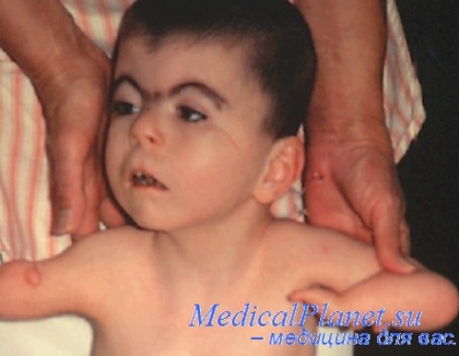
There are also a number congenital genetic diseases accompanied by intellectual impairment and characteristic appearance. The most common and well-known is Down syndrome. Such children are mentally retarded, but in general they can develop and learn about the world, with right approach and work with them, albeit slower than others. Usually such people are able to serve themselves on their own, do simple work and live normally under the supervision of relatives.
Patau Syndrome- the heaviest congenital disease, 95% of children suffering from it die in the first years of life, and those who managed to survive suffer from deep idiocy. Patau syndrome includes whole line malformations - physical and mental underdevelopment, malformations of the heart, visual impairment, cysts in thyroid gland and kidneys, disrupting their work, underdevelopment of the reproductive system.
Causes of congenital malformations
Reasons and mechanisms the occurrence of almost half and deformities are still unknown to scientists. However, there are a number of common factors, which absolutely can provoke the appearance of mutations - changes in genes.
First on the list factors stand chemical compounds. Alcohol, nicotine and narcotic substances adversely affect the development of the embryo, especially in the first trimester of pregnancy.
The same can be said about medicines , the influence of many of them on the formation of the fetus is not fully understood. A striking example of this is the "thalidomide disaster" that occurred between 1956 and 1962. It was caused by the drug "Thalidomide", which was recommended to pregnant women as a sedative and sleeping pill. The result was the birth of 12,000 "thalidomide children" around the world, suffering from underdevelopment upper limbs, eyes and facial muscles.
Relate to reception vitamins is also necessary in the strictest way, since excess has teratogenic effect- causes developmental anomalies, as well as a serious lack of vitamins and trace elements.
Row infectious diseases
, such as rubella, cytomegalovirus and some others, can be transmitted from mother to fetus and cause the development of deformities in it, lead to death.
radioactive radiation negatively affects the genetic apparatus of the mother and child, causing congenital anomalies.
All eggs women are laid and formed in the ovaries during her embryonic development, they are no longer formed during life and are not updated as male spermatozoa. This means that all eggs are exposed to negative impacts from the side environment, wrong image life, alcohol, nicotine, malnutrition. All this can lead to a violation of the genetic apparatus of the egg and the development of an unhealthy child from it. Keep this in mind and protect yourself and your future children from unnecessary negative influences- Prevention is better than cure.
As a result of a developmental defect, the fetus may die even in the embryonic period or soon after birth due to its inability to independent, out of touch with the mother's organism, existence. With some types of malformations of organs that are not vital (cleft lip, malformations of the uterus), occurs to one degree or another proper development child.
Fetal deformities do not always complicate the course of childbirth. This is most often seen in hydrocephalus and double malformations.
Hydrocephalus(hydrocephalia) - dropsy of the head. Severe hydrocephalus is rare. It occurs as a result of excessive (up to 5 l) accumulation cerebrospinal fluid in the cranial cavity, almost usually in the cerebral ventricles, extremely rarely in the subarachnoid space, and also due to its difficult outflow. The bones of the skull, under the influence of increased intracranial pressure, diverge and sometimes become thinner to the thickness of parchment paper, their fibrous joints (fontanelles and sutures) stretch, and therefore the head volume increases significantly. The brain undergoes a sharp atrophy from pressure.
If the fetus dies long before birth, intracranial pressure decreases, the head turns into a soft-elastic bladder, which can independently pass through the birth canal during childbirth. With a live fetus, a large taut-elastic head is infringed in the pelvis, resulting in overstretching of the lower segment of the uterus. If this is not helped, uterine rupture will occur. At breech presentation, more often than usual observed in hydrocephalus, the need for prompt assistance in childbirth arises when a subsequent head is inserted into the entrance to the small pelvis.
Childbirth, in addition, is often complicated by the weakness of the birth forces, bleeding from the uterus in the succession and early postpartum periods, infection.
Hydrocephalus is recognized collectively the following signs: big size go-. dexterity, not inserted into the pelvis, despite the good tribal activity, thinning of the bones of the skull (when pressing on them with a finger, the sound of a crunch of parchment is obtained), their mobility, the presence of wide fontanelles and sutures.
With cephalic presentation and with sufficient opening of the uterine os, the skull is punctured and fluid is released. In the future, childbirth is provided with an independent course, if there are no indications for their acceleration. In the presence of the latter, the fetus is removed using a cranioclast.
Hydrocephalus is sometimes seen in association with rare disease fetus - common dropsy of the fetus (hydrops foetus universalis congenitalis). There is a sharp puffiness of the fetus. The edema is sometimes so pronounced that cracks form on the skin, from which fluid flows. Such children die, if not in childbirth, then soon after them. Currently, this disease is considered as erythroblastosis.
(module direct4)
Anencephaly(lanencephalia) is one of the most common fetal malformations. The fetus lacks a vault of the skull and most of the brain; the front part of the head is well developed. With anencephaly, the fetus has characteristic appearance: a small head is located directly on the shoulder girdle, the ears lie on the shoulders due to the raised face, frontal and parietal part heads are missing eyeballs protruding, tongue protrudes from the semi-open mouth.
Anencephaly are born dead or die in the first hours after birth.
With this type of deformity, childbirth proceeds without any difficulty. The diagnostic difficulties that arise in this case are important: anencephala is mistaken for a fetus in the breech presentation. Vaginal examination without much difficulty allows us to exclude the latter.
Anencephaly is often accompanied by polyhydramnios and spina bifida.
Double deformities. Conjoined twins result from the fusion of identical twins. This pathology may occur in the absence of fertilized egg septa between twins (monoamniotic twins). In such cases, due to the incomplete separation of the egg between the twins, there may be a connection, as a result of which they are bound friend with a friend in some parts of the body. Double deformities can also occur as a result of incomplete splitting of the germ; at the same time, fruits develop with a doubling of entire body parts - two heads with a common body, two bodies with one head, doubling or tripling the number of limbs, etc.
If each of the fused twins is developed equally (or almost equally), they speak of symmetrical deformity. Otherwise, when one of the freaks lags far behind the other in its development, they speak of asymmetric deformity.
A typical example of asymmetric malformation is a heartless malformation (acardiacus): the heart of one of the twins in identical twins due to the general placental circulation works for the heart of the second twin, which entails a gradual atrophy of the heart, part blood vessels, lungs and upper limbs of the latter. Often, there is also a lack of a head; the whole fruit turns into a shapeless mass covered with skin.
The heartless freak is usually born without difficulty shortly after the birth of a normally developed twin.
Incomparably more practical value have symmetrical deformities, often leading to severe complications childbirth requiring surgical intervention.
All double deformities (fused twins and incompletely separated twins) can be divided into two groups depending on the place of union.
The first group includes twins fused with the ends of the fetal body - heads (craniopagus) or buttocks. These freaks, if they are located during childbirth in one row, easily pass through the birth canal and usually do not complicate the course of childbirth.
The second group consists of twins fused with the body - chest (thoracopagus), as well as two-headed freaks (dicephalus), having a common body and two heads. Ischiopagi and thoracopagi can continue to live even after birth, remaining undivided. Such freaks included the so-called " Siamese twins who lived to 63 years of age. Twins can in some cases be separated by surgery.
Two-headed malformations, like some other types of malformations in which the fusion is excessively intimate, present great difficulties, both diagnostic and therapeutic.
Childbirth in most cases proceeds with a sharp violation biomechanism. In each individual case, it is necessary to find a more careful and Right way for childbirth.
A relatively common type of deformities are cerebral hernias.
A hernia of the brain (meningocele) is usually observed in the region of the nose in the presence of a hole in it, as well as in the region of the large and small fontanel. Such freaks die in the first hours or days after birth. Difficulties in childbirth do not cause.
Spina bifida (spina bifida) occurs due to a bifurcation of the posterior arches of the vertebrae, more often in lumbar spine.
Through the hernial opening protrude in the form of a cyst meninges, and sometimes spinal cord covered with thin skin.
Children born with spina bifida often suffer later on with paralysis of the limbs (mostly lower), urinary incontinence and other serious illnesses.
Cerebral hernias do not violate the biomechanism of childbirth. After the birth of a child, it is necessary to cover the hernial protrusion with a soft sterile cloth moistened with sterile vaseline. Further Help consists in the operation of reducing the hernia into the spinal canal and closing hernial opening. You can count on the success of the operation only in mild cases of this deformity.
Cerebral hernias are often accompanied by other fetal deformities.
Of the anomalies of the fetus, it should be noted cleft lip, cleft palate, hernia umbilical ring(with entry into the hernial sac of the omentum, liver, intestines), clubfoot, multi-toedness, infection anus and others. Some of these anomalies require immediate surgical treatment(an occlusion of the anus, hernia of the umbilical ring), in other cases, the operation is performed later, when the child gets stronger (hare lip, cleft palate), in third cases, it is required orthopedic treatment(clubfoot).
