Drawing of a prokaryotic. Who are eukaryotes and prokaryotes: comparative characteristics of cells of different kingdoms
Prokaryotic cells- these are the most primitive, very simply structured organisms that retain the features of deep antiquity. TO prokaryotic(or prenuclear) organisms include bacteria and blue-green algae (cyanobacteria). Based on the similarity of structure and sharp differences from other cells, prokaryotes are classified into the independent kingdom of crushed cells.
Let's look at the structure prokaryotic cell using bacteria as an example. The genetic apparatus of a prokaryotic cell is represented by the DNA of a single circular chromosome, is located in the cytoplasm and is not delimited from it by a membrane. This analogue of the nucleus is called a nucleoid. DNA does not form complexes with proteins and therefore all genes that are part of the chromosome “work”, i.e. information is continuously read from them.
Prokaryotic cell surrounded by a membrane separating the cytoplasm from the cell wall, formed from a complex, highly polymeric substance. There are few organelles in the cytoplasm, but numerous small ribosomes are present (bacterial cells contain from 5,000 to 50,000 ribosomes).
 The cytoplasm of a prokaryotic cell is penetrated by membranes that form the endoplasmic reticulum; it contains ribosomes that carry out protein synthesis.
The inner part of the cell wall of a prokaryotic cell is represented by a plasma membrane, the protrusions of which into the cytoplasm form mesosomes, which are involved in the construction of cell walls, reproduction, and are the site of DNA attachment. Respiration in bacteria occurs in mesosomes, and in blue-green algae in cytoplasmic membranes.
Many bacteria deposit reserve substances inside the cell: polysaccharides, fats, polyphosphates. Reserve substances, when included in metabolism, can prolong the life of a cell in the absence of external energy sources.
(1-cell wall, 2-outer cytoplasmic membrane, 3-chromosome (circular DNA molecule), 4-ribosome, 5-mesosome, 6-invagination of the outer cytoplasmic membrane, 7-vacuoles, 8-flagella, 9-stacks of membranes, in which photosynthesis occurs)
As a rule, bacteria reproduce by dividing in two. After cell elongation, a transverse partition is gradually formed, which is laid in the direction from the outside to the inside, then the daughter cells diverge or remain connected in characteristic groups - chains, packets, etc. The bacterium E. coli doubles its number every 20 minutes.
Bacteria are characterized by spore formation. It begins with the detachment of part of the cytoplasm from the mother cell. The detached part contains one genome and is surrounded by a cytoplasmic membrane. Then a cell wall, often multilayered, grows around the spore. In bacteria, the sexual process occurs in the form of an exchange of genetic information between two cells. The sexual process increases the hereditary variability of microorganisms.
Most living organisms are united in the superkingdom of eukaryotes, which includes the kingdom of plants, fungi and animals. Eukaryotic cells are larger prokaryotic cells, consist of a surface apparatus, a nucleus and a cytoplasm.
Lesson type: combined.
Methods: verbal, visual, practical, problem-search.
Lesson Objectives
Educational: deepen students’ knowledge of the structure of eukaryotic cells, teach them to apply them in practical classes.
Developmental: improve students’ abilities to work with didactic material; develop students' thinking by offering tasks for comparing prokaryotic and eukaryotic cells, plant cells and animal cells, identifying similar and distinctive features.
Equipment: poster “Structure of the cytoplasmic membrane”; task cards; handout (structure of a prokaryotic cell, a typical plant cell, structure of an animal cell).
Interdisciplinary connections: botany, zoology, human anatomy and physiology.
Lesson Plan
I. Organizational moment
Checking readiness for the lesson.
Checking the list of students.
Communicate the topic and objectives of the lesson.
II. Learning new material
Division of organisms into pro- and eukaryotes
The cells are extremely varied in shape: some are round in shape, others look like stars with many rays, others are elongated, etc. Cells also vary in size - from the smallest, difficult to distinguish in a light microscope, to perfectly visible to the naked eye (for example, the eggs of fish and frogs).
Any unfertilized egg, including the giant fossilized dinosaur eggs that are kept in paleontological museums, was also once living cells. However, if we talk about the main elements of the internal structure, all cells are similar to each other.
Prokaryotes (from lat. pro- before, earlier, instead of and Greek. karyon– nucleus) are organisms whose cells do not have a membrane-bound nucleus, i.e. all bacteria, including archaebacteria and cyanobacteria. The total number of prokaryotic species is about 6000. All the genetic information of a prokaryotic cell (genophore) is contained in a single circular DNA molecule. Mitochondria and chloroplasts are absent, and the functions of respiration or photosynthesis, which provide the cell with energy, are performed by the plasma membrane (Fig. 1). Prokaryotes reproduce without a pronounced sexual process by dividing in two. Prokaryotes are capable of carrying out a number of specific physiological processes: they fix molecular nitrogen, carry out lactic acid fermentation, decompose wood, and oxidize sulfur and iron.
After an introductory conversation, students review the structure of a prokaryotic cell, comparing the main structural features with the types of eukaryotic cells (Fig. 1).
Eukaryotes - these are higher organisms that have a clearly defined nucleus, which is separated from the cytoplasm by a membrane (karyomembrane). Eukaryotes include all higher animals and plants, as well as unicellular and multicellular algae, fungi and protozoa. Nuclear DNA in eukaryotes is contained in chromosomes. Eukaryotes have cellular organelles bounded by membranes.
Differences between eukaryotes and prokaryotes
– Eukaryotes have a real nucleus: the genetic apparatus of the eukaryotic cell is protected by a membrane similar to the membrane of the cell itself.
– Organelles included in the cytoplasm are surrounded by a membrane.
Structure of plant and animal cells
The cell of any organism is a system. It consists of three interconnected parts: shell, nucleus and cytoplasm.
In your studies of botany, zoology, and human anatomy, you have already become familiar with the structure of different types of cells. Let's briefly review this material.
Exercise 1. Based on Figure 2, determine which organisms and tissue types the cells numbered 1–12 correspond to. What determines their shape?
Structure and functions of organelles of plant and animal cells
Using Figures 3 and 4 and the Biology Dictionary and Textbook, students complete a table comparing animal and plant cells.
Table. Structure and functions of organelles of plant and animal cells
Cell organelles |
Structure of organelles |
Function |
Presence of organelles in cells |
|
plants |
animals |
|||
Chloroplast |
It is a type of plastid |
Colors plants green and allows photosynthesis to occur. |
||
Leukoplast |
The shell consists of two elementary membranes; internal, growing into the stroma, forms a few thylakoids |
Synthesizes and accumulates starch, oils, proteins |
||
Chromoplast |
Plastids with yellow, orange and red colors, the color is due to pigments - carotenoids |
Red, yellow color of autumn leaves, juicy fruits, etc. |
||
Occupies up to 90% of the volume of a mature cell, filled with cell sap |
Maintaining turgor, accumulation of reserve substances and metabolic products, regulation of osmotic pressure, etc. |
|||
Microtubules |
Composed of the protein tubulin, located near the plasma membrane |
They participate in the deposition of cellulose on cell walls and the movement of various organelles in the cytoplasm. During cell division, microtubules form the basis of the spindle structure |
||
Plasma membrane (PMM) |
Consists of a lipid bilayer penetrated by proteins immersed at varying depths |
Barrier, transport of substances, communication between cells |
||
Smooth EPR |
System of flat and branching tubes |
Carries out the synthesis and release of lipids |
||
Rough EPR |
It got its name because of the many ribosomes located on its surface. |
Protein synthesis, accumulation and transformation for release from the cell to the outside |
||
Surrounded by a double nuclear membrane with pores. The outer nuclear membrane forms a continuous structure with the ER membrane. Contains one or more nucleoli |
Carrier of hereditary information, center for regulating cell activity |
|||
Cell wall |
Consists of long cellulose molecules arranged in bundles called microfibrils |
External frame, protective shell |
||
Plasmodesmata |
Tiny cytoplasmic channels that penetrate cell walls |
Unite protoplasts of neighboring cells |
||
Mitochondria |
ATP synthesis (energy storage) |
|||
Golgi apparatus |
Consists of a stack of flat sacs called cisternae, or dictyosomes |
Synthesis of polysaccharides, formation of CPM and lysosomes |
||
Lysosomes |
Intracellular digestion |
|||
Ribosomes |
Consist of two unequal subunits - |
Site of protein biosynthesis |
||
Cytoplasm |
Consists of water with a large number of dissolved substances containing glucose, proteins and ions |
It houses other cell organelles and carries out all processes of cellular metabolism. |
||
Microfilaments |
Fibers made from the protein actin, usually arranged in bundles near the surface of cells |
Participate in cell motility and change in shape |
||
Centrioles |
May be part of the cell's mitotic apparatus. A diploid cell contains two pairs of centrioles |
Participate in the process of cell division in animals; in zoospores of algae, mosses and protozoa they form basal bodies of cilia |
||
Microvilli |
Plasma membrane protrusions |
They increase the outer surface of the cell; microvilli collectively form the cell border |
||
conclusions
1. The cell wall, plastids and central vacuole are unique to plant cells.
2. Lysosomes, centrioles, microvilli are present mainly only in the cells of animal organisms.
3. All other organelles are characteristic of both plant and animal cells.
Cell membrane structure
The cell membrane is located outside the cell, separating the latter from the external or internal environment of the body. Its basis is the plasmalemma (cell membrane) and the carbohydrate-protein component.
Functions of the cell membrane:
– maintains the shape of the cell and gives mechanical strength to the cell and the body as a whole;
– protects the cell from mechanical damage and the entry of harmful compounds into it;
– carries out recognition of molecular signals;
– regulates the metabolism between the cell and the environment;
– carries out intercellular interaction in a multicellular organism.
Cell wall function:
– represents an external frame – a protective shell;
– ensures the transport of substances (water, salts, and molecules of many organic substances pass through the cell wall).
The outer layer of animal cells, unlike the cell walls of plants, is very thin and elastic. It is not visible under a light microscope and consists of a variety of polysaccharides and proteins. The surface layer of animal cells is called glycocalyx, performs the function of direct connection of animal cells with the external environment, with all the substances surrounding it, but does not play a supporting role.
Under the glycocalyx of the animal cell and the cell wall of the plant cell there is a plasma membrane bordering directly on the cytoplasm. The plasma membrane consists of proteins and lipids. They are arranged in an orderly manner due to various chemical interactions with each other. Lipid molecules in the plasma membrane are arranged in two rows and form a continuous lipid bilayer. Protein molecules do not form a continuous layer; they are located in the lipid layer, plunging into it to different depths. Molecules of proteins and lipids are mobile.
Functions of the plasma membrane:
– forms a barrier separating the internal contents of the cell from the external environment;
– provides transport of substances;
– provides communication between cells in the tissues of multicellular organisms.
Entry of substances into the cell
The surface of the cell is not continuous. In the cytoplasmic membrane there are numerous tiny holes - pores, through which, with or without the help of special proteins, ions and small molecules can penetrate into the cell. In addition, some ions and small molecules can enter the cell directly through the membrane. The entry of the most important ions and molecules into the cell is not passive diffusion, but active transport, requiring energy expenditure. The transport of substances is selective. Selective permeability of the cell membrane is called semi-permeability.
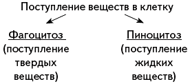
By phagocytosis Large molecules of organic substances, such as proteins, polysaccharides, food particles, and bacteria enter the cell. Phagocytosis occurs with the participation of the plasma membrane. At the point where the surface of the cell comes into contact with a particle of any dense substance, the membrane bends, forms a depression and surrounds the particle, which is immersed inside the cell in a “membrane capsule”. A digestive vacuole is formed, and organic substances entering the cell are digested in it.
Amoebas, ciliates, and leukocytes of animals and humans feed by phagocytosis. Leukocytes absorb bacteria, as well as a variety of solid particles that accidentally enter the body, thus protecting it from pathogenic bacteria. The cell wall of plants, bacteria and blue-green algae prevents phagocytosis, and therefore this route of entry of substances into the cell is not realized in them.
Drops of liquid containing various substances in a dissolved and suspended state also penetrate into the cell through the plasma membrane. This phenomenon was called pinocytosis. The process of fluid absorption is similar to phagocytosis. A drop of liquid is immersed in the cytoplasm in a “membrane package”. Organic substances that enter the cell along with water begin to be digested under the influence of enzymes contained in the cytoplasm. Pinocytosis is widespread in nature and is carried out by cells of all animals.
III. Reinforcing the material learned
What two large groups are all organisms divided into based on the structure of their nucleus?
Which organelles are characteristic only of plant cells?
Which organelles are unique to animal cells?
How does the structure of the cell membrane of plants and animals differ?
What are the two ways substances enter a cell?
What is the significance of phagocytosis for animals?
There are only two types of organisms on Earth: eukaryotes and prokaryotes. They differ greatly in their structure, origin and evolutionary development, which will be discussed in detail below.
In contact with
Signs of a prokaryotic cell
Prokaryotes are also called prenuclear. A prokaryotic cell does not have other organelles that have a membrane membrane (endoplasmic reticulum, Golgi complex).
Also characteristic of them are the following:
- without a shell and does not form bonds with proteins. Information is transmitted and read continuously.
- All prokaryotes are haploid organisms.
- Enzymes are located in a free state (diffusely).
- They have the ability to form spores under unfavorable conditions.
- The presence of plasmids - small extrachromosomal DNA molecules. Their function is the transfer of genetic information, increasing resistance to many aggressive factors.
- The presence of flagella and pili - external protein formations necessary for movement.
- Gas vacuoles are cavities. Due to them, the body is able to move in the water column.
- The cell wall of prokaryotes (namely bacteria) consists of murein.
- The main methods of obtaining energy in prokaryotes are chemo- and photosynthesis.
These include bacteria and archaea. Examples of prokaryotes: spirochetes, proteobacteria, cyanobacteria, crenarchaeotes.
Attention! Despite the fact that prokaryotes lack a nucleus, they have its equivalent - a nucleoid (a circular DNA molecule devoid of shells), and free DNA in the form of plasmids.
Structure of a prokaryotic cell
Bacteria
Representatives of this kingdom are among the most ancient inhabitants of the Earth and have a high survival rate in extreme conditions.
There are gram-positive and gram-negative bacteria. Their main difference lies in the structure of the cell membrane. Gram-positive have a thicker shell, up to 80% consists of a murein base, as well as polysaccharides and polypeptides. When stained with Gram, they give a violet color. Most of these bacteria are pathogens. Gram-negatives have a thinner wall, which is separated from the membrane by the periplasmic space. However, such a shell has increased strength and is much more resistant to the effects of antibodies.
Bacteria play a very important role in nature:
- Cyanobacteria (blue-green algae) help maintain the required level of oxygen in the atmosphere. They form more than half of all O2 on Earth.
- They promote the decomposition of organic remains, thereby taking part in the cycle of all substances, and participate in the formation of soil.
- Nitrogen fixers on legume roots.
- They purify water from waste, for example, from the metallurgical industry.
- They are part of the microflora of living organisms, helping to maximize the absorption of nutrients.
- Used in the food industry for fermentation. This is how cheeses, cottage cheese, alcohol, and dough are produced.
Attention! In addition to their positive significance, bacteria also play a negative role. Many of them cause deadly diseases, such as cholera, typhoid fever, syphilis, and tuberculosis.

Bacteria
Archaea
Previously, they were combined with bacteria into the single kingdom of Drobyanok. However, over time, it became clear that archaea have their own individual path of evolution and are very different from other microorganisms in their biochemical composition and metabolism. There are up to 5 types, the most studied are euryarchaeota and crenarchaeota. The features of archaea are:
- most of them are chemoautotrophs - they synthesize organic substances from carbon dioxide, sugar, ammonia, metal ions and hydrogen;
- play a key role in the nitrogen and carbon cycle;
- participate in digestion in humans and many ruminants;
- have a more stable and durable membrane shell due to the presence of ether bonds in glycerol-ether lipids. This allows archaea to live in highly alkaline or acidic environments, as well as high temperatures;
- the cell wall, unlike bacteria, does not contain peptidoglycan and consists of pseudomurein.

Structure of eukaryotes
Eukaryotes are a superkingdom of organisms whose cells contain a nucleus. Apart from archaea and bacteria, all living things on Earth are eukaryotes (for example, plants, protozoa, animals). Cells can vary greatly in their shape, structure, size and functions. Despite this, they are similar in the basics of life, metabolism, growth, development, ability to irritate and variability.
Eukaryotic cells can be hundreds or thousands of times larger than prokaryotic cells. They include the nucleus and cytoplasm with numerous membranous and non-membranous organelles. Membranous ones include: endoplasmic reticulum, lysosomes, Golgi complex, mitochondria,. Non-membrane: ribosomes, cell center, microtubules, microfilaments.

Structure of eukaryotes
Let's compare eukaryotic cells from different kingdoms.
The superkingdom of eukaryotes includes the following kingdoms:
- protozoa. Heterotrophs, some capable of photosynthesis (algae). They reproduce asexually, sexually and in a simple way into two parts. Most lack a cell wall;
- plants. They are producers; the main method of obtaining energy is photosynthesis. Most plants are immobile and reproduce asexually, sexually and vegetatively. The cell wall is made of cellulose;
- mushrooms. Multicellular. There are lower and higher. They are heterotrophic organisms and cannot move independently. They reproduce asexually, sexually and vegetatively. They store glycogen and have a strong cell wall made of chitin;
- animals. There are 10 types: sponges, worms, arthropods, echinoderms, chordates and others. They are heterotrophic organisms. Capable of independent movement. The main storage substance is glycogen. The cell wall consists of chitin, just like in fungi. The main method of reproduction is sexual.
Table: Comparative characteristics of plant and animal cells
| Structure | plant cell | animal cell |
| Cell wall | Cellulose | Consists of the glycocalyx - a thin layer of proteins, carbohydrates and lipids. |
| Core location | Located closer to the wall | Located in the central part |
| Cell center | Exclusively in lower algae | Present |
| Vacuoles | Contains cell sap | Contractile and digestive. |
| Spare substance | Starch | Glycogen |
| Plastids | Three types: chloroplasts, chromoplasts, leucoplasts | None |
| Nutrition | Autotrophic | Heterotrophic |
Comparison of prokaryotes and eukaryotes

 The structural features of prokaryotic and eukaryotic cells are significant, but one of the main differences concerns the storage of genetic material and the method of obtaining energy.
Prokaryotes and eukaryotes photosynthesize differently. In prokaryotes, this process takes place on membrane outgrowths (chromatophores), arranged in separate stacks. Bacteria do not have a fluorine photosystem, so they do not produce oxygen, unlike blue-green algae, which produce it during photolysis. The sources of hydrogen in prokaryotes are hydrogen sulfide, H2, various organic substances and water. The main pigments are bacteriochlorophyll (in bacteria), chlorophyll and phycobilins (in cyanobacteria).
Of all the eukaryotes, only plants are capable of photosynthesis. They have special formations - chloroplasts, containing membranes arranged in grana or lamellae. The presence of photosystem II allows the release of oxygen into the atmosphere during the process of photolysis of water. The only source of hydrogen molecules is water. The main pigment is chlorophyll, and phycobilins are present only in red algae.
The main differences and characteristic features of prokaryotes and eukaryotes are presented in the table below.
Table: Similarities and differences between prokaryotes and eukaryotes
| Comparison | Prokaryotes | Eukaryotes |
| Appearance time | More than 3.5 billion years | About 1.2 billion years |
| Cell sizes | Up to 10 microns | From 10 to 100 µm |
| Capsule | Eat. Performs a protective function. Associated with the cell wall | Absent |
| Plasma membrane | Eat | Eat |
| Cell wall | Composed of pectin or murein | Yes, except animals |
| Chromosomes | Instead there is circular DNA. Translation and transcription take place in the cytoplasm. | Linear DNA molecules. Translation takes place in the cytoplasm, and transcription in the nucleus. |
| Ribosomes | Small 70S-type. Located in the cytoplasm. | Large 80S-type, can attach to the endoplasmic reticulum and be located in plastids and mitochondria. |
| Membrane-enclosed organoid | None. There are membrane outgrowths - mesosomes | There are: mitochondria, Golgi complex, cell center, ER |
| Cytoplasm | Eat | Eat |
| None | Eat | |
| Vacuoles | Gas (aerosomes) | Eat |
| Chloroplasts | None. Photosynthesis takes place in bacteriochlorophylls | Present only in plants |
| Plasmids | Eat | None |
| Core | Absent | Eat |
| Microfilaments and microtubules. | None | Eat |
| Division methods | Constriction, budding, conjugation | Mitosis, meiosis |
| Interaction or contacts | None | Plasmodesmata, desmosomes or septa |
| Types of cell nutrition | Photoautotrophic, photoheterotrophic, chemoautotrophic, chemoheterotrophic | Phototrophic (in plants) endocytosis and phagocytosis (in others) |
Differences between prokaryotes and eukaryotes
Similarities and differences between prokaryotic and eukaryotic cells
Conclusion
Comparing a prokaryotic and eukaryotic organism is a rather labor-intensive process that requires consideration of many nuances. They have much in common with each other in terms of structure, ongoing processes and properties of all living things. The differences lie in the functions performed, methods of nutrition and internal organization. Anyone interested in this topic can use this information.
The prokaryotic cell is much simpler than animal and plant cells. On the outside, it is covered with a cell wall that performs protective, formative and transport functions. The rigidity of the cell wall is provided by murein. Sometimes the bacterial cell is covered on top with a capsule or mucous layer.
The protoplasm of bacteria, like that of eukaryotes, is surrounded plasma membrane. Saccular, tubular or lamellar invaginations of the membrane contain mesosomes involved in the respiration process, bacteriochlorophyll and other pigments. The genetic material of prokaryotes does not form a nucleus, but is located directly in the cytoplasm. Bacterial DNA is a single circular molecule, each of which consists of thousands and millions of nucleotide pairs. The genome of a bacterial cell is much simpler than that of the cells of more developed creatures: on average, bacterial DNA contains several thousand genes.
Absent in prokaryotic cells endoplasmic reticulum, A ribosomes float freely in the cytoplasm. Prokaryotes do not have mitochondria; Their functions are partially performed by the cell membrane.
Prokaryotes
Bacteria are the smallest of organisms with a cellular structure; their sizes range from 0.1 to 10 microns. A typical printing point can accommodate hundreds of thousands of medium-sized bacteria. Bacteria can only be seen through a microscope, which is why they are called microorganisms or microbes; microorganisms are being studied microbiology . The branch of microbiology that studies bacteria is called bacteriology . This science began Anthony van Leeuwenhoek in the 17th century.
Bacteria - the oldest known organisms. Traces of the vital activity of bacteria and blue-green algae (stromatolites) belong to the Archean and date back to 3.5 billion years old.Due to the possibility of gene exchange between representatives of different species and even genera, it is quite difficult to systematize prokaryotes. A satisfactory taxonomy of prokaryotes has not yet been constructed; all existing systems are artificial and classify bacteria according to some group of characteristics, without taking into account their phylogenetic relationship. Previously, bacteria along with mushrooms And algae included in the subkingdom of lower plants. Currently, bacteria are classified as a separate superkingdom of prokaryotes. The most common classification system is Bergey system, which is based on the structure of the cell wall.
At the end of the 20th century, scientists discovered that cells of a relatively little-studied group of bacteria - archaebacteria – contain rRNA, different in structure from both the r-RNA of prokaryotes and the r-RNA of eukaryotes. The structure of the genetic apparatus of archaebacteria (presence introns and repeating sequences, processing, form ribosomes) brings them closer to eukaryotes; on the other hand, archaebacteria also have typical features of prokaryotes (absence of a nucleus in the cell, presence of flagella, plasmids and gas vacuoles, rRNA size, nitrogen fixation). Finally, archaebacteria differ from all other organisms in the structure of their cell wall, the type of photosynthesis, and some other characteristics. Archaebacteria are capable of existing in extreme conditions (for example, in hot springs at temperatures above 100 ° C, in the ocean depths at a pressure of 260 atm, in saturated salt solutions (30% NaCl)). Some archaebacteria produce methane, others use sulfur compounds to produce energy.
Apparently, archaebacteria are a very ancient group of organisms; "extreme" possibilities indicate the conditions characteristic of the Earth's surface in Archean era. It is believed that archaebacteria are closest to the hypothetical “pro-cells” that subsequently gave rise to all the diversity of life on Earth.
Recently it has become clear that there are three main types rRNA, presented, respectively, the first - in eukaryotic cells, the second - in the cells of real bacteria, as well as in mitochondria And chloroplasts eukaryotes, the third - in archaebacteria. Research in molecular genetics has forced us to take a fresh look at the theory of the origin of eukaryotes. It is now believed that three different branches of prokaryotes evolved simultaneously on ancient Earth - archaebacteria, eubacteria and urkaryotes , characterized by different structures and different methods of obtaining energy. Urkaryotes, which were essentially the nuclear-cytoplasmic component of eukaryotes, were subsequently included as symbionts representatives of various groups of eubacteria, which turned into mitochondria and chloroplasts of future eukaryotic cells.
Thus, the class rank previously allocated for archaebacteria is clearly insufficient. Currently, many researchers tend to divide prokaryotes into two kingdoms: archaebacteria and real bacteria (eubacteria ) or even separate archaebacteria into a separate superkingdom Archaea.
The classification of real bacteria is given in scheme.
IN bacterial cell There is no nucleus, chromosomes are freely located in the cytoplasm. In addition, the bacterial cell lacks membrane organelles: mitochondria, EPS, Golgi apparatus etc. The outside of the cell membrane is covered with a cell wall.
Most bacteria move passively, using water or air currents. Only some of them have movement organelles - flagella . Prokaryotic flagella are very simple in structure and consist of the flagellin protein, which forms a hollow cylinder with a diameter of 10–20 nm. They screw into the medium, propelling the cell forward. Apparently, this is the only structure known in nature that uses the wheel principle.
Based on their shape, bacteria are divided into several groups:
cocci (have a round shape);
bacilli (have a rod-shaped form);
spirilla (have the shape of a spiral);
vibrios (have the shape of a comma).
Based on the method of respiration, bacteria are divided into aerobes (most bacteria) and anaerobes (causative agents of tetanus, botulism, gas gangrene). The former need oxygen to breathe; for the latter, oxygen is useless or even poisonous.
Bacteria reproduce by dividing approximately every 20 minutes (under favorable conditions). DNA is replicated, with each daughter cell receiving its own copy of the parent DNA. Transfer of DNA between non-dividing cells is also possible (through the capture of “naked” DNA, using bacteriophages or by conjugation , when bacteria are connected to each other by copulatory fimbriae), however, an increase in the number of individuals does not occur. Reproduction is prevented by the sun's rays and the products of their own vital activity.The behavior of bacteria is not particularly complex. Chemical receptors record changes in the acidity of the environment and the concentration of various substances: sugars, amino acids, oxygen. Many bacteria respond to changes in temperature or light, and some bacteria can sense the Earth's magnetic field.
Under unfavorable conditions, the bacterium becomes covered with a dense shell, the cytoplasm is dehydrated, and vital activity almost ceases. In this state, bacterial spores can remain in a deep vacuum for hours and tolerate temperatures from –240 °C to +100 °C.
Figure 1 - Image of a prokaryotic cell

Figure 4 - Structure of the flagellum of gram-negative bacteria.
1 - thread; 2 - hook; 3 - basal body; 4 - rod; 5 - L-ring; 6 - P-ring; 7 - S-ring; 8 - M-ring; 9 - CPM; 10 - periplasmic space; 11 - peptidoglycan layer; 12 - outer membrane
The structure of the cells of lower prokaryotes is much simpler (Fig. 1). Moreover, the different structure of the nuclear apparatus is not the only feature that distinguishes a eukaryotic cell from a prokaryotic one.
One of the main structural components of a prokaryotic cell is cell membrane (Fig. 2, 3). The cell membrane of bacteria includes complex molecular complexes consisting of proteins, polysaccharides and fat-like substances. Being rigid, it serves as a kind of skeleton of the cell, giving it a certain shape. The cell membrane of prokaryotes forms a kind of barrier to the passage of solutes from the environment into the cell. Cyanobacteria cells are covered with an elastic pectin shell. In some types of bacteria, a layer of mucus forms on the surface of the cell, forming a kind of case - capsule .
The surface structures of the cells of many bacteria include flagella - organs of movement that are long, very thin filaments, spiral, wavy or curved (Fig. 4).

Figure 3 - Cell wall of gram-negative bacteria (A) and the structure of the lipopolysaccharide molecule (B).
A. Cell wall of gram-negative bacteria 1 - cytoplasmic membrane; 2 - peptidoglycan layer; 3 - periplasmic space; 4 - protein molecules; 5 - phospholipid; 6 - lipopolysaccharide.
B. The structure of the lipopolysaccharide molecule 1 - lipid A; 2 - internal polysaccharide core; 3 - outer polysaccharide core; 4 - O-antigen
The length of the flagella can be many times greater than the length of the body of the bacterium. The number and location of flagella are a characteristic feature of the species. Some types of bacteria have one flagellum ( monotrichs ), in others the flagella are located in bundles at one or both ends of the cell ( lophotrichs ), others have one flagellum at both ends of the cell ( amphitrichs ), in the fourth they cover the entire surface of the cell ( peritrichous ).
The cytoplasmic membrane is closely adjacent to the shell. It has selective permeability - it allows certain substances to enter the cell and remove certain substances from it. Thanks to this ability, the membrane plays the role of an organelle that concentrates nutrients inside the cell and facilitates the removal of waste products to the outside. Inside the cell there is always an increased osmotic pressure compared to the environment. The cytoplasmic membrane ensures its permanence. In addition, it is the site of localization of a number of enzyme systems, in particular redox enzymes associated with energy production (in eukaryotes they are located in mitochondria). Unlike eukaryotic cells, a prokaryotic cell is not divided into compartments. Prokaryotic cells have neither the Golgi complex nor mitochondria, and there is no directional movement of the cytoplasm in them. The phenomena of pinocytosis and phagocytosis are not characteristic of prokaryotes. Of the organelles, only ribosomes are similar to the ribosomes of eukaryotes.
Many bacterial cells have special membrane structures - mesosomes formed as a result of the retraction of the cytoplasmic membrane into the cell. Their role has not yet been fully clarified. There are assumptions about the participation of mesosomes in the most important intracellular processes of cell division, synthesis of cell membrane substances, and in energy metabolism.
