The sympathetic part of the autonomic. Sympathetic nervous system
The sympathetic division is part of the autonomic nervous tissue, which, together with the parasympathetic, ensures the functioning of internal organs, chemical reactions responsible for the vital activity of cells. But you should know that there is a metasympathetic nervous system, a part of the vegetative structure, located on the walls of organs and capable of contracting, contacting directly with the sympathetic and parasympathetic, making adjustments to their activity.
The internal environment of a person is under the direct influence of the sympathetic and parasympathetic nervous system.
The sympathetic division is located in the central nervous system. Spinal nerve tissue carries out its activities under the control of nerve cells located in the brain.
All elements of the sympathetic trunk, located on two sides from the spine, are directly connected with the corresponding organs through the nerve plexuses, while each has its own plexus. At the bottom of the spine, both trunks in a person are combined together.
The sympathetic trunk is usually divided into sections: lumbar, sacral, cervical, thoracic.
The sympathetic nervous system is concentrated near the carotid arteries of the cervical region, in the thoracic - cardiac and pulmonary plexus, in the abdominal cavity solar, mesenteric, aortic, hypogastric.
These plexuses are divided into smaller ones, and from them impulses move to the internal organs.
The transition of excitation from the sympathetic nerve to the corresponding organ occurs under the influence of chemical elements - sympathins, secreted by nerve cells.
They supply the same tissues with nerves, ensuring their interconnection with the central system, often having a directly opposite effect on these organs.
The influence exerted by the sympathetic and parasympathetic nervous systems can be seen from the table below:
 Together they are responsible for cardiovascular organisms, digestive organs, respiratory structures, excretion, smooth muscle function of hollow organs, control metabolic processes, growth, and reproduction.
If one begins to predominate over the other, symptoms of increased excitability of sympathicotonia (the sympathetic part predominates), vagotonia (the parasympathetic predominates) appear.
Sympathicotonia manifests itself in the following symptoms: fever, tachycardia, numbness and tingling in the limbs, increased appetite without the appearance of being deprived of weight, indifference to life, restless dreams, fear of death without a cause, irritability, distraction, decreased salivation, and also sweating, migraine appears.
In humans, when the increased work of the parasympathetic department of the vegetative structure is activated, increased sweating appears, the skin feels cold and wet to the touch, a decrease in heart rate occurs, it becomes less than 60 beats in 1 minute, fainting, salivation and respiratory activity increase. People become indecisive, slow, prone to depression, intolerant.
The parasympathetic nervous system reduces the activity of the heart, has the ability to dilate blood vessels.
Functions
The sympathetic nervous system is a unique design of an element of the autonomic system, which, in the event of a sudden need, is able to increase the body's ability to perform work functions by collecting possible resources.
As a result, the design carries out the work of organs such as the heart, reduces blood vessels, increases the ability of muscles, frequency, strength of the heart rhythm, performance, inhibits the secretory, suction capacity of the gastrointestinal tract.

The SNS maintains such functions as the normal functioning of the internal environment in an active position, being activated during physical effort, stressful situations, illness, blood loss, and regulates metabolism, for example, an increase in sugar, blood clotting, and others.
It is most fully activated during psychological upheavals, by producing adrenaline (enhancing the action of nerve cells) in the adrenal glands, which enables a person to respond faster and more efficiently to sudden factors from the outside world.
Adrenaline is also able to be produced with an increase in load, which also helps a person to better cope with it.
After coping with the situation, a person feels tired, he needs to rest, this is due to the sympathetic system, which has most fully used up the body's capabilities, due to an increase in body functions in a sudden situation.
The parasympathetic nervous system performs the functions of self-regulation, protection of the body, and is responsible for emptying a person.
Self-regulation of the body has a restorative effect, working in a calm state.
The parasympathetic part of the activity of the autonomic nervous system is manifested by a decrease in the strength and frequency of the heart rhythm, stimulation of the gastrointestinal tract with a decrease in glucose in the blood, etc.
Carrying out protective reflexes, it relieves the human body of foreign elements (sneezing, vomiting, and others).
The table below shows how the sympathetic and parasympathetic nervous systems act on the same elements of the body. 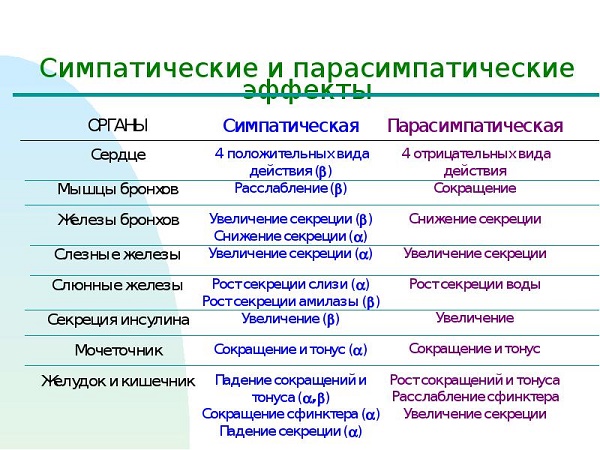
Treatment
If you notice signs of increased sensitivity, you should consult a doctor, as this can cause a disease of an ulcerative, hypertensive nature, neurasthenia.
Only a doctor can prescribe the correct and effective therapy! There is no need to experiment with the body, since the consequences, if the nerves are in a state of excitability, are a rather dangerous manifestation not only for you, but also for people close to you.
When prescribing treatment, it is recommended, if possible, to eliminate factors that excite the sympathetic nervous system, whether it be physical or emotional stress. Without this, no treatment is likely to help, after drinking a course of medicine, you will get sick again.
You need a cozy home environment, sympathy and help from loved ones, fresh air, good emotions.
First of all, you need to make sure that nothing raises your nerves.
The drugs used in the treatment are basically a group of potent drugs, so they should be used carefully only as directed or after consulting a doctor.
The prescribed drugs usually include: tranquilizers (Phenazepam, Relanium and others), antipsychotics (Frenolone, Sonapax), hypnotics, antidepressants, nootropic drugs and, if necessary, cardiac drugs (Korglikon, Digitoxin) ), vascular, sedative, vegetative preparations, a course of vitamins.
It is good when using physiotherapy, including physiotherapy exercises and massage, you can do breathing exercises, swimming. They help to relax the body.
In any case, ignoring the treatment of this disease is categorically not recommended, it is necessary to consult a doctor in a timely manner, to conduct the prescribed course of therapy.
The sympathetic division of the autonomic nervous system is divided into central and peripheral parts. The central part of the sympathetic nervous system includes suprasegmental and segmental centers.
Nadsegmental centers are determined in the cerebral cortex, basal ganglia, limbic system, hypothalamus, reticular formation, cerebellum.
Central segmental centers - in the lateral intermediate nuclei of the lateral horns of the spinal cord, starting from VIII to L II segments.
The peripheral part of the sympathetic nervous system includes vegetative nodes of the I and II order.
Nodes of the first order (paravertebral or paravertebral), there are 20-25 pairs of them, they form a sympathetic trunk.
Nodes of the second order (prevertebral) - celiac, superior mesenteric, aorto-renal.
In the sympathetic (Fig. 18) trunk, there are: cervical, thoracic, lumbar, sacral, coccygeal sections.
The cervical region of the sympathetic trunk is represented by 3 nodes: upper, middle and lower, as well as their internodal branches.
The autonomic nerves that come from the sympathetic trunk are sent to the blood vessels, as well as to the organs of the head and neck.
Sympathetic nerves form plexuses around the carotid and vertebral arteries.
Along the course of the arteries of the same name, these plexuses are sent to the cranial cavity, where they give branches to the vessels, the meninges of the brain and the pituitary gland.
From the carotid plexus, fibers go to the lacrimal, sweat, salivary glands, to the muscle that dilates the pupil, to the ear and submandibular nodes.
The organs of the neck receive sympathetic innervation through the laryngeal-pharyngeal plexus. from all three cervical nodes.
From each of the cervical nodes in the direction of the chest cavity depart the upper, middle and lower cardiac nerves, involved in the formation of the heart plexus.
In the thoracic region of the sympathetic trunk, there are up to 10-12 nodes. From 2 to 5 thoracic nodes depart the thoracic cardiac branches involved in the formation of the cardiac plexus.
Thin sympathetic nerves also depart from the thoracic nodes to the esophagus, lungs, thoracic aorta, forming the esophageal, pulmonary, and thoracic aortic plexus.
From the fifth to the ninth thoracic node departs a large splanchnic nerve, and from 10 and 11 - a small splanchnic nerve. Both nerves contain mainly preganglionic fibers that transit through the sympathetic nodes. Through the diaphragm, these nerves enter the abdominal cavity and end at the neurons of the celiac (solar) plexus.
from the solar plexus postganglionic fibers go to the vessels, stomach, intestines and other organs of the abdominal cavity.
The lumbar sympathetic trunk consists of 3-4 nodes. Branches depart from them to the largest visceral plexus - solar, as well as to the abdominal aortic plexus.
The sacral section of the sympathetic trunk is represented by 3-4 nodes, from which sympathetic nerves depart to the organs of the small pelvis (Fig. 18).

Rice. 18. The structure of the sympathetic division of the autonomic nervous system (S.V. Saveliev, 2008)
parasympathetic nervous system
In the parasympathetic nervous system, there are three foci of exit of fibers from the substance of the brain and spinal cord: mesencephalic, bulbar and sacral.
Parasympathetic fibers are usually components of the spinal or cranial nerves.
Parasympathetic ganglia are located in the immediate vicinity of the innervated organs or in themselves.
The parasympathetic division of the autonomic nervous system is divided into central and peripheral parts. The central part of the parasympathetic nervous system includes suprasegmental and segmental centers.
The central (cranial) section is represented by nuclei III, VII, IX, X pairs of cranial nerves and parasympathetic nuclei of the sacral segments of the spinal cord.
The peripheral section includes: preganglionic fibers in the composition of the cranial nerves and sacral spinal nerves (S 2 -S 4), cranial autonomic nodes, organ plexuses, postganglionic plexuses ending on the working organs.
In the parasympathetic nervous system, the following vegetative nodes are distinguished: ciliary, pterygopalatine, submandibular, sublingual, ear (Fig. 19).
The ciliary node is located in the eye socket. Its size is 1.5-2mm. Preganglionic fibers go to it from the nucleus of Yakubovich (III pair), postganglionic - as part of the ciliary nerves to the muscle that narrows the pupil.
Ear knot, 3-4 mm in diameter, located in the region of the outer base of the skull near the foramen ovale. Preganglionic fibers come to it from the lower salivary nucleus and as part of the glossopharyngeal, and then the tympanic nerves. The latter penetrates into the tympanic cavity, forming the tympanic plexus, from which a small stony nerve is formed, containing preganglionic fibers to the ear node.
Postganglionic fibers (axons of parasympathetic neurons of the ear node) go to the parotid gland as part of the ear-temporal nerve.
Pterygopalatine node (4-5 mm ) located in the pit of the same name.
The preganglionic fibers go to the pterygopalatine ganglion from the superior salivary nucleus, located in the operculum of the bridge, as part of the facial nerve (intermediate). In the canal of the temporal bone, a large stony nerve departs from the facial nerve, it connects with the deep stony nerve (sympathetic), forming the nerve of the pterygoid canal.
After leaving the pyramid of the temporal bone, this nerve enters the pterygopalatine fossa and comes into contact with the neurons of the pterygopalatine ganglion. Postganglionic fibers come from the pterygopalatine ganglion, join the maxillary nerve, innervating the mucous membrane of the nose, palate, and pharynx.
Part of the preganglionic parasympathetic fibers from the superior salivary nucleus, which are not included in the large stony nerve, form a string tympani. The drum string emerges from the pyramid of the temporal bone, joins the lingual nerve and, in its composition, goes to the submandibular and hyoid nodes, from which postganglionic fibers begin to the salivary glands.
Nervus vagus - the main collector of parasympathetic nerve pathways. Preganglionic fibers from the dorsal nucleus of the vagus nerve go along numerous branches of the vagus nerve to the organs of the neck, chest and abdominal cavities. They end on the neurons of the parasympathetic ganglions, periorganic and intraorganic autonomic plexuses.
For parenchymal organs, these nodes are near-organ or intraorgan, for hollow organs - intramural.
The sacral part of the parasympathetic nervous system is represented by pelvic ganglions scattered throughout the visceral plexuses of the pelvis. Preganglionic fibers originate from the sacral parasympathetic nuclei of the II-IV sacral segments of the spinal cord, exit them as part of the anterior roots of the spinal nerves and branch off from them in the form of pelvic splanchnic nerves. They form a plexus around the pelvic organs (rectum and sigmoid colon, uterus, fallopian tubes, vas deferens, prostate, seminal vesicles).
In addition to the sympathetic and parasympathetic nervous systems, the existence of a metasympathetic nervous system has been proven. It is represented by nerve plexuses and microscopic nodes in the walls of hollow organs with motor skills (stomach, small and large intestines, bladder, etc.). These formations differ from parasympathetic mediators (purine bases, peptides, gamma-aminobutyric acid). Nerve cells of metasympathetic nodes are capable of generating nerve impulses without the participation of the central nervous system and sending them to smooth myocytes, causing movement of the organ wall or its part.


Rice. 19. The structure of the parasympathetic division of the autonomic nervous system (S.V. Saveliev, 2008)
Sympathetic department according to its main functions, it is trophic. It provides an increase in oxidative processes, an increase in respiration, an increase in the activity of the heart, i.e. adapts the body to the conditions of intense activity. In this regard, the tone of the sympathetic nervous system prevails during the day.
Parasympathetic department performs a protective role (constriction of the pupil, bronchi, decrease in heart rate, emptying of the abdominal organs), its tone prevails at night ("the kingdom of the vagus").
The sympathetic and parasympathetic divisions also differ in mediators - substances that carry out the transmission of nerve impulses in synapses. The mediator in sympathetic nerve endings is norepinephrine. mediator of parasympathetic nerve endings acetylcholine.
Along with the functional ones, there are a number of morphological differences between the sympathetic and parasympathetic divisions of the autonomic nervous system, namely:
Parasympathetic centers are separated, located in three parts of the brain (mesencephalic, bulbar, sacral), and sympathetic - in one (thoracolumbar region).
The sympathetic nodes include nodes of the I and II order, the parasympathetic nodes are of the III order (final). In this connection, the preganglionic sympathetic fibers are shorter, and the postganglionic ones are longer than the parasympathetic ones.
The parasympathetic division has a more limited area of innervation, innervating only the internal organs. The sympathetic department innervates all organs and tissues.
Sympathetic division of the autonomic nervous system
The sympathetic nervous system consists of a central and a peripheral division.
Central department represented by the intermediate-lateral nuclei of the lateral horns of the spinal cord of the following segments: W 8, D 1-12, P 1-3 (thoracolumbar region).
Peripheral department sympathetic nervous system are:
nodes I and II order;
internodal branches (between the nodes of the sympathetic trunk);
connecting branches are white and gray, associated with the nodes of the sympathetic trunk;
visceral nerves, consisting of sympathetic and sensory fibers and heading to the organs, where they end with nerve endings.
The sympathetic trunk, paired, is located on both sides of the spine in the form of a chain of nodes of the first order. In the longitudinal direction, the nodes are interconnected by internodal branches. In the lumbar and sacral regions, there are also transverse commissures that connect the nodes of the right and left sides. The sympathetic trunk extends from the base of the skull to the coccyx, where the right and left trunks are connected by one unpaired coccygeal node. Topographically, the sympathetic trunk is divided into 4 sections: cervical, thoracic, lumbar and sacral.
The nodes of the sympathetic trunk are connected to the spinal nerves by white and gray connecting branches.
white connecting branches consist of preganglionic sympathetic fibers, which are axons of cells of the intermediate-lateral nuclei of the lateral horns of the spinal cord. They separate from the trunk of the spinal nerve and enter the nearest nodes of the sympathetic trunk, where part of the preganglionic sympathetic fibers is interrupted. The other part passes the node in transit and through the internodal branches reaches the more distant nodes of the sympathetic trunk or passes to the nodes of the second order.
As part of the white connecting branches, sensitive fibers also pass - the dendrites of the cells of the spinal nodes.
White connecting branches go only to the thoracic and upper lumbar nodes. The preganglionic fibers enter the cervical nodes from below from the thoracic nodes of the sympathetic trunk through the internodal branches, and into the lower lumbar and sacral - from the upper lumbar nodes also through the internodal branches.
From all nodes of the sympathetic trunk, part of the postganglionic fibers joins the spinal nerves - gray connecting branches and as part of the spinal nerves, sympathetic fibers are sent to the skin and skeletal muscles in order to ensure the regulation of its trophism and maintain tone - this somatic part sympathetic nervous system.
In addition to the gray connecting branches, visceral branches depart from the nodes of the sympathetic trunk to innervate the internal organs - visceral part sympathetic nervous system. It consists of: postganglionic fibers (processes of cells of the sympathetic trunk), preganglionic fibers that passed through the nodes of the first order without interruption, as well as sensory fibers (processes of cells of the spinal nodes).
cervical The sympathetic trunk often consists of three nodes: top, middle and bottom.
T op e n i n i n g n o d lies in front of the transverse processes of the II-III cervical vertebrae. The following branches depart from it, which often form plexuses along the walls of blood vessels:
Internal carotid plexus(along the walls of the artery of the same name ) . A deep stony nerve departs from the internal carotid plexus to innervate the glands of the mucous membrane of the nasal cavity and palate. A continuation of this plexus is the plexus of the ophthalmic artery (for the innervation of the lacrimal gland and the muscle that dilates the pupil ) and plexuses of cerebral arteries.
External carotid plexus. Due to the secondary plexuses along the branches of the external carotid artery, the salivary glands are innervated.
Laryngo-pharyngeal branches.
Superior cervical cardiac nerve
M e d i n i o n c h i n g n o d e located at the level of the VI cervical vertebra. Branches extend from it:
Branches to the inferior thyroid artery.
Middle cervical cardiac nerve entering the heart plexus.
L i n i n g e n i n g n o d e located at the level of the head of the 1st rib and often merges with the 1st thoracic node, forming the cervicothoracic node (stellate). Branches extend from it:
Inferior cervical cardiac nerve entering the heart plexus.
Branches to the trachea, bronchi, esophagus, which, together with the branches of the vagus nerve, form plexuses.
Thoracic sympathetic trunk consists of 10-12 nodes. The following branches depart from them:
Visceral branches depart from the upper 5-6 nodes for innervation of the organs of the chest cavity, namely:
Thoracic cardiac nerves.
Branches to the aorta that form the thoracic aortic plexus.
Branches to the trachea and bronchi participating together with the branches of the vagus nerve in the formation of the pulmonary plexus.
Branches to the esophagus.
5. Branches depart from the V-IX thoracic nodes, forming great splanchnic nerve.
6. From X-XI chest nodes - small splanchnic nerve.
The splanchnic nerves pass into the abdominal cavity and enter the celiac plexus.
Lumbar sympathetic trunk consists of 4-5 nodes.
The visceral nerves depart from them - splanchnic lumbar nerves. The upper ones enter the celiac plexus, the lower ones enter the aortic and inferior mesenteric plexuses.
sacral department The sympathetic trunk is represented, as a rule, by four sacral nodes and one unpaired coccygeal node.
Depart from them splanchnic sacral nerves entering the upper and lower hypogastric plexuses.
PREVERTEBRAL NODES AND VEGETATIVE PLEXES
The prevertebral nodes (nodes of the second order) are part of the autonomic plexuses and are located in front of the spinal column. On the motor neurons of these nodes, preganglionic fibers end, which passed without interruption the nodes of the sympathetic trunk.
Vegetative plexuses are located mainly around the blood vessels, or directly near the organs. Topographically, the vegetative plexuses of the head and neck, chest, abdominal and pelvic cavities are distinguished. In the head and neck region, sympathetic plexuses are located mainly around the vessels.
In the chest cavity, sympathetic plexuses are located around the descending aorta, in the region of the heart, at the gates of the lung and along the bronchi, around the esophagus.
The most significant in the chest cavity is cardiac plexus.
In the abdominal cavity, sympathetic plexuses surround the abdominal aorta and its branches. Among them, the largest plexus is distinguished - the celiac ("brain of the abdominal cavity").
celiac plexus(solar) surrounds the origin of the celiac trunk and superior mesenteric artery. From above, the plexus is limited by the diaphragm, on the sides by the adrenal glands, from below it reaches the renal arteries. The following are involved in the formation of this plexus: nodes(nodes of the second order):
Right and left celiac nodes semilunar shape.
Unpaired superior mesenteric node.
Right and left aorto-renal nodes located at the site of origin of the renal arteries from the aorta.
Preganglionic sympathetic fibers come to these nodes, which switch here, as well as postganglionic sympathetic and parasympathetic and sensory fibers passing through them in transit.
In the formation of the celiac plexus are involved nerves:
Great and small splanchnic nerves, extending from the thoracic nodes of the sympathetic trunk.
Lumbar splanchnic nerves - from the upper lumbar nodes of the sympathetic trunk.
Branches of the phrenic nerve.
Branches of the vagus nerve, consisting mainly of preganglionic parasympathetic and sensory fibers.
The continuation of the celiac plexus are secondary paired and unpaired plexuses along the walls of the visceral and parietal branches of the abdominal aorta.
The second most important in the innervation of the abdominal organs is abdominal aortic plexus, which is a continuation of the celiac plexus.
From the aortic plexus inferior mesenteric plexus, braiding the artery of the same name and its branches. Here is located
pretty big knot. The fibers of the inferior mesenteric plexus reach the sigmoid, descending and part of the transverse colon. The continuation of this plexus into the pelvic cavity is the superior rectal plexus, which accompanies the artery of the same name.
The continuation of the abdominal aortic plexus downwards are the plexuses of the iliac arteries and arteries of the lower limb, as well as unpaired superior hypogastric plexus, which at the level of the cape is divided into the right and left hypogastric nerves, which form the lower hypogastric plexus in the pelvic cavity.
In education inferior hypogastric plexus vegetative nodes of the II order (sympathetic) and III order (periorgan, parasympathetic), as well as nerves and plexuses are involved:
1. splanchnic sacral nerves- from the sacral part of the sympathetic trunk.
2.Branches of the inferior mesenteric plexus.
3. splanchnic pelvic nerves, consisting of preganglionic parasympathetic fibers - processes of cells of the intermediate-lateral nuclei of the spinal cord of the sacral region and sensory fibers from the sacral spinal nodes.
PARASYMPATIC DEPARTMENT OF THE AUTONOMIC NERVOUS SYSTEM
The parasympathetic nervous system consists of a central and a peripheral division.
Central department includes nuclei located in the brain stem, namely in the midbrain (mesencephalic region), the pons and medulla oblongata (bulbar region), as well as in the spinal cord (sacral region).
Peripheral department presented:
preganglionic parasympathetic fibers passing in the III, VII, IX, X pairs of cranial nerves, as well as in the composition of the splanchnic pelvic nerves.
nodes of the III order;
postganglionic fibers that terminate in smooth muscle and glandular cells.
Parasympathetic part of the oculomotor nerve (IIIpair) represented by an accessory nucleus located in the midbrain. Preganglionic fibers are part of the oculomotor nerve, approach the ciliary ganglion, located in the orbit, they are interrupted there and the postganglionic fibers penetrate the eyeball to the muscle that narrows the pupil, providing a pupil response to light, as well as to the ciliary muscle, which affects the change in the curvature of the lens.
Parasympathetic part of the interfacial nerve (VIIpair) represented by the upper salivary nucleus, which is located in the bridge. The axons of the cells of this nucleus pass as part of the intermediate nerve, which joins the facial nerve. In the facial canal, parasympathetic fibers are separated from the facial nerve in two portions. One portion is isolated in the form of a large stony nerve, the other - in the form of a drum string.
Greater stony nerve connects with the deep stony nerve (sympathetic) and forms the nerve of the pterygoid canal. As part of this nerve, the preganglionic parasympathetic fibers reach the pterygopalatine node and end on its cells.
Postganglionic fibers from the node innervate the glands of the mucous membrane of the palate and nose. A smaller part of the postganglionic fibers reaches the lacrimal gland.
Another portion of preganglionic parasympathetic fibers in the composition drum string joins the lingual nerve (from the III branch of the trigeminal nerve) and, as part of its branch, approaches the submandibular node, where they are interrupted. The axons of the ganglion cells (postganglionic fibers) innervate the submandibular and sublingual salivary glands.
Parasympathetic part of the glossopharyngeal nerve (IXpair) represented by the lower salivary nucleus located in the medulla oblongata. Preganglionic fibers exit as part of the glossopharyngeal nerve, and then its branches - tympanic nerve, which penetrates the tympanic cavity and forms the tympanic plexus, which innervates the glands of the mucous membrane of the tympanic cavity. Its continuation is small stony nerve, which emerges from the cranial cavity and enters the ear canal where the preganglionic fibers are interrupted. Postganglionic fibers are sent to the parotid salivary gland.
Parasympathetic part of the vagus nerve (Xpair) represented by the dorsal nucleus. Preganglionic fibers from this nucleus as part of the vagus nerve and its branches reach the parasympathetic nodes (III
order), which are located in the wall of internal organs (esophageal, pulmonary, cardiac, gastric, intestinal, pancreatic, etc. or at the gates of organs (liver, kidneys, spleen). The vagus nerve innervates the smooth muscles and glands of the internal organs of the neck, thoracic and abdominal cavity to the sigmoid colon.
The sacral division of the parasympathetic part of the autonomic nervous system represented by the intermediate-lateral nuclei II-IV of the sacral segments of the spinal cord. Their axons (preganglionic fibers) leave the spinal cord as part of the anterior roots, and then the anterior branches of the spinal nerves. They are separated from them in the form pelvic splanchnic nerves and enter the lower hypogastric plexus for innervation of the pelvic organs. Part of the preganglionic fibers has an ascending direction for the innervation of the sigmoid colon.
Sympathetic nervous system (from the Greek sympathes - sensitive, susceptible to influence)
part of the autonomic nervous system of vertebrates and humans, consisting of sympathetic centers, right and left border sympathetic trunks located along the spine, ganglia (nodes) and nerve branches connecting the ganglia to each other, with the spinal cord and with effectors (See Effectors). Border sympathetic trunk - a chain of ganglia connected by internodal commissures; lies (right or left) on the vertebral bodies; each ganglion is also connected to one of the spinal nerves (See Spinal Nerves). S.'s fibers of n. With. innervates all organs and tissues of the body without exception. S.'s centers of n. With. located in the thoracic and lumbar segments of the spinal cord. The sympathetic nuclei that form the lateral horns of the gray matter of the spinal cord are present only in 15-16 segments (from the last cervical or 1st thoracic to the 3rd lumbar segment). These nuclei are considered as a working apparatus, subordinate to suprasegmental formations, which are localized in the medulla oblongata (See. Medulla oblongata) and the Hypothalamus e, controlled by the cerebral cortex. A special place in the physiology of S. n. With. and coordination of the processes controlled by it is occupied by the cerebellum. S. n. With. - efferent system that conducts impulses to various internal organs. Most authors deny the existence of their own afferent fibers in S. n. With. However, a number of works provide evidence of their existence. In an abdominal cavity S.'s fibers of n. With. pass in the composition of the large, small and lumbar celiac nerves. Afferent nerves that conduct impulses from internal organs are represented in the cerebral cortex and subcortical ganglia. Sympathetic nerve impulses from the central nervous system to the executive organs follow a two-neuron path. The first neuron is located in the lateral horns of the spinal cord. The axons (processes) of the first neuron (preganglionic fibers) exit the spinal cord through the ventral roots of the corresponding segments and enter the mixed spinal nerves, from which, as part of the white connecting branches, they reach the corresponding node of the border sympathetic trunk, where some of the fibers end in synapses (See Synapses) on effector neurons; at the same time, each preganglionic fiber is in contact with a large number of nerve cells (up to 30). Another part of the preganglionic fibers passes through the nodes of the border sympathetic trunk, without ending on its cells, and together with other fibers forms a number of nerves: large and small celiac, lumbar celiac, entering the prevertebral sympathetic nodes. Some preganglionic fibers pass without interruption through these nodes, reaching the working organ, in the nerve nodes of the walls of which they make a break. The second effector neuron is located in the peripheral sympathetic nodes, its processes (postganglionic fibers) enter the innervated organ. The second neuron is located in the paravertebral (paravertebral) ganglia or in the prevertebral (prevertebral) ganglia (solar plexus nodes, inferior mesenteric node, and others located at a great distance from the central nervous system, near the internal organs). Postganglionic fibers enter the spinal nerve through the gray connecting branches, in its composition they reach the innervated organ. Consequently, the interruption of each efferent sympathetic pathway in the arc that closes in the spinal cord occurs only once: either in the node of the borderline sympathetic trunk, or in nodes remote from the spine. Along with the sympathetic arc, which closes in the spinal cord, there are also short sympathetic reflex arcs, which close in the peripheral sympathetic ganglia (solar plexus, caudal mesenteric). The rate of conduction of excitation in sympathetic pre- and especially postganglionic fibers is many times less than in somatic, i.e. bodily, and is about 1-3 m/s. To cause effects in sympathetic fibers, a much greater force of irritation is required. Arising in S. n. With. excitation, as a rule, involves a large number of neurons, so the effects of irritation are not localized in any particular organ, but cover wide areas. The reactions that follow in response to irritation of sympathetic fibers are characterized by a relatively slow and prolonged character, as well as a slow, prolonged attenuation of ongoing processes. A number of substances (ganglioblockers, ergot preparations) suppress the effects of S.'s excitation of n. With. Some chemicals have the same effect on organs and tissues as irritation of sympathetic nerves. This is due to the fact that when the sympathetic nerves are irritated, substances of a similar action are released by the terminal formations of postganglionic sympathetic fibers (see Mediators). At the endings of all preganglionic fibers, as well as postganglionic, innervating sweat glands, the mediator Acetylcholine is formed, at the endings of postganglionic fibers (with the exception of innervating sweat glands) - Norepinephrine. The influence of the sympathetic and parasympathetic nervous systems on the activity of an organ is often opposite. When sympathetic fibers innervating various organs are irritated, typical effects occur: acceleration and intensification of heart contractions, pupil dilation and blurred lacrimation, contraction of smooth muscle fibers (pilomotors) that raise hair, secretion of sweat glands, poor secretion of thick saliva and gastric juice, inhibition of contractions and weakening of the tone of the smooth muscles of the stomach and intestines (excluding the area of the ileocecal sphincter), relaxation of the muscles of the bladder and inhibition of contractions of the obturator sphincter, expansion of the coronary vessels of the heart, narrowing of the small arteries of the abdominal organs and skin, small arteries of the lungs and brain, changes in the excitability of receptors, and also various parts of the central nervous system, an increase in the strength of contractions of a tired skeletal muscle, an increase in its excitability and a change in mechanical properties. Neurons S. n. N of pages, influencing the executive organs, are in a state of constant tonic excitation as a result of the interaction of unconditioned and conditioned reflexes carried out by the higher parts of the central nervous system. Tonic impulses S. n. With. are extremely important for maintaining the constancy of the internal environment of the body (homeostasis a). Through sympathetic fibers and centers, a reflex relationship is provided between all internal organs. The reflexes involving S.'s action of n. N of page, can arise at irritation of both visceral, and somatic nerves. So, with viscero-visceral reflexes, excitation arises and ends in the internal organs (irritation of the peritoneum causes a slowdown in cardiac activity). With visceromotor reflexes, excitation from the internal organs passes to the skeletal muscles (irritation of the peritoneum increases the tone of the abdominal muscles). Animals with completely removed borderline sympathetic trunks and ganglia (desympathized) outwardly differ little from normal ones, but under certain loads (muscular work, cooling, etc.) they are less enduring. This indicates that S. n. N of page, rendering on a functional condition of fabrics the regulating action, adapts (adapts) them to performance of functions in the given conditions (see. Adaptive and trophic function ). S. n. With. stimulates mainly the processes associated with the release of energy in the body, with vigorous activity. Physiological displays of emotions (See. Emotions) are connected mainly with S.'s excitation of n. With. A. D. Nozdrachev. Great Soviet Encyclopedia. - M.: Soviet Encyclopedia.
1969-1978
.
See what the "Sympathetic nervous system" is in other dictionaries:
SYMPATIC NERVOUS SYSTEM- see Autonomic nervous system. Big psychological dictionary. Moscow: Prime EUROZNAK. Ed. B.G. Meshcheryakova, acad. V.P. Zinchenko. 2003 ... Great Psychological Encyclopedia
SYMPATIC NERVOUS SYSTEM, one of two parts of the AUTONOMOUS NERVOUS SYSTEM, its second part is the PARASYMPATIC NERVOUS SYSTEM. Both systems are involved in the work of SMOOTH MUSCLES (involuntarily contracting). The sympathetic nervous system... ... Scientific and technical encyclopedic dictionary
Big Encyclopedic Dictionary
A department of the autonomic nervous system that regulates the activity of the heart, lungs, intestines, gonads, and other organs that do not depend (or depend to a very small extent) on the will of a person. It used to be seen as a place of sympathy and love… Philosophical Encyclopedia
Anatomy of the innervation of the autonomic nervous system. Systems: sympathetic (in red) and parasympathetic (in blue) Sympathetic nervous system (from Greek ... Wikipedia
In invertebrates, little has been studied so far. In higher worms, ganglion cells and nerve fibers are found in various parts of the intestine, probably of sympathetic significance, but their relationship to the central system has not been elucidated. At the highest ... ... Encyclopedic Dictionary F.A. Brockhaus and I.A. Efron
Part of the autonomic nervous system, including nerve cells of the thoracic and upper lumbar spinal cord and nerve cells of the border sympathetic trunk, solar plexus, mesenteric nodes, the processes of which innervate all organs ... encyclopedic Dictionary
Sympathetic part of the autonomic nervous system
The central division of the sympathetic part of the autonomic nervous system consists of numerous multipolar cells, neurocytes multipolares, located in the lateral intermediate (gray) substance of the spinal cord, extending from the 8th cervical to the 2nd-3rd lumbar segments (see Fig.,) and forming together the sympathetic center.
The peripheral section of the sympathetic part of the autonomic [autonomous] nervous system consists of the right and left sympathetic trunks and nerves extending from these trunks, as well as plexuses formed by nerves and nodes that lie outside or inside the organs.
Each sympathetic trunk, truncus sympathicus (Fig.,; see Fig.,), is formed by nodes of the sympathetic trunk, ganglia trunci sympathici, which are interconnected by internodal branches, rr. interganglionares.
The right and left sympathetic trunks lie on the corresponding sides of the spinal column from the level of the base of the skull to the top of the coccyx, where, ending, they are connected unpaired knot, ganglion impar.
The nodes of the sympathetic trunk are a combination of a different number of nerve cells ( neurocytes gangliae autonomicae), have a different size and are predominantly spindle-shaped. Along the sympathetic trunk, there are single intrastem nerve cells or small intermediate nodes, ganglia intermedia, most often on the cervical and lumbar connecting branches. The number of nodes of the sympathetic trunk, with the exception of the cervical region, basically corresponds to the number of spinal nerves.
Distinguish 3 cervical ganglion, ganglia cervicalia, 10–12 thoracic nodes, ganglia thoracica, 4–5 lumbar nodes, ganglia lumbalia, 4 sacral node, ganglia sacralia, and one unpaired knot, ganglion impar. The latter lies on the anterior surface of the coccyx, uniting both sympathetic trunks.
Two kinds of branches depart from each node of the sympathetic trunk: connecting branches and branches that go to the autonomic (autonomous) plexuses (see Fig.,).
In turn, two types of connecting branches are distinguished: white connecting branches and gray connecting branches.
Each white connecting branch, r. communicans albus, is a set prenodular nerve fibers, neurofibrae preganglionares connecting the spinal cord with the sympathetic ganglion. It contains myelinated nerve fibers (processes of nerve cells of the lateral horns of the spinal cord) that pass through the anterior root to the cells of the node of the sympathetic trunk or, having passed it, to the cells of the node of the autonomic plexus. These fibers, because they terminate at the ganglion cells, are called prenodular nerve fibers.
The lateral horns are located only in the range from the 8th cervical to the 2nd-3rd lumbar segments of the spinal cord. Therefore, the prenodal fibers for those nodes of the sympathetic trunks that are located above and below the level of the indicated segments, i.e., for the neck, lower parts of the lumbar and the entire sacral region, follow in the internodal branches of the sympathetic trunk.
Each gray connecting branch, r. communicans griseus, is a branch that connects the sympathetic trunk with the spinal nerve. It contains non-myelinated nerve fibers, neurofibrae nonmyelinatae(processes of cells of the node of the sympathetic trunk), which are sent to the spinal nerve and are part of its fibers, reaching the glands and blood vessels of the soma.
These fibers, since they start from the cells of the nodes, are called post-nodular nerve fibers, neurofibrae postganglionares.
The branches leading to the autonomic plexuses are different in the nodes of the cervical, thoracic, lumbar and sacral sections of the sympathetic trunk.
