Damage to the oral mucosa with pemphigus. Damage to the oral mucosa: key factors in making the diagnosis of pemphigus vulgaris
Pemphigus is a very serious dermatological disease that can lead to serious consequences and complications. The development of pemphigus in newborns is especially dangerous; in this case, even death cannot be ruled out.
People of all ages are susceptible to this disease, however, in most cases it is observed in adults 40-60 years old. Children suffer from this disease extremely rarely. Since the disease is an autoimmune pathology, it is difficult to treat and is chronic.
The disease is expressed by the appearance of blisters filled with exudate on the skin and mucous membrane. New growths merge with each other and quickly spread throughout the body.
Causes of the disease
Specific causes of the occurrence of vesicles have not been identified, but provoking factors are identified that influence the development of the disease, namely:

An autoimmune cause of the disease is also being considered. In children, the cause of the disease can be viruses and bacteria, the main one being Staphylococcus aureus.
You can become infected through airborne droplets or by using the patient’s belongings.
Types of pemphigus
First of all, the pathological process of the disease is classified into:
- true pemphigus (acantholic);
- benign pemphigus (neacantholic).
True pemphigus, depending on the manifestation of the pathological process, is classified into the following subtypes:

Symptoms of the disease
Regardless of the type, the disease has similar symptoms. The disease develops very rapidly, so at the first manifestations you should immediately consult a doctor.
Pemphigus is characterized by a wavy course, but a rapid spread of foci and a progressive course. There are remissions of varying degrees and durations.
Pemphigus in the mouth does not appear in the form of ulcers immediately, several days after infection. The first symptoms may be malaise, fever and redness of the throat. In such cases, the disease is classified as a simple cold, and treatment is prescribed incorrectly.
Only the manifestation of ulcers and blisters in the oral cavity indicates the development of a pathological process. In this case, the patient experiences bad breath.
Specifics of pemphigus of the oral mucosa
According to medical dental statistics, the oral mucosa is most often affected by pemphigus vulgaris. It is this type of disease that first affects the mucous membrane of the mouth and larynx, and then spreads to the face and body.
 The spread of blisters on the skin may begin several months after the first blisters appear in the mouth, or they may appear immediately, after one or two days.
With a strong immune system, timely and competent treatment, the spread of blisters over the skin may not begin.
Erosion that appears on the mucous membrane does not bleed, but due to constant friction and contact with food, it quickly opens.
In this case, when examining the oral cavity, it is rarely possible to detect the presence of blisters. In place of the blisters, oval or round ulcers remain, which take a long time to heal. As a rule, erosions of the oral mucosa heal without scars.
However, without proper treatment, erosions do not heal, but on the contrary, they increase and merge into large affected areas. Most often, ulcers are localized on the inside of the cheeks, the lower surface of the tongue and the palate. A white coating appears on the ulcers, which can be easily removed by the dentist using a medical spatula.
The patient's pain is quite strong, especially intense when talking and eating. Ulcers become infected quite quickly. If the oral cavity is not sanitized, the likelihood of infection of erosions and the addition of additional fungal and viral infections increases. A dental examination is required.

With oral pemphigus, the patient often suffers from stomatitis. In addition to the mouth, the disease can spread to other organs covered with mucous membrane: the larynx, digestive tract and others.
Diagnosis and severity of the disease
Since at the initial stage of the development of the disease the symptoms are similar to many other dermatological diseases, to accurately determine the diagnosis it is necessary to conduct a comprehensive examination, namely:
- examination of the patient - the nature of the lesion and location;
- studying the patient’s medical history and the presence of concomitant diseases;
- performing Nikolsky's test, which will distinguish pemphigus from similar pathological processes;
- carrying out histological and cytological studies;
- the use of immunological methods that confirm or refute the autoimmune nature of the disease.
As the blisters develop and spread, the disease is characterized by varying degrees of severity: 
- Lightweight. The pathological process appears on the mucous membranes or skin gradually with a minimum number of foci.
- Average. With a moderate course of the disease, an increase in the spread of blisters on the skin and mucous membrane is observed.
- Heavy. Most of the skin and oral cavity are affected. The ulcers merge into large lesions. Complications and pathologies begin.
What are the methods of treatment and prevention?
Without proper competent treatment, the outcome of the disease is unfavorable. Almost two years later, if the disease is advanced, death can occur.
Self-medication for pemphigus is strictly prohibited.
The treatment process is quite complex and lengthy. Due to the rapid development of the disease, the functioning of many organs is disrupted, which can lead to the appearance of additional diseases.
Drug treatment
While taking any drug, you should see a doctor and monitor your blood pressure and sugar levels by regularly taking laboratory blood and urine tests.
Drug therapy includes taking the following drugs:
- antibiotics - used for purulent infectious processes, contribute to the death of the pathogen;
- corticosteroid drugs - help reduce the manifestations and speed of spread of blisters over the skin;
- cytostatics - used to reduce the possibility of complications.

The dose of the drug and duration of administration are selected by the doctor based on the severity of the disease. After a course of treatment, the patient may be prescribed corticosteroid drugs to prevent relapse. Many medications have side effects.
Local treatment
Local therapy plays only a supporting role in drug treatment. During the treatment of pemphigus of the oral mucosa, it is advisable to follow the following recommendations as local therapy:

Local treatment and folk remedies will only help temporarily reduce the symptoms of the disease and reduce pain, but will not cure the disease.
Prevention methods
Patients who have been diagnosed with this disease should be registered with a dermatologist. The disease requires constant medical supervision and compliance with preventive measures:
- observe a gentle work schedule and moderate physical activity;
- to refuse from bad habits;
- avoid exposure to sunlight on the skin;
- avoid stressful situations;
- monitor the cleanliness of bed linen and underwear.
In the case of an advanced form of pemphigus and the absence of timely and competent treatment, the prognosis of the disease is unfavorable. Various infections may occur, as a result of which the patient may die from complications.
Possible complications and consequences
In the case of a complicated course of the disease, a large area of the skin or mucous membrane is affected, ulcerative erosions are formed, which become covered with crusts during the healing process.

The most dangerous complication is infection of damaged skin or the addition of secondary infections.
Untimely treatment can lead to infection and the development of deadly diseases.
For children, the complications and consequences of pemphigus pose a great threat to their further development. The disease has a detrimental effect on the child’s immune system and his health.
The most severe consequences arise due to the lack of timely diagnosis and necessary treatment, namely:
- the patient’s general condition deteriorates significantly and the protective immune system decreases;
- bubbles of varying densities spread quickly, causing the patient severe pain and inconvenience;
- dense crusts of a large area are formed, resembling lichen in appearance;
- Serious secondary diseases develop, for example, heart failure, cerebral edema and many others.
Any form of pemphigus is a dangerous and serious pathology that should not be neglected. The disease develops very quickly, so any delay is fraught with serious consequences and complications that can lead to death. At the first symptoms of the disease, you need to urgently undergo examination and monitor the course of the disease.
Pemphigus is a chronic autoimmune disease characterized by the appearance of a special type of blisters on the surface of previously healthy skin and mucous membranes. Among the types of pemphigus can be distinguished: vulgar, vegetative, erythematous and foliate.Pemphigus can be diagnosed if acantholytic cells are detected, which are detected in a smear taken or as part of blisters in the epidermis itself (during histological examination). To treat pemphigus, glucocorticosteroids are first used (a whole course of treatment is prescribed). The latter always goes well with extracorporeal hemocorrection (plasmophoresis, cryoapherosis, hemosorption).
What it is?
Pemphigus is a serious disease that affects the human skin. As a result of its progression, pathological blisters are formed on the skin and mucous membranes, filled with exudate inside. This process begins due to the stratification of the epithelium. Pathological foci can merge and tend to grow rapidly.
Causes
The reasons for the development of pemphigus have not yet been fully studied. One of the main causes of pemphigus is a violation of autoimmune processes, thereby the cells become antibodies to the immune system.
Violation of cell structure is subject to the influence of external factors, as well as aggressive environmental conditions. As a result, the communication between cells is disrupted, which leads to the formation of bubbles. The incidence rate in people with a hereditary predisposition is much higher.
Mechanism of bubble formation
Human skin can be figuratively described as a water-spring “mattress” covered with a kind of “wall”. The “mattress” does not participate in the formation of bubbles - only the top layer, the epidermis, suffers.
The epidermal layer consists of 10-20 cell layers, which look like bricks under a microscope. The “bricks” of the second layer of the epidermis are connected to each other by peculiar “bridges”. On top of the “wall” there are layers of cells that are no longer quite similar to cells, reminiscent of applied cream. These are scales, corneocytes, necessary for protection from mechanical, chemical and physical damage.
If, under the influence of internal or external causes, antibodies are formed that destroy the “bridges” - desmosomes between the cells of the basal layer (this is called acantholysis and can be seen under a microscope), this is true pemphigus. If tissue fluid penetrates between the basal and upper layers of the epidermis without destroying the “bridges,” it is pemphigoid. Viral pemphigus also occurs without destruction of desmosomes.
Classification
Types of non-acantholytic pemphigus:
- Non-acantholytic pemphigus is benign. Pathological elements are formed exclusively in the human oral cavity. Upon examination, inflammation of the mucous membrane, as well as its slight ulceration, can be detected.
- Bullous form of non-acantholytic pemphigus. This is a benign disease that develops in both adults and children. Blisters form on the skin, but there are no signs of acantholysis. These pathological elements can spontaneously disappear without scarring.
- Cicatricial non-acantholytic pemphigus. This pemphigoid is called pemphigus of the eye in the medical literature. Most often it is diagnosed in women who have crossed the 45-year age limit. A characteristic symptom is damage to the visual apparatus, skin and oral mucosa.
Classification of true pemphigus:
- Erythematous form. This pathological process combines several diseases. Its symptoms are similar to seborrheic dermatitis, an erythematous variant of systemic lupus, as well as true pemphigus. Erythematous pemphigus in adults and children is very difficult to treat. It is worth noting that the disease is diagnosed not only in people, but also in some animals. A characteristic symptom is the appearance of red spots on the skin of the body and face, covered with crusts on top. Simultaneously with this symptom, seborrheic manifestations appear on the scalp.
- Pemphigus vulgare. This type of pathology is diagnosed in patients more often. Blisters form on the skin, but there are no signs of inflammation. If pemphigus is not treated on time, pathological elements can spread throughout the entire skin. It is worth noting that they can merge and form large lesions.
- Pemphigus foliaceus. This form received its name due to the characteristics of the pathological elements. Blisters form on the human skin, which practically do not rise above the epidermis (not tense). Crusts form on top of them, which tend to layer on top of each other. The effect of sheet material folded in stacks is created.
- Brazilian pemphigus. There are no restrictions regarding gender and age. Cases of its development have been recorded in both young children and elderly people aged 70 to 80 years. It is also possible that it may progress in middle-aged people. It is worth noting that this variety is endemic and is therefore found only in Brazil.
View photos
[collapse]
Symptoms
Considering that experts have identified several different types of this pathology, the symptoms of each of them will be very specific. Of course, there are a number of general trends and signs inherent in all types of the disease. This may include, for example, the wave-like course of the pathological process.
Periods of exacerbation alternate with the transition of pemphigus to a calmer stage, when the main symptoms subside or completely disappear. An important factor for the patient will be the fact that in the absence of timely diagnosis and prescription of an effective course of treatment, there is a high risk of developing severe conditions aggravated by concomitant diseases.
- The presence of crusts, ranging from pale pink soft to red dense;
- There is a deterioration in the general condition;
- Decreased immune response of the body;
- Formation of bubbles of varying densities;
- Also, in severe cases, separation of the layers of the epidermis is noted, and it can occur both in the lesion and away from it.
- Damage and ulcers of the mucous membrane of the mouth, nasopharynx or genitals;
- Pain when performing the act of swallowing or when eating;
- Bad breath, indicating damage to the mucous membranes;
- Hypersalivation or, in other words, increased salivation;
- In the seborrheic form, characteristic yellowish or brown-brown crusts appear on the scalp.
- Bubbles vary in appearance, ranging from flat to thin-walled, which burst with a slight touch. In their place, erosions and, subsequently, crusts form.
- In severe cases, an eroded surface of the skin may form in place of the blisters. Their feature is a tendency towards peripheral growth. Over time, such erosions occupy a large surface of the skin, causing pain and inconvenience to the patient.
- In children, manifestations of pemphigus are localized over the entire surface of the skin, including the limbs.
Experts say that with this disease, both a pure form of the pathological process and mixed forms that smoothly transform into one another can be observed. Therefore, the symptoms and signs of pemphigus in a given person may vary and indicate the presence of several types of disease.
What does pemphigus look like: photo
The photo below shows how the disease manifests itself in humans.
Click to view
[collapse]
Diagnostics
Experts say that a correct diagnosis can be made based on a comprehensive examination of the patient, which includes several important stages:
- Examination of the patient for the presence of a clinical picture. At this point, the doctor establishes the nature of the lesions, their localization, the degree of development of the disease, etc.
- Cytological analysis necessary to establish the presence of acantholic cells in smears of biomaterial.
- Carrying out the Nikolsky test, which allows to differentiate pemphigus from similar pathological processes.
- Method of direct immunofluorescence. This study allows us to detect the presence of immunoglobulin in the intercellular substance of the epidermis.
- A histological study, which is based on a technique for detecting crevices and other damage within the epidermis.
Only the totality of all the results makes it possible to make an accurate diagnosis and prescribe an effective course of treatment, leading to the patient’s recovery.
Treatment of viral pemphigus
Treatment of viral pemphigus involves the use of the following systemic drugs:
- cytostatics stop the division of immune cells: Sandimmune, Azathioprine, Methotrexate;
- antiviral: Viferon, Laferon, Cycloferon;
- glucocorticosteroids: Dexamethasone, Prednisolone;
- antipyretics: Ibuprofen, Paracetamol, Nimesil, Mefenamic acid;
- antihistamines relieve itching: Cetrin, Diazolin, Fenistil.
For external treatment of affected skin areas, the following may be prescribed:
- antimicrobial local anesthetics for irrigating the oral cavity if viral pemphigus has affected the child’s mucous membranes: Forteza, Orasept;
- antiseptics: Chlorhexidine, Methylene blue, Miramistin;
- combination preparations of antiseptics and anesthetics: Oflokain, pharmaceutical talkers;
- antipruritic lotions made from nettle juice, aloe, and walnut oil.
Since children with this diagnosis are usually treated in a hospital setting, to enhance the therapeutic course, therapeutic procedures can be carried out aimed at clearing the blood of antibodies:
- plasmapheresis - replacement of the liquid part of the blood with similar solutions without microbes, immune complexes and antibodies;
- hemosorption using a carbon filter.
Only a doctor can tell how to treat viral pemphigus, because in each individual case it can acquire some special features. As for other forms of pemphigus, the therapeutic course for them is also determined individually.
How to treat other forms of pemphigus?
The treatment process for pemphigus is quite complicated. Therefore, self-medication of this type of disease is under no circumstances acceptable. The disease progresses rapidly, affecting large areas of the skin, which leads to disruption of the internal organs.
Treatment of pemphigus is mandatory in a dermatological hospital. First of all, corticosteroid drugs, cytostatics and other drugs are prescribed to alleviate the course of the disease and the life expectancy of patients.
The drugs must first be taken in large doses. At the same time, pay attention to blood and urine sugar levels, monitor blood pressure and observe personal hygiene rules. With frequent changes of bed linen and underwear, secondary infection is prevented.
View photos

[collapse]
Medicines for the treatment of pemphigus
The patient is advised to take glucocorticoids in high doses. The following drugs can be used for this:
- Metipred;
- Prednisolone;
- Dexamethasone;
- Polcortolon.
When symptoms begin to regress, the doses of these drugs are gradually reduced to the minimum effective. Patients with pathologies of the gastrointestinal tract are prescribed long-acting glucocorticoids:
- Metipred-depot;
- Diprospan;
- Depo-Medrol.
Treatment with hormonal drugs can cause a number of complications, but they are not a reason to discontinue corticosteroids. This is explained by the fact that refusal to take them can lead to relapses and progression of pemphigus.
Possible complications during treatment:
- acute psychosis;
- arterial hypertension;
- depressive states;
- insomnia;
- increased excitability of the nervous system;
- steroid diabetes;
- thrombosis;
- obesity;
- angiopathy;
- erosions or ulcers of the stomach and/or intestines.
If the patient’s condition sharply worsens while taking corticosteroids, the following measures may be recommended:
- diet: limiting fats, carbohydrates and table salt, introducing more protein and vitamins into the diet;
- drugs to protect the gastric mucosa: Almagel, etc.
In parallel with glucocorticoids, cytostatics and immunosuppressants are prescribed to increase the effectiveness of therapy and the possibility of reducing doses of hormonal agents.
The following medications can be used for this:
- Sandimmune;
- Methotrexate;
- Azathioprine.
To prevent electrolyte imbalance, the patient is recommended to take calcium and potassium supplements. And for secondary infection of erosions - antibiotics or antifungal agents.
The ultimate goal of drug therapy is to make the rash disappear.
Preventive measures
There are no specific measures to prevent the development of pathology. The higher the level of immune protection, the less chance of dermatological diseases.
- control the nature of chronic diseases;
- strengthen immunity;
- maintain personal hygiene;
- Healthy food.
Measures to prevent pemphigus in newborns:
- change your underwear more often;
- Caring for newborns with pustular skin lesions is prohibited;
- Take regular care of your child’s skin;
- strengthen the immune system of weakened children;
- daily wet cleaning and ventilation of the room are required.
If you notice any rashes on the skin, the formation of pustules and blisters, immediately contact a dermatologist.
Forecast
The prognosis for acantholytic pemphigus is conditionally unfavorable. On the one hand, in the absence of effective treatment, there is a high probability of complications and death.
On the other hand, patients with pemphigus are forced to take glucocorticosteroids for a long time, and sometimes for life, which is fraught with the development of side effects. But hasty refusal of drugs leads to immediate relapse of the disease. Glucocorticosteroids do not eliminate the cause of the disease, but inhibit the pathological process and prevent its progression.
A chronic disease of an autoimmune nature, which manifests itself through the formation of blisters on the skin and mucous membranes, is called pemphigus. This pathology has several stages of progression.
The child's body is fragile and susceptible to many diseases. A disease in which not water but purulent blisters form on the child’s body is called streptoderma. You can read more about this disease in the article on the topic of streptoderma in children, photo.
Symptomatic manifestations of the disease:
- blisters in the mucous membranes of the eyes, mouth or genitals;
- the appearance of an unpleasant odor in areas of affected skin;
- the formation of colorless bubbles inside;
- after the vesicles rupture, ulcers appear.
Most often, signs of the disease are localized on the mucous membranes in the area:
- groin areas;
- nasal cavity;
Mechanism of bubble formation
Human skin can be figuratively described as a water-spring “mattress” covered with a kind of “wall”. The “mattress” does not participate in the formation of bubbles - only the top layer, the epidermis, suffers.

The epidermal layer consists of 10-20 cell layers, which look like bricks under a microscope. The “bricks” of the second layer of the epidermis are connected to each other by peculiar “bridges”.
On top of the “wall” there are layers of cells that are no longer quite similar to cells, reminiscent of applied cream. These are scales, corneocytes, necessary for protection from mechanical, chemical and physical damage.
Cause of pemphigus
Until now, doctors have not been able to determine what causes the blisters. There are autoimmune, toxic, bacterial, viral, neurogenic theories.

It has been proven that the cause of the destruction of intercellular connections is the aggression of the body’s own (autoimmune process), but what provokes it is unknown.
Possible root causes of the formation of pemphigus are disturbances in the functioning of the child’s immune system. As a result, the immune system reacts to its own cellular structures.
But damage to the integrity of the skin occurs under the influence of retroviruses or aggressive environmental conditions. Bubbles are formed due to disturbances in metabolic processes between cells.
The main factors provoking the disease are:
- diseases of the nervous system;
- disruption of the body's metabolic processes;
- diseases of endocrine organs;
- changes in the structure of enzymes;
- exposure to harmful factors.
 Reproduction of microorganisms under the dermis
Types of pemphigus
Depending on whether acantholysis (destruction of intercellular connections) occurs in the skin, all pemphigus is divided into true (acantholytic) and non-acantholytic (pemphigoid). In both cases, blisters appear on the patient’s body, which are very similar to each other.

Currently, doctors distinguish the following forms:
- pemphigus vulgaris;
- pemphigus vegetans;
- Brazilian pemphigus is endemic and is found only in some peoples of Brazil;
- pemphigus foliaceus;
- erythematous pemphigus;
- paraneoplastic pemphigus – provoked by cancer;
- chronic congenital familial pemphigus (also known as Hailey-Hailey disease);
- Dühring's dermatosis herpetiformis;
- cicatricial pemphigoid;
- bullous pemphigoid.
The group of blistering dermatoses includes the following diseases:
pemphigus classic or pemphigus;
pemphigus ocular or cicatricial pemphigoid;
bullous pemphigoid;
Dühring's dermatitis herpetiformis.
Pemphigus is a fairly common disease, since one of the varieties of its forms is viral. A sick person can easily infect a healthy person who has a weak immune system during this period. The incubation period is only 3 to 6 days. Both men and women are equally likely to get sick. Depending on the stage of development of the disease, there are 4 main stages of pemphigus:
- initial stage - characterized by multiple rashes in the form of blisters with clear liquid, on no more than two parts of the body;
- stage of active spread of the disease (generalization) - the general condition worsens, signs of dehydration are recorded, rashes appear on three or more anatomical areas of the body;
- temporary weakening or disappearance of the main symptoms, in particular, after a course of glucocorticosteroids, which have an immunosuppressive effect;
- repeated exacerbation of pemphigus - observed in the chronic, most common form.
Pemphigus as a dermatological disease has not been fully studied to this day. Doctors and scientists cannot determine the main reasons for its origin, but they have already been able to accurately identify two main varieties: acantholytic or true pemphigus and non-acantholytic or benign pemphigus.
Each of them is divided into several subspecies. Thus, the acantholytic form is divided into 4 key types:.
- Vulgar is the most common. Blisters, as the main symptom of the disease, are localized on the back and chest, as well as on the oral mucosa. In this case, the initially formed single foci gradually spread throughout the cavity and can merge with each other. After opening the bubble, a bright red erosion forms. Severe pain makes it difficult to eat.
With this disease, transformation from one form to another is often observed.
There are several types of pemphigus:
Vulgar, which occurs most often. Its main symptoms are blisters on the mucous membrane of the gums, cheeks, and palate. They quickly burst, and in their place painful red erosions are formed, which are bordered by the remains of a blister. Sometimes these wounds become covered with a whitish coating. Over time, blisters appear on the skin of the chest and back of a sick person. Moreover, they can be of different sizes. The blisters contain clear serous fluid. After a few days they dry out and become crusty. In some cases, the blisters burst and red erosions appear in their place. Medical history is important in treating this disease. Pemphigus vulgaris often appears in those whose parents suffered from this disease. Once a hereditary connection is established, it will be easier for the doctor to prescribe the most effective type of therapy.
Erymatous, in which blisters first appear on the skin. They form on the face, chest, neck, and scalp. At the beginning of the disease they are seborrheic in nature. The bubbles have clear boundaries, and their surface is covered with yellow-brown crusts. When they are separated, the eroded surface of the skin is revealed. Erymatous pemphigus is a disease that experts differentiate from seborrheic dermatitis or lupus erythematosus.
Leaf-shaped, which manifests itself as erythema-squamatous rashes. With it, thin-walled blisters appear on previously affected areas of the skin. Once opened, a red, eroded surface is exposed. When it dries, lamellar crusts form. With this form, blisters may reappear directly on them. Because of this, a thick layered crust forms on the skin. There is a constant separation of exudate.
Vegetative, which is characterized by a sluggish course. With it, blisters most often affect the skin around openings on the body and in the area of skin folds. After opening them, erosions with a foul odor remain. Vegetations (pathological growths of tissue) appear on them, which are covered with serous-purulent plaque.
Symptoms
Pemphigus in adults is a chronic disease with an undulating course, that is, it is characterized by alternating periods of extinction of clinical manifestations and exacerbations of the disease. A characteristic sign of the disease is the appearance of blisters (bulls).
Bubbles can be localized on the mucous membranes of the mouth, upper respiratory tract, external genitalia, and skin. There are several forms of pemphigus:
- Ordinary (vulgar);
- Vegetative;
- Leaf-shaped;
- Erythematous (seborrheic);
- Brazilian.
Pemphigus vulgaris
This is the most common form of pemphigus in adults. It usually begins unnoticed, for no apparent reason.
The disease manifests itself with the appearance of blisters on the mucous membrane of the mouth, red border of the lips, nose, and nasopharynx. The patient experiences pain when swallowing food and saliva, and when talking.
In addition, there is increased salivation and, characteristically, bad breath. Patients often turn to a dentist or otolaryngologist with such symptoms and are unsuccessfully treated for stomatitis, rhinitis or laryngitis.
As can be assumed from what was written earlier, so many forms of the disease have been identified due to the wide variety of manifestations and course of the disease. Basic information on each variety will be briefly presented below.

Pemphigus vulgaris (acantholytic)
In patients, against the background of absolutely normal epithelium, large blisters form in the oral cavity and in the pharynx area. They quickly open spontaneously or as a result of damage, revealing a large area of erosion, along the edges of which fragments of epithelium remain.
Erosions on the lips are covered with large hemorrhagic crusts. For several months, patients may experience exclusively lesions in the oral mucosa. They turn to dentists, who often mistake pemphigus for stomatitis.
Afterwards, blisters appear on the previously unchanged skin of the back and chest. Particularly large elements under their own weight can take on a pear-shaped shape. The contents of the bladder are transparent serous.
Pemphigus vegetans (in the oral cavity)
With this form, blisters can appear not only on the oral mucosa and open areas of the body, but also in the armpits, in the groin, near natural openings, and in the folds under the mammary glands.
After their opening, erosions form, in place of which growths of the papillomatous type gradually form (hence the name). They gradually resolve on their own, leaving behind an area of hyperpigmentation.
If proper measures are not taken, infectious complications quickly develop, which worsens the patient's condition.
Pemphigus foliaceus
It got its name because of the appearance of the bubbles. They are very flat, but occupy a large area. There is relatively little content inside, which is why the elements are quite sluggish.
Whether a person has become infected or not after contacting a sick person will not be visible immediately, but after 3-10 days of the incubation period. Next, children develop general signs indicating that the child is sick:
- weakness;
- fast fatiguability;
- drowsiness;
- loss of appetite;
- may be: runny nose, sore throat, headache, cough, and sometimes loose stool.
Pemphigus is divided into several types: viral, common, vegetative, foliaceous and seborrheic.
Viral pemphigus is a common, harmless skin disease caused by an enterovirus. Most often, such pemphigus is observed in children in autumn or spring and goes away after a week.
Infection occurs, for example, during sneezing, and symptoms appear after a few days. Manifestations of the disease can be seen in the mouth (making it difficult to eat) and on the extremities.
Thin-shelled blisters appear that may rupture. The child feels weak and has a fever, and may have a sore throat.
No special treatment is required, except for treating wounds with disinfectants and avoiding spicy and spicy foods to avoid irritation of the mucous membrane.
Pemphigus vulgaris begins acutely and, as a rule, begins with damage to the oral cavity. This symptom is the only manifestation of the disease for a long time.
The patient observes the appearance of single bubbles or a small number of them in the tongue area. Due to mechanical damage, the shell of the bubbles is gradually damaged and opened, forming bright red erosions.
They are so painful that a person cannot chew and swallow food. Later, deep cracks appear in the corners of the mouth, which further complicate the course of the disease.
After 3-5 months, blisters appear on other parts of the body. They can be of various sizes, with serous or cloudy contents.
The rash covers increasingly large areas of the skin, forming large lesions. Opened blisters leave painful erosions, and later secondary pigmented spots.
Scars form rarely and only against the background of associated infection or damage to the basement membrane.
The main symptom of this disease is blisters that appear throughout the body on healthy areas of the skin and mucous membranes. Their size rarely exceeds three centimeters in diameter.
At the initial stage, pemphigus disease manifests itself through white or transparent rashes, which become cloudy and filled with blood over time. In some cases, the contents of the blisters spill out, but most often they dry out, forming a crust of the contents.
Despite the common symptom in the form of a blistering rash, different diseases have their own, characteristic only for them, manifestations.
Pemphigus
This is a classic version of pemphigus. The essence of the disease is the appearance of blisters from exfoliated epidermis on the inflamed skin. Mostly people over 35 years of age get sick; children very rarely get sick.
The first rashes appear on the mucous membrane of the oral cavity, in the pharynx. Gradually the rash spreads to the entire body.
Pemphigus vesicles on the mucous membrane are very thin and burst easily. In their place, erosive changes form.
As a result, eating and even talking are very difficult due to pain.
Pemphigus vesicles on the skin are more durable, but they also open with the formation of erosions. They occupy vast areas. Then the erosions are covered with dense crusts, after the removal of which foci of pigmentation remain.
Note. The disease can have a benign course, in which the patient’s condition practically does not worsen. There is also a malignant course with severe intoxication, exhaustion and dehydration.

According to the characteristics of symptoms, four forms are distinguished:
pemphigus vulgaris;
pemphigus foliaceus;
seborrheic pemphigus, or erythematous;
vegetative.
With pemphigus vegetans, the rash tends to be located in skin folds, around the natural openings of the body and the navel. After the blisters open, instead of erosions, skin growths appear - vegetations.
They have a grayish color. Papillomas can merge and form extensive lesions.
The growths produce abundant fluid. Patients experience severe pain and itching.
The leaf-shaped form of pemphigus is often found in children. Bubbles with this option consist of several layers of the epidermis. After opening them, scaly crusts form on the skin.
Important. This disease lasts for many years, gradually spreading to the entire skin, including the scalp. The larger the lesions, the worse the patient's condition.
A type of pemphigus foliaceus is Brazilian or epidemic pemphigus. It often affects all members of the same family. The disease is widespread in South America. There is a high probability that this pemphigus is infectious, but its causative agent has not yet been identified.
Seborrheic pemphigus is called Senir-Usher syndrome. By its origin, this is true pemphigus - it can develop into other variants of pemphigus.
The main part of the rashes is localized on the skin. If bubbles appear on the mucous membrane, this is an unfavorable sign.
The blisters are practically invisible; they immediately become covered with yellow crusts, as with seborrhea.
Pemphigus eye

The disease is typical for women over 50 years of age. First, conjunctivitis develops - unilateral or bilateral.
Then, against the background of the inflamed conjunctiva, thin blisters form. After opening them, adhesions are formed, which lead to the fusion of the eyelids with each other.
The eyeball becomes motionless and blindness develops.
Note. In addition to the conjunctiva, pemphigus vesicles appear on the oral mucosa. There they are dense and tense. After opening them, deep painful erosions form.
Bullous pemphigoid
Bubbles appear on symmetrical areas of the body - on the sides of the torso, the inner surfaces of the thighs. The background may be unchanged or hyperemic skin. Some blisters contain hemorrhagic contents. The rash is accompanied by itching.
The blisters can merge and reach several centimeters in diameter, forming bullae.

Dühring's dermatitis
Often, the development of pemphigus begins with the mucous membranes (mouth, pharynx). It is very difficult to detect them in a timely manner, because these bubbles burst very quickly. After their accidental opening, only erosions remain, which hurt and have a characteristic bright red color. If treatment is not started, the bubbles grow and merge. At this stage of the disease, the following symptoms are observed:
- foul odor from the mouth;
- decreased appetite due to pain;
- erosions on the oral mucosa.
Bubbles will begin to appear on the epidermis several months after their formation on the oral mucosa. Very rarely, redness of the dermis around the bladder may be observed. It is like a thin rim. Rashes with this pathology are focal in nature. The rash usually appears in the following areas:
- inguinal folds;
- back;
- axillary areas;
- breast.
Bubbles that form in the active phase of the disease are located inside the epidermis. At the same time, the skin around remains unchanged.
They have a very soft and thin shell through which a transparent liquid can be seen. If it turns whitish, it means there is a bacterial infection.
After a few days, erosion forms in the focal lesions, and the bubble opens. It has been repeatedly noted that the patient emits a specific smell, similar to rotten apples.
To confirm the diagnosis, you can conduct a kind of experiment: if you pull the membrane of the bladder or move two areas of skin near it, a detachment of the epidermis by 1 - 2 millimeters will become noticeable.
Large bubbles may take on a pear shape due to the gravity of the contents. Soreness of pemphigus foci is not always noted, as is itching.
However, the resulting erosions always cause a lot of unpleasant sensations. Due to the inflammatory process, the ulcers are bordered by a red rim, then covered with a crust.
It disappears on its own after a few days.
The symptoms described above appear approximately 3-4 days after the end of the incubation period. The duration of which is from 3 to 6 days. The first signs indicating the initial stage of the disease are:
- general deterioration of condition, weakness;
- increased body temperature;
- deterioration of mood and appetite;
- rarely - cough, runny nose, migraine.
When rashes occur in the oral cavity, pain is inevitable. The pain intensifies when the blisters come into contact with cold, hot, sour and spicy foods.
The condition becomes especially severe after opening of the formations. Often, a rash in the mouth causes nausea and vomiting.
In most cases, the cervical and submandibular lymph nodes are enlarged.
If the pemphigus rash is localized on the extremities: fingers, hands, feet, a few days after the onset of the disease, the nails are most likely to begin to crumble and peel off.
Interestingly, this symptom does not cause pain. After 2-3 weeks, a new nail plate grows, so there are no external traces of the transferred pemphigus.
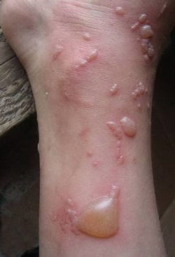
There are several main types of pemphigus, and each of them has its own symptoms.
Pemphigus vulgaris
 Photo: pemphigus vulgaris on the forearm
 This form of pemphigus is characterized by the presence of blisters throughout the body. Their shell is thin, sinks in the center, and the purulent contents are cloudy. Bubbles appear first in the oral cavity and cause an unnecessary visit to the dentist.
Diagnostics
If acantholytic pemphigus is suspected, a cytological examination of a smear from the ulcer is performed. During the examination, the laboratory technician can detect acantholytic epidermal Tzanck cells, which indicate the presence of pemphigus in the patient.
A histological examination of the skin area can also be performed. With pemphigus, intercellular edema in the epidermis, acantholytic disturbances of integrity, and blisters are detected.
Using an immunological study, deposits of IgG and IgA can be detected between the epidermal cells in the area of the bladder.
The diagnosis is made based on:

Diagnosis of pemphigus The diagnosis of true pemphigus is established based on the following methods.
The diagnosis is based on a characteristic clinical picture. Confirmation is a histological examination of the epidermis. If acantholysis is detected, the diagnosis of pemphigus is considered reliable.
Pemphigoids - bullous and cicatricial - are diagnosed based on the characteristic clinical picture. Histological examination of the epidermis does not reveal acantholysis of the stratum spinosum.
Dühring's dermatitis is diagnosed based on the typical clinical picture. Of the specific methods, the Jadasson test is used - when an iodine solution is applied to the skin, the number of rashes increases.

Mechanical symptoms indicating acantholysis are considered particularly significant. Specialists can perform the following procedures:
At the initial stage of this disease, only an experienced specialist can determine that it is pemphigus. The medical history in this case can cover a very long period of time, since its course can be quite long.
At the initial stages, the clinical manifestations of pemphigus are uninformative, so the doctor carefully interviews the patient.
To confirm the diagnosis, laboratory tests are performed:
Pricytological, during which acantholytic cells are detected in fingerprint smears.
Histological. With its help, the intraepidermal location of blisters is detected.
A dermatologist can cure pemphigus. However, depending on the course of the disease, consultation with additional specialists may be necessary.
Often this is a surgeon or infectious disease specialist. For a complete diagnosis, only an examination by a dermatologist is sufficient.
But at the same time, the specialist excludes all possible diseases, according to the clinical manifestation of the disease.
For a detailed diagnosis of a disease in a child, you may need:
- General blood analysis.
- Histological analysis of the contents of the vesicles.
- Immunological research.
- Carrying out an antibiogram.
A dermatologist diagnoses and treats skin pathologies. During an external examination, it is difficult to suspect pemphigus in the early stages, so the patient undergoes biochemical tests:
- Blood analysis
The platelet count determines the general state of health and immunity.
- Analysis of urine
An increased content of leukocytes and protein breakdown products indicates the presence of an extensive inflammatory process.
Laboratory technicians culture a urine sample into a culture medium. The pathogen begins to actively reproduce with the formation of colonies. Pemphigus enterovirus can be identified by the shape and color of the colony.
Treatment
- Since the exact cause of the disease is unknown, treatment is symptomatic.
- Glucocorticoids (prednisolone) are the drug of choice, without which patients die. Complex regimens are used depending on the severity of symptoms. When a positive effect is achieved, the dose is reduced very slowly. They try to achieve a minimum at which new bubbles do not appear - it is taken on an ongoing basis.
- Cytostatic agents (azathioprine, cyclophosphamide) are combined with glucocorticoids in order to prescribe less of the latter, which reduces the likelihood and severity of side effects.
- Antibiotics and antifungals are used when a secondary infection occurs. They are prescribed depending on what flora is detected.
- Ointments with corticosteroids and antiseptic solutions for mouth rinsing are used locally.

Plasmapheresis allows you to clear the body of autoantibodies and can be used to prevent relapses. At the same time, you should monitor blood pressure, blood sugar levels, and regularly check internal organs, since glucocorticoids and cytostatics, unfortunately, are highly likely to provoke complications.
You may also be interested in: Every woman has encountered this - candidiasis (thrush); find out the causes, symptoms, treatment, diet on our website. Lice is not just a dirty word - what are pediculosis and lice, where do they come from and for what reason, prevention and control measures by various means Lichen sclerosus - women at risk: symptoms, treatment and prognosis for girls.
http://idermatolog. net/bolezni-soedinitelnoi-tkani/skleroatroficheskij-lihen.
htmlVitiligo is not a death sentence: symptoms, photos, modern treatment methods, read on our website.
Treatment for pemphigus begins after diagnosis. This is done based on an examination and conversation with the patient’s parents or the patient himself, if he is an adult.
The diagnosis is performed either by an infectious disease specialist or a dermatologist (a joint examination of these two specialists is more often used). After the examination, it is necessary to donate blood from a vein to check for antibodies to the enterovirus, but treatment is prescribed immediately, since the diagnosis of the virus will last at least 2 weeks.
Therapy is as follows:
- If the blisters itch, antihistamines are prescribed: Fenistil, Erius, Zodak, Suprastin. In case of severe itching, 2 of these drugs (for example, Suprastin and Erius) can be combined without exceeding the daily dosage.
- In case of severe itching of the rash, general treatment is supplemented by treating the blisters with local antihistamines: “Psilo-balm”, “Fenistil-gel”.
- To relieve the pain of the rash and reduce body temperature, Nurofen, Paracetamol, and Nise are used (the latter only for adults). Aspirin or acetylsalicylic acid should not be used by children!
- Apply a special diet: exclude from your diet hot, smoked, sour and spicy foods and drinks that will irritate the inflamed oral mucosa. Also exclude hot food, give preference to those dishes and drinks that feel more harmonious when cold (okroshka, compotes, ice cream, fruit ice).
- Rinse your mouth with antiseptic solutions: an aqueous solution of furacillin, chlorhexidine. For adults, you can use Orasept, Strepsils spray with lidocaine and other sprays containing an antiseptic and anesthetic.
- Blisters on the skin can be treated with fucorcin or a solution of brilliant green.
In some cases, infectious disease doctors prescribe antiviral drugs. For children this is “Viferon” or “Laferon” in suppositories, for adults - “Cycloferon” in tablets or “Laferon” in the form of intramuscular injections.
To relieve itching, in addition to official medicine, folk remedies are successfully used:
The main treatment for this disease is taking corticosteroid hormonal drugs, such as prednisolone. The dosage is 80-100 mg/day to relieve the disease and 200 mg/day to treat advanced cases.
The effect of taking the medication will be noticeable within two weeks after starting treatment. Then the dose is reduced to 5 mg/day to avoid relapse. In addition to prednisolone, urbazone, triamcinolone or metypred are also used.
Hormone therapy often causes complications, such as obesity, diabetes, gastric ulcers, hypertension, thromboembolism, pancreatitis and decreased immunity. Therefore, in order to avoid the occurrence of concomitant diseases, you should follow a special diet rich in vitamins and protein, limit the consumption of salt and carbohydrates.
Potassium chloride (3 g/day) and, if necessary, antibiotics are also prescribed.
In addition, medicinal baths with a weak solution of potassium permanganate or oak bark, external use of ointments and brilliant green, and various oils to soften the skin are used.
Painkillers such as novocaine and disinfectants such as hydrogen peroxide are used.
Patients must be under the supervision of a doctor and undergo blood and urine tests once a month. It is necessary to avoid physical activity and adhere to the regime. Climate change and self-medication are not advisable.
True pemphigus, which is an autoimmune disease, can be treated with hormonal drugs.
Important. Since the disease is fatal without proper therapy, there are no contraindications to the use of corticosteroids in this case. The benefits of their use far outweigh the risk of side effects.
The main drugs used in the treatment of pemphigus are Prednisolone and Dexamethasone. They start taking them with high doses, then there is a gradual decrease to a maintenance dose - one at which the appearance of fresh rashes will not be observed.

Cytostatic agents are prescribed simultaneously with corticosteroids. Usually this is Methotrexate or Cyclosporine. A long-acting corticosteroid is also used - Diprospan in injections.
Local treatment of pemphigus in adults is of secondary importance. Bubbles and erosions are treated with antiseptics - brilliant green solution, fucorcin. For extensive rashes, baths with potassium permanganate are used.
Solcoseryl, a paste with a regenerating effect, is applied to the mucous membranes. Apply rinses with antiseptics. Thorough sanitation of the oral cavity must be ensured.
Treatment of dermatitis herpetiformis involves the use of DDS - diamine diphenyl sulfone. The drug is taken in courses. A diet excluding gluten products is indicated. Local treatment is the same as for true pemphigus.
Treatment of viral pemphigus in adults involves the use of antiviral drugs - orally and locally. The drug is determined by the doctor after examining the contents of the blisters.
The only effective way to treat this disease is through the use of medications. As an auxiliary method, you can use a therapeutic one.
Therapeutic
Along with the use of medications, extracorporeal hemocorrection is prescribed. To purify blood, the following are most often used:
Medication
Drug therapy involves the use of the following groups of drugs:
Therapy for pemphigus of any etiology always begins with taking loading doses of hormones such as Prednisolone, Dexamethasone and the like. The dosage is determined by the attending physician individually, calculating it based on the severity of the disease.
Treatment with corticosteroids is very long-term and can last for several months. The patient takes a loading dose until the formed blisters and erosions begin to heal and disappear.
After which the amount of the drug is slowly reduced to a certain minimum.
Acute forms of pemphigus are often treated with corticosteroid hormones.
Very often doctors prescribe drugs:
- prednisolone;
- polcortolon;
- metipred;
- dexamethasone.
Contraindications to the use of hormones are peptic ulcers of the stomach or duodenum. In cases where such indications are present, hormonal drugs are administered intramuscularly.
Dangerous complications of pemphigus are meningitis and encephalopathy - damage to brain cells and (or) its membranes, leading to human death.
The following therapy is carried out:
- Antiviral drugs: Cycloferon (350 rubles), Lavomax (730 rubles), Acyclovir (25 rubles).
- Anti-inflammatory and painkillers: Nimesulide (100 rubles), Ibuprofen (40 rubles).
- Antihistamines: Loratadine (20 rubles), Zodak (125 rubles).
- Disinfecting solutions: Miramistin (230 rubles), Chlorhexidine bigluconate (12 rubles).
- External ointments: Acyclovir (20 r.), Solcoseryl (250 r.).
The course of treatment for pemphigus takes about 2 months, but the likelihood of relapse remains. After completion of therapy, the patient is recommended to strengthen his immune system.
To strengthen it and prevent the disease, smoked and fatty foods should be excluded from the diet. Walking in the fresh air and doing physical exercise are remarkably helpful in reducing the risk of relapse.
Folk remedies for pemphigus
Thanks to alternative medicine, it is impossible to get rid of such a dermatological disease as pemphigus, but it is possible to alleviate the condition of painful rashes. The following recipes will help reduce inflammation and speed up the healing process of formed wounds:

- soak napkins with juice from fresh nettle leaves and apply to the erosion or wound. This compress has a wound-healing, hemostatic and analgesic effect;
- the same can be done from the juice of green aloe leaves, the effect will be identical;
- mix onion, garlic, salt, pepper and honey in equal proportions and simmer in the oven for at least 15 minutes. Cool the resulting viscous slurry and lubricate the opened bubbles. In addition to healing wounds, the product helps to draw out purulent contents;
- Pour 2 tablespoons of chopped meadow clover flower heads into one glass of boiling water and leave for about 2 hours. After which the decoction can be used to wash the resulting erosions due to pemphigus, which will facilitate their speedy healing.
As a preventive measure against pemphigus, the simplest recommendations are effective: wash your hands as often as possible - after using the toilet, visiting public places, going outside; monitor your immunity, avoid overwork; respond promptly to signs of any disease and do not neglect the full course of treatment; adhere to a healthy diet and active lifestyle.
Complications
Enteroviral pemphigus is a disease that is usually mild, but in the presence of a weakened immune system it can be complicated:

- meningitis - inflammation of the membranes of the brain. In most cases, it has a mild course, ending with recovery;
- encephalitis - inflammation of the brain. It develops rarely and can occur in varying degrees of severity;
- pneumonia;
- myocarditis – inflammation of the heart muscle, which without proper treatment can result in heart failure. The cause of myocarditis is that the sequence of antigens that myocardial cells display to the immune system (as almost all cells do) is in a particular region identical to those of the Coxsackie virus, which causes pemphigus. The immune system “thinks” that the myocardium is a microbe and begins to attack it.
Developing in the first three months of pregnancy, viral pemphigus can cause miscarriage. Under the influence of this virus, severe fetal malformations can form, due to which artificial premature birth will have to be induced.
Due to the large number of side effects of glucocorticoids, serious complications are possible. Long-term use of these drugs may cause:
Prevention
There are no vaccines or serums for enterovirus - there are so many strains that it is impossible to guess which one you will come into contact with. If you or your child has been in contact with a person with viral pemphigus, in order to reduce the chance that you will get sick, you need to eat well for the next week, enriching your diet with a sufficient amount of vitamin foods (fruits, vegetables, natural freshly squeezed juices, raisins).
It is also worth consulting with your doctor about whether you can take calcium supplements, and, if this does not harm your health, drink Calcium-D3 or Calcium gluconate in an age-appropriate dosage for 3-7 days.

If there is a history of pemphigus, it is necessary to take maintenance therapy in the form of hormones. Healthy people need to monitor blood and urine sugar levels and maintain normal blood pressure.
To prevent viral pemphigus, you should wash your hands often with soap and practice personal hygiene.
After eliminating the signs of the disease, you should think about a number of preventive measures that are necessary to prevent relapses. They are:
- monitoring the condition of the dermis;
- taking vitamins, calcium, potassium;
- monitoring the occurrence of adverse reactions after taking medications;
- control (regular) sugar levels in urine and blood;
- blood pressure control;
- control over prothrombin.
Pemphigus vulgaris in the oral cavity
People who have been diagnosed with pemphigus should be regularly monitored by a dermatologist. All information about the course of the disease and methods of its treatment are reflected in the patient’s medical history.
Those who have recovered from pemphigus are recommended to work with a gentle work schedule. You should also be careful about physical activity and avoid exposing the skin surface to sunlight. It is necessary to change underwear and bed linen frequently.
The best way to avoid pemphigus in a child is to follow preventive recommendations.
The main preventive measures for pemphigus are:
- Follow the doctor's instructions.
- Do not interrupt treatment with hormonal drugs.
- Eliminate exposure to provoking factors.
Pemphigus in children of any age requires mandatory and precise implementation of drug therapy. As well as correction of the child’s nutrition and lifestyle.
Post Views: 797
In this article:
Pemphigus is a disease that is not fully understood by medicine; in fact, it is a group of diseases. They are considered autoimmune, that is, associated with an attack by the body's immune cells on its own tissues.
These are dangerous and potentially fatal pathologies, which are commonly called vesiculobullous (vesicular). Another name for pemphigus disease. This group of pathologies is also called acantholytic pemphigus. The Latin name is pemphigus acantholiticus, hence the variety of “names”.
Cause of the disease
In order not to throw up our hands at all on this issue, let’s say that most likely the cause of acantholytic pemphigus lies in a malfunction of the immune system. As a result of such a malfunction, the cells of one’s own body become antibodies (that is, hostile cells, like bacteria, cells of someone else’s blood, for example). For some forms of this disease the cause remains unknown (for example, Brazilian pemphigus).
For other forms, today it is believed that epidermal cells are structurally damaged with the participation of external negatively influencing agents:
- retroviruses;
- environmental factors;
- hyperinsolation;
- other aggressive external factors.
Bubbles in this disease appear because intercellular connections are disrupted. The immune system perceives some proteins in the body (desmogleins) as hostile. And destroys them. These proteins can be called the “glue” that binds the individual scales of the epidermis together. It turns out that lymphocytes, destroying the “glue,” “unstick” the epidermis.
Note to patient:
The danger of the disease is its chronic course with constant progression of symptoms (worsening of the condition). This means that individual bubbles merge, the lesion enlarges, and the patient’s body loses protein and fluid. “Unsticking” the epidermis significantly reduces its barrier qualities. That is, the skin becomes easily permeable to bacteria, viruses, fungi and toxins, which can cause sepsis or heart defects.
We do not know what additional risk factors are for acantholytic pemphigus, but it has been established that individuals with a negative genetic status (hereditary predisposition) are more likely to develop this disease.
Classification of the disease and stages of its course
Traditionally, acantholytic pemphigus is divided into :
- Ordinary or vulgar (vulgar is the translation of the word ordinary);
- Erythematous (seborrheic) or Senir-Usher syndrome;
- Vegetative;
- Leaf-shaped (exfoliative);
- Other types of illness.
We remind you that we are talking about true pemphigus, that is, an autoimmune disease, a clear manifestation of which is blisters filled with exudate (liquid released) on the mucous membrane and skin. The disease has many faces. Pemphigus may look like in Figure 1 and like in Figure 2 or 3, and also very differently.
Figure No. 1 True pemphigus

Figure No. 2. Pemphigus

Figure No. 3 Acantholytic pemphigus

Pemphigus vulgaris
This type of disease accounts for the vast majority of reported cases. It is characterized by the appearance of bubbles. The debut of the disease is the formation of blisters on the mucous membrane of the nose and mouth, with further wave-like spread over the entire surface of the epidermis, not excluding the groin area and armpits.
In the early stages, the patient may feel satisfactory and not pay due attention to the rash. Patients complain:
- for pain during long conversations and eating;
- increased sweating;
- as well as a specific putrid odor from the mouth.
Patients do not notice the vesicles because they are small and have a thin cap (the cap is the thin part of the epidermis that covers the fluid cavity and the bottom of the vesicle). Such elements quickly burst. Erosion forms in their place. It's quite painful.
Figure No. 4. Pemphigus vulgaris, elements on the oral mucosa

Figure 5. Erosion on the mucous membrane of the palate with pemphigus vulgaris

In this relatively mild form, the disease can last from three months to a year. Further, without taking medication solutions, the disease can progress, “occupying” more and more new areas of the skin with bubbles.
Figure No. 6. Epithelization of erosions on the skin in pemphigus vulgaris

At the same time, the element itself increases in diameter, reaching the size of a walnut. They are filled with transparent or bloody contents.
After the bubbles open, the liquid flows out and dries, and the erosion epithelializes. If the bubble is not damaged, but regresses due to the drying out of the contents, the surface of the affected area remains covered with a dark crust, which is rejected over time.
The process is aggravated by general weakness, fever, headaches and loss of fluid.
The Nikolsky symptom is of diagnostic value for this disease:
If you pull a piece of tire from a burst bladder, the epidermis (its upper layers) peels off in areas of healthy skin outside the bottom of the burst element.
Seborrheic pemphigus
Unlike vulgar, they appear in approximately 8-10% of the total number of recorded cases. Typical for this variant of the disease:
- the appearance of butterfly-shaped erythema on the patient’s cheeks;
- “butterfly” is covered with gray-yellow crusts.
Unlike lupus erythematosus, to which this variant of the disease is very similar, the crusts are easily removed. Nikolsky's symptom is positive. And over time, the outbreak becomes larger. And it already imitates seborrheic eczema.
Figure 7. Severe form of seborrheic pemphigus

Pemphigus vegetans
Fixed in 5-7% of cases. the bubbles are smaller than with ordinary pemphigus and are localized on the mucous membranes and folds. They especially “love” the mucous membrane of the anal area, axillary areas, navel and inguinal folds.
Unlike the vulgar form, non-traditional epithelial erosion is formed at the site of the opening of the blisters. The erosive bottom of the opened blisters forms soft vegetations (tissue growths). The growths are covered with fluid, often containing pus. Small pustules form around the opened vesicle. Therefore, the smell of an opened fire is extremely unpleasant.
Figure No. 8. Vegetative form of the disease, damage to the axillary region

Nikolsky's symptom is positive, but only near the lesion itself. The disease is benign. Patients complain of pain and burning when walking, associated with the location of the rash. They may complain of chronic fatigue and weakness.
Figure No. 9. Severe pemphigus vegetans

Pemphigus foliaceus
This form of pathology is characterized by the formation of flaccid thin-walled blisters that quickly dry out to form crusts resembling puff pastry. Or they open up, forming crusty leaves. Hence the name of the disease. For the patient, this is one of the unpleasant forms because:
- rashes appear on previously affected areas;
- erosions are covered with entire piles of crusts;
- between the crusts the epidermis is torn (looks like a wet crack);
- exudate constantly oozes from the cracks.
This type of pemphigus primarily affects the skin, but in particularly advanced cases, or weakened immunity, it can also affect the mucous lining of the oral cavity. In women, isolated cases of damage to the vaginal mucosa were recorded.
Figure No. 10. Leaf-shaped pemphigus

With pemphigus foliaceus, there is a high probability of developing sepsis, and this, outside the inpatient conditions of a dispensary, guarantees the death of the patient. The duration of treatment for the disease is individual. And it depends on the state of the immune system and the general health of the patient. But it usually takes 2-3 months to eliminate symptoms of this severity with medical help. In case of complications leading to sepsis (if the patient survives), from six months or more.
Other forms of pemphigus
Pemphigus herpetiformis is characterized by the formation of herpetic-like rashes along with elements characteristic of the leaf-shaped form of the disease. Diagnosis is made based on histology.
Risk 11. Pemphigus herpetiformis

The eosinophilic form is characterized by the presence of eosinophils in fingerprint smears. Sometimes drug-induced pemphigus occurs, caused by taking certain medications (antibiotics). Most likely, it is caused not by autoimmune processes, but by the biochemistry of the body.
There is also a rare form of pemphigus - IgA-dependent, which is benign. Refers to intraepidermal dermatoses. The blisters are flaccid and are more often localized on the extremities and in the folds.
Paraneoplastic pemphigus usually occurs against the background of cancer or chemotherapy. She looks like a vulgar one. It is characterized by damage to both the skin and mucous membranes.
There are other types of this disease, rare and fortunately not all of them occur in our country, for example, Brazilian.
Diagnostics
The active phase of pemphigus is easily identified by the appearance of characteristic blisters. Depending on the type of disease, formations can have different sizes, densities and localizations, but are always filled with liquid exudate during development.
Side signs of the disease are general weakness and fever.
In order to reliably identify the disease at an early stage, a dermatologist must conduct a series of immunological and cytological studies. Check the fingerprint smear for the presence of acantholytic cells, as well as histological analysis to identify the location among the layers of the epidermis of the vesicle. Often, before the blisters, a deep red rash appears on the patient’s skin.
It is very important that if you suspect pemphigus, you must undergo the “Nikolsky test”, which with one hundred percent probability will separate pemphigus from symptomatically similar diseases.
Treatment
The process of successful treatment of pemphigus is quite complex even in a modern medical center. Self-medication is absolutely unacceptable here and can even lead to death. Those suffering from the consequences of pemphigus, even in its mildest, seborrheic form, must undergo medical observation by a dermatologist.
Patients are prescribed a calm regimen without active physical activity, a complete elimination of stress, sleep that is clearly limited by hours, and contact with salt water on the skin is strictly contraindicated (swimming in the sea will have to be avoided). All patients, regardless of the type of disease, are required to follow a hypoallergenic diet: that is, completely exclude hard foods, canned food, pickles, sweets and extractive substances from consumption.
In order to avoid secondary infection, patients are recommended to constantly change their bed linen.
Patients are prescribed high dosages of cytostatics and glucocorticosteroids (as open manifestations of the disease heal, the dosage level is reduced), potassium, calcium and ascorbic acid. When the crisis is over, the doctor, ignoring contraindications, may prescribe Prednisolone orally and Betamethasone for external use.
When treating all types of pemphigus, methods of treating blood outside the patient’s body are used, they are called extracorporeal hemocorrection. And this:
- membrane plasmapheresis;
- hemosorption;
- cryoapheresis;
- other methods.
Neutral antiseptic solutions and traditional aniline dyes can be used as accompanying preparations.
Risk group
Symptoms of pemphigus can appear at any age: from infancy to old age. But statistically, people in the age category 40-45+ are most often susceptible to the disease. People with a hereditary predisposition to the disease are also at increased risk.
Prevention
There are no specific measures. Prevention of the disease is simple and universal. It consists of strict adherence to basic hygiene principles, the absence of bad habits, a healthy diet high in protein and an active, mobile lifestyle, regardless of age.
Consequences
Obvious damage to the epidermis, and associated aesthetic inconveniences, psychological trauma, secondary infections and a sharp decrease in the quality and sometimes life expectancy. Since the immune mechanism itself is affected, and the disease can last for years: all organs and systems of the body are affected, but especially the heart, lungs, kidneys and liver.
Pemphigus (pemphigus) is a dermatological disease characterized by the appearance of blisters (bullas) on the skin and mucous membranes. This is a fairly rare and very serious disease. It is often experienced by adults between the ages of thirty and sixty, although the disease can occur at any age.
Causes of pemphigus
Pemphigus is caused by autoimmune disorders. With this disease, autoantibodies are formed to special proteins - desmogleins. This substance connects nearby epidermal cells. When autoantibodies attack desmogleins, their function is disrupted. Because of this, the epidermal cells are already loosely connected and separate from each other. The epidermis becomes porous, easily exfoliates, and is susceptible to the penetration of microorganisms. As a result, bubbles with serous fluid form on the surface of the skin, after opening which ulcers are exposed. The destruction of the connection between epidermal cells is called acantholysis, which is why pemphigus is called acantholytic.
Acantholytic pemphigus (pemphigus) and neonatal pemphigus are completely different diseases. While pemphigus is caused by autoimmune disorders, neonatal pemphigus is caused by.

Pemphigus symptoms
Pemphigus in adults is a chronic disease with an undulating course, that is, it is characterized by alternating periods of extinction of clinical manifestations and exacerbations of the disease. A characteristic sign of the disease is the appearance of blisters (bulls).
Bubbles can be localized on the mucous membranes of the mouth, upper respiratory tract, external genitalia, and skin. There are several forms of pemphigus:
- Ordinary (vulgar);
- Vegetative;
- Leaf-shaped;
- Erythematous (seborrheic);
- Brazilian.
Pemphigus vulgaris

This is the most common form of pemphigus in adults. It usually begins unnoticed, for no apparent reason. The disease manifests itself with the appearance of blisters on the mucous membrane of the mouth, red border of the lips, nose, and nasopharynx. The patient experiences pain when swallowing food and saliva, and when talking. In addition, increased salivation and, characteristically, are noted. Often, patients go to a dentist or otolaryngologist with such symptoms and are unsuccessfully treated for, or.
After a few months (from three to twelve), the pathological process begins to actively spread to the skin. Transparent flabby blisters with serous fluid inside appear on the skin, which quickly open, literally in the first day. In their place, erosions with a bright pink and moist, and therefore shiny surface are exposed. The serous fluid flowing from the bulla shrinks into crusts. Bubbles can appear in any area, without any favorite location. A typical sign of acantholytic pemphigus is a positive Nikolsky sign. With this symptom, rubbing the skin in the affected area or on a healthy fragment leads to peeling of the epidermis.
 With a moderate process, the general condition of the person remains unchanged. However, pemphigus can become generalized, when extensive blisters and ulcers form on the skin. In this case, your health sharply deteriorates, weakness, loss of appetite, and weight loss occur. The skin with pemphigus is extremely susceptible to microorganisms. There is a possibility of pathogenic microflora joining with serious consequences.
Erythematous (seborrheic) pemphigus

Erythematous pemphigus is rare. The peculiarity of this type of pemphigus is that the first rashes are localized on the skin, namely in areas with a large accumulation of sebaceous glands: face, scalp, back, chest. Erythematous (red) spots with clear boundaries form on the skin. Externally, this clinical picture resembles seborrheic eczema.
Soon, blisters appear on these erythematous spots, and brown or yellowish crusts can be seen on their surface. When the crusts are peeled away, erosion with a wet surface is exposed. The disease can occur in a limited form for several years, or it can spread further through the skin. The oral mucosa is rarely affected.
Pemphigus foliaceus

Pemphigus foliaceus is very similar to common pemphigus. This form is isolated separately due to the fact that acantholysis occurs in the upper layers of the epidermis.
Blisters can appear on any part of the body, but are often localized in the face and scalp. The lid of the bubbles is very thin and breaks at the slightest irritation. After opening them, the serous fluid flows out, and layered scaly crusts are formed covering the ulcer.
Damage to the oral mucosa is quite rare in this form of pemphigus. In this case, the organ of vision may be affected with the formation of purulent conjunctivitis.
Pemphigus vegetans
This type of pemphigus can be benign for a long time (the rash occupies limited areas of the skin) and does not affect general well-being. Blisters appear mainly on the mucous membrane of the mouth, around the nose, anus, on the genitals, as well as in large folds of skin (axillary, inguinal, in women under the breasts).
This type of pemphigus is characterized by the appearance of soft growths at the bottom of the erosion - vegetations covered with a serous coating. Vegetations emit an unpleasant odor and can reach a centimeter in size.
In general, the identification of various forms of pemphigus is quite arbitrary. After all, one type of pemphigus may resemble another. In addition, it is possible to transform one form into another.
Brazilian pemphigus

In its clinical manifestations it resembles pemphigus foliaceus. This disease occurs among residents of Brazil, Northern Argentina, Peru, Bolivia, mainly in tropical areas. Improved living conditions and deforestation led to a decrease in morbidity. Doctors suggest that Brazilian pemphigus is probably infectious in nature, and its activity is maintained by environmental factors. The disease often affects several family members, with women being more susceptible.
Flat blisters appear on the skin, which then crack to form scaly crusts. The patient may complain of a burning sensation of the skin, which is why Brazilian pemphigus is called “wild fire.”
Diagnostics
If acantholytic pemphigus is suspected, a cytological examination of a smear from the ulcer is performed. During the examination, the laboratory technician can detect acantholytic epidermal Tzanck cells, which indicate the presence of pemphigus in the patient.
As an external treatment, blisters on the skin are treated with solutions of aniline dyes (fucorcin). To treat the oral cavity, rinsing with solutions with an anti-inflammatory effect is practiced.
The prognosis for acantholytic pemphigus is conditionally unfavorable. On the one hand, in the absence of effective treatment, there is a high probability of complications and death. On the other hand, patients with pemphigus are forced to take glucocorticosteroids for a long time, and sometimes for life, which is fraught with the development of side effects. But hasty refusal of drugs leads to immediate relapse of the disease. Glucocorticosteroids do not eliminate the cause of the disease, but inhibit the pathological process and prevent its progression.
Grigorova Valeria, medical observer
