Blood supply to the hip joint. Collateral circulation in the hip joint
The hip joint is the largest joint in the human musculoskeletal system, connecting the lower limbs to the body. Takes an active part in movement and maintaining balance with a vertical position of the body. Despite its strength, the hip joint is one of the most vulnerable parts of the human skeleton, as it experiences stress every day when walking, running and doing physical exercise.
Anatomy of the human hip joint
The hip joint is a large spherical joint with several axes of rotation, formed by the articular surface of the head of the femur and the acetabulum of the iliac bone of the pelvis. The structure of the hip joints in women and men is not fundamentally different.
In fact, the hip joint consists of a neck and head, covered with cartilage tissue, the femoral bone, the acetabulum and the acetabular lip that deepens it, located inside the capsule. The joint capsule of the hip joint is a hollow formation that limits its internal cavity. The walls of the capsule consist of three layers:
- external – dense fibrous tissue;
- middle – connective tissue fibers;
- internal – synovial membrane.
The synovial membrane lining the joint capsule from the inside produces a serous secretion that serves to lubricate the articular surfaces during movement, reducing their friction against each other.
Articular ligaments
The ligamentous apparatus of the hip joint provides rotation, supination, and mobility of the lower extremities in the longitudinal and transverse direction; it is formed by several structures: 
- The iliofemoral ligament is the largest and strongest of all, holding and ensuring mobility of the hip joint. It originates near the anterior inferior spine of the pelvic bone, and then diverges in a fan-shaped manner, attaching in bundles to the femur along the intertrochanteric line. Included in the group of muscles and ligaments responsible for balance and keeping the body in an upright position. Another function of the ligament is to inhibit hip extension.
- Ischiofemoral - one end is attached to the ischium; passing inside the trochanteric fossa, the other end is woven into the articular capsule. Inhibits the adducting movements of the hip.
- Pubofemoral - originates on the anterior surface of the pubic bone and is woven into the articular capsule. Responsible for inhibiting hip movements performed in a direction transverse to the axis of the body.
- Circular ligament - located inside the joint capsule, originates from the anterior edge of the ilium and loops around the head of the femur.
- Femoral head ligament – located inside the joint capsule, protecting the blood vessels of the femoral head.
Muscles of the hip joint
The hip joint has several axes of rotation:
- frontal (transverse),
- sagittal (antero-posterior),
- longitudinal (vertical).
 Movements of the joint along the frontal axis provide flexion and extension movements of the hip. The muscles responsible for hip flexion are:
- straight,
- comb,
- iliopsoas,
- tailoring,
- wide.
Hip extension is provided by antagonist muscles:
- two-headed,
- semitendinosus,
- semimembranous,
- gluteus maximus.
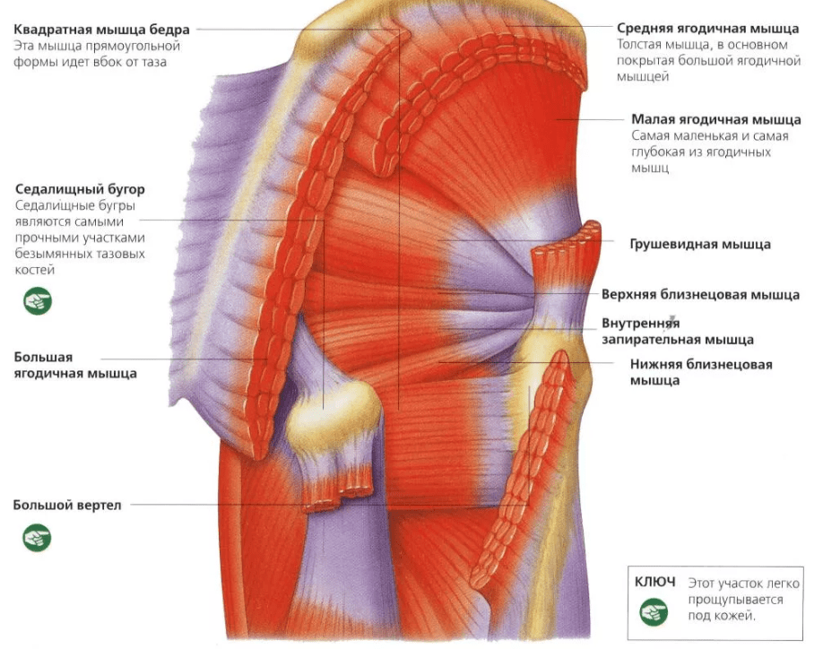
The adducting and abducting movements of the hip are performed along the sagittal axis. The following are responsible for hip abduction:
- pear-shaped,
- twin,
- obturator internus muscle.
The casting is carried out:
- adductor magnus,
- comb,
- thin,
- adductor brevis and longus muscles.
 The longitudinal axis of rotation is necessary for hip rotation, as well as for pronation and supination of the joint. These functions are carried out:
- square,
- gluteus maximus,
- iliopsoas,
- pear-shaped,
- twin,
- tailoring,
- obturator externus and obturator internus muscles.
Blood supply of hip joint
 The blood supply to the hip joint is provided;
- ascending branch of the lateral femoral artery,
- round ligament artery
- acetabular branch of the obturator artery,
- branches of the inferior and superior gluteal arteries,
- deep branch of the medial femoral artery,
- branches of the external iliac artery,
- branches of the inferior hypogastric artery.
The importance of these arteries for providing blood supply to the hip joint varies. The main supply is provided by the deep branch of the medial femoral artery. The outflow of blood from the joint and surrounding tissues is provided by the branches of the femoral, hypogastric and iliac veins.
Innervation and lymphatic drainage of the hip joint
 The innervation of the hip joint is carried out through the branches of the femoral, obturator, sciatic, inferior gluteal, and genital nerve trunks.
Periarticular neurovascular formations and nerve roots of the periosteum also take part in the innervation.
The lymphatic drainage of the joint passes through deep lymphatic vessels leading to the pelvic lymph nodes and internal sinuses.
Functions of the hip joint
One of the main functions of the hip joint is the connection of the lower limbs with the body. In addition, the joint plays an important role in ensuring their movement, performing the following functions:
- supports,
- bending,
- extension,
- rotations,
- pronation,
- supination,
- leads,
- adduction of legs.
Possible causes of pain in the hip joint
Daily stress, injuries, age-related changes, inflammatory and infectious processes in the tissues of the joint and its surroundings can cause pain.
Injuries
Injuries are one of the most common causes of hip pain. The severity of symptoms is directly related to the severity of the injuries received.
The mildest injury to a joint is a bruise caused by a blow or falling on its side. Symptoms of a bruise are pain in the hip area, swelling and redness, temporary lameness.
 A more severe injury to the hip joint is a dislocation, which can be the result of a strong blow, for example, in a traffic accident, a fall from a height, a sharp jerk, or excessive movement. Symptoms of a dislocation are:
- sharp pain that gets worse when you try to move your leg or lean on it;
- swelling and redness of tissue in the area of the damaged joint;
- formation of an extensive hematoma in the thigh area;
- visually discernible deformities, protrusion on the thigh at the site of ligament separation;
- forced rotational position of the limb;
- loss of functionality of the affected leg.
The most severe injury is considered to be a fracture of the femoral neck. In young and middle-aged people, such injuries are relatively rare and occur as a result of severe blows received in a car accident or fall from a height. The vast majority of hip fractures occur in older people.
The bone tissue of older people loses its strength as a result of hormonal and age-related changes that accelerate the process of calcium leaching. A fracture can occur with minor physical impact or even spontaneously, in the absence of any external causes.
 Symptoms of a femoral neck fracture:
- pain in the groin area;
- loss of function of the injured limb, inability to lean on it;
- forced rotational position of the leg outward;
- shortening of the injured limb relative to the healthy one, visually discernible in the supine position;
- “stuck heel” syndrome - the inability to lift a leg straightened at the knee from a supine position;
- swelling and redness of tissues.
Inflammatory and degenerative diseases
One of the most common causes of pain in the hip joint is inflammatory processes in the tissues.
Arthritis- inflammation of joint tissue caused by autoimmune reactions, chronic injuries, bacterial or viral infections. The disease can affect either one or both joints, manifesting itself as pain that intensifies after exercise and with prolonged exposure to a stationary position, limited mobility, swelling, redness of the tissues, and a local increase in temperature.
 Arthrosis hip joint, or coxarthrosis, is a chronic, steadily progressing disease accompanied by degenerative changes in tissues. The causes of development can be injuries, genetic predisposition, endocrine disorders. In the early stages, pain in the joint area is the only symptom; as the disease progresses, it leads to dysfunction of the joint and, ultimately, to its complete destruction.
Bursitis– an inflammatory process that develops in the synovial cavity of the trochanteric bursa of the joint. The causes of development may be chronic injuries, as well as complications of inflammatory diseases of the joint. A characteristic symptom of the pathology is pain in the subgluteal region and on the back of the thigh, which intensifies when running or walking.
Tendinitis– inflammation of the ligaments that stabilize the joint. The cause of the development of the disease in most cases is inadequately high loads and regular microtraumas of the connective tissue. As a result of the formation of microtears in the fibers, scars are formed, and when pathogenic microorganisms enter them, an inflammatory process develops.
Systemic connective tissue diseases
Systemic connective tissue diseases mostly develop as a result of pathological autoimmune reactions or genetic disorders; in this case, several joints are involved in the pathological process.
 Gout- pathological accumulation of uric acid salts in organs and tissues, causing inflammation of the joints and the formation of tophi - specific lumps in the area of the affected joints.
Ankylosing spondylitis, or ankylosing spondylitis, is a genetically determined disease, in the early stages manifested by pain and a decrease in the range of motion, and in the later stages leading to ankylosis - complete loss of mobility - of the affected joints.
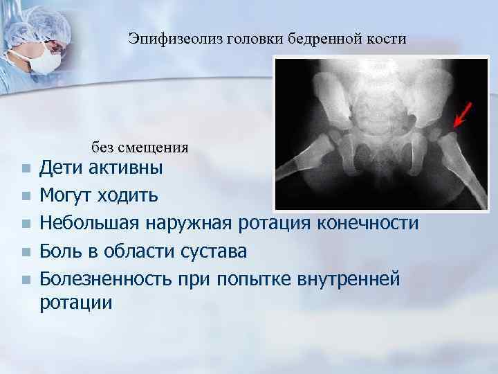
Epiphysiolysis– a disease whose development mechanisms are based on endocrine disorders, presumably of a hereditary nature. The main symptom of the pathology is displacement and slippage of the femoral head from the acetabulum, accompanied by forced outward rotation of the limb, changes in gait, lameness and chronic pain in the hip joint.
Diagnostics
Treatment of diseases of the hip joint is impossible without making an accurate diagnosis, since there are many reasons for the development of pain and impaired mobility, and each pathology requires its own tactics and choice of treatment methods. At the initial stage of diagnosis, the specialist conducts an examination and collects an anamnesis, and also prescribes a number of instrumental and laboratory tests to clarify the clinical picture:
- X-rays can reveal the integrity of bone structures and the presence of foci of tissue changes;
- ultrasound examination detects changes in soft and cartilaginous tissues;
- MRI and CT help to obtain the most accurate picture of the affected area for layer-by-layer study;
- arthroscopy and examination of effusion - pathological fluid accumulating in the synovial capsule.
Prevention of diseases and injuries of the hip joint
 Injuries and diseases of the hip joint are the most common orthopedic pathologies that can be encountered by both professional athletes and people who are as far away from sports as possible. Compliance with a number of preventive measures will help minimize the risk of complications.
The anatomy of the human hip joint (HJ) is interesting because of its significant modification throughout evolution, which can be seen when compared with non-upright mammals. Maintaining body weight in an upright position required special mechanics of this joint, which cast a shadow on the structure of the joint.
The hip joint is the connecting link between the torso and lower limbs. It is a strong and spherical joint. Its structure is aimed at maintaining stability and performing a large number of movements in it.

Important! The hip joint is the second most mobile in the human body.
Bone anatomy - what connects and how
The head of the femur has the shape of a sphere located on the “pedicle” - its neck. Its entire surface is covered with articular cartilage, thickening in areas of increased exposure to body weight on the lower limb. The exception is the place of attachment of the own ligament of the femoral head, namely its fovea (fovea for the ligament of the femoral head).
The acetabulum (English, acetabulum), in turn, the second main component of the joint, is a hemisphere covered over most of its length with cartilage tissue. This reduces the friction of the head on the pelvic bone.

In the photo - intra-articular surfaces - head and cavity (fossa)
The depression is a consequence of the connection of the three bones of the pelvis - the ilium, the ischium and the pubis. It consists of a semilunar-shaped rim, protruding slightly upward, covered with cartilage, and being the articular part of the joint, as well as the surface of the acetabulum, which has the same shape.
Attached to the rim is the acetabular labrum, which in appearance resembles a lip, which is how it got its name. Through it, the surface area of a given cavity increases by approximately 10%. The part of the acetabulum that does not participate in the formation of the joint is called the fossa, and is made entirely of the ischium.
Due to the presence of a complete connection between the femoral head and the pelvic bones, the structure of the hip joint allows it to remain one of the most stable joints. The congruence of the articular surfaces is most complete at a position of flexion at the joint at 90°, abduction of the lower limb at 5° and external rotation at 10°. It is in this position that the axis of the pelvis coincides with the axis of the head of the femur and forms a straight line.
Joint capsule and its ligaments
The stability of the hip joint is further strengthened by covering the entire length of the joint with two layers of capsule - a loose outer fibrous layer and an inner synovial membrane.

The hip ligaments are compacted parts of the fibrous layer of the capsule, which are spirally stretched between the pelvic bones and the thigh, thereby strengthening this connection.
The structure of the human hip joint, especially its ligamentous apparatus, determines the complete insertion of the head into the acetabulum during its extension by rewinding the spiral ligaments that tighten the fibrous capsule; problems in this place can occur. Thus, the congruence of the joint during its extension is produced through passive movements of its articular surfaces.
The tense ligaments of the fibrous capsule limit excessive extension, which is why the full vertical position is 10-20° short, however, it is this slight difference in angle that increases the stability of this joint.
The structure of the hip joint includes three internal ligaments:
- Iliofemoral ligament. It is located in front and slightly upward, stretching between the lower anterior iliac spine and the intertrochanteric line of the femur distally.
It is believed that this ligament is the strongest in the body. Its job is to limit hyperextension of the hip joint in a standing position. - Pubofemoral ligament(English, pubofemoral ligament). It extends from the obturator ridge, going down and laterally to connect with the fibrous capsule. Intertwined with the medial part of the iliofemoral ligament, it is also involved in limiting excessive extension of the joint, but to a greater extent prevents hip hyperabduction (too much abduction).
- Ischiofemoral ligament. Localized on the posterior surface of the joint. It is the weakest of all three ligaments. It spirals around the neck of the femur, attaching to the base of the greater trochanter.

A major role in gait is played by the hip joint, the structure of which is supported precisely by the above-described ligaments and muscle frame, which ensure its structural integrity. Their work is interconnected, where the disadvantage of some elements is offset by the advantage of others. More details about this can be found in the video in this article.
Thus, the work of the ligamentous and muscular apparatus is balanced. The medial hip flexors, located in front, are weaker than the medial rotators, but their function is strengthened by the anterior internal ligaments of the thigh (pubofemoral and iliofemoral), which are much stronger and denser than the posterior ligament of the joint.
The only ligament that performs almost no function in relation to strengthening the joint is the ligament of the femoral head. Its weak fibers are directed from the fossa located in the center of the femoral head to the acetabular notch. Its work consists largely of creating protection for the vessel (artery of the head of the femur) stretching between its fibers.
The fatty tissue that fills the fossa of the acetabulum, together with the ligament, is covered with a synovial membrane. This adipose tissue compensates for the lack of congruence of the articular surfaces by changing its shape during movements.
Movements in the joint
This:
- flexion and extension;
- abduction and adduction;
- medial and lateral rotation;
- rotation.
All of the movements described above are extremely important, as they ensure such daily human activity as getting out of bed, holding the body in an upright position, sitting, if you have problems with performing these simple actions, please read.
The anatomy of the hip joint is rich in muscles that allow the above-described functions of the hip joint to be realized.
These include:
- iliopsoas muscle - the strongest flexor of the lower limb;
- the adductor magnus muscle is its synergist;
- simultaneous flexion and adduction of the limb is ensured by the piriformis and gracilis muscles;
- The gluteus minimus and medius muscles serve simultaneously as abductors and medial rotators;
- The gluteus maximus plays the role of the main extensor, participating in the transition of the body from a bent position in the hip joint to an extended one (standing up).
Blood supply
The head and neck of the femur are supplied by branches of the medial and lateral circumflex artery, the deep femoral artery, and the own artery of the femoral head. In adulthood, the medial circumflex femoral artery is considered the most important source of blood supply to the femoral head and proximal neck.
Attention! In old age, the blood supply to the head and proximal neck of the femur is reduced, which causes a high incidence of trauma to this area and difficulty in healing fractures, which is why complete or partial replacement of the joint is often required to restore its mobility.

Among other things, recovery from a hip fracture is long and requires the patience and desire of the patient, but more importantly, the full implementation of all the techniques suggested by the instructions developed by the rehabilitation doctor. The lesson plan is developed individually and requires the efforts of the patient.
Important! Only a doctor can diagnose problems in the hip joint and prescribe appropriate treatment. If symptoms appear that indicate a violation of full movements in this joint, contact an orthopedic traumatologist.
Go to the contents of the Bulletin of the Russian Scientific Research Center for Reconstruction and Research of the Ministry of Health of the Russian Federation N8.
Current section: Radiation diagnostics
Modern data on the anatomy and blood supply of the hip joint, clinical picture and diagnosis of its inflammatory-necrotic lesions.
Khisametdinova G.R., Federal State Institution “RNTsRR Rosmedtekhnologii” Moscow.
The main task of early diagnosis of Perthes disease, aseptic necrosis of the femoral head of another origin, is to detect the stage of vascular disorders, when, if adequate measures are taken, the process can reverse. Ultrasound examination with Dopplerography, which allows assessing regional blood supply in various pathologies of the hip joints in children, is an important method for assessing the effectiveness and adequacy of treatment, load regulation and functional therapy.
Key words: hip joint, diagnosis, blood supply Khisametdinova G. R.
The modern knowledge about anatomy and blood supply of hip joint in clinics and diagnostics of its inflammatory-necrotic lesions
Federal State Enterprise Russian Scientific Center of Roentgenoradiology (Russian Medical Technologies Department)
The main purpose of the early diagnostics of Pertes’ disease and of other hip bone aseptic necrosis is the detection of their vascular stage, when adequate therapy may cause resolution of the disease. Sonographic investigation with Doppler techniques assesses regional blood supply in different pathology of hip joint in children, and evaluates the effectiveness and adequacy of the treatment to adjust load and functional therapy.
Keywords: hip joint, diagnostics, blood supply Contents:
Etiology, classification and clinic of Legg-Calvé-Perthes disease and aseptic
necrosis of the femoral head of another origin.
Ultrasound methods for studying the hemodynamics of the hip joint. Ultrasound research methods for a number of pathologies of the hip joint. Bibliography.
Embryogenesis, anatomy and blood supply of the hip joint.
The hip joint is the largest joint in humans. The embryogenesis of the hip joint is of significant interest in terms of substantiating the congenital predisposition to various pathological conditions. In a number of diseases of the hip joint that are detected in young children, there is a single mechanism of disruption of embryogenesis during the formation of the musculoskeletal system, which leads, in the process of growth and formation of the musculoskeletal structures of the hip joint, to a violation of their spatial relationship.
All elements of the hip joint are formed from a single scleroblastoma mass. The skin and its derivatives develop from the ectodermal layer, and cartilage, bones, tendons, ligaments and capsule develop from the mesodermal layer. Already at the end of the 4th week of gestation, the buds of the lower limbs are determined in the embryo in the form of vascularized mesenchymal nuclei. Between the 6th and 7th weeks, the first cartilaginous elements appear, and in the hip joint, the 3 cartilaginous elements of the femur combine into a cartilaginous formation (“hemitasis-hemitavis”) and create a flat acetabulum. Between the acetabulum and the cartilaginous elements of the thigh, the future joint space is still filled with connective tissue. At this stage, the cartilaginous lip is already recognized as compacted connective tissue.
At the 7th week of intrauterine development, when the embryo is about 1 cm long, the glenoid cavity, ligament of the femoral head, joint capsule and joint space appear (Fig. 1). The femoral diaphysis ossifies, resulting in the formation of a bony diaphyseal tube and a medullary space. Bone anlages are formed from precartilaginous cells. By this time, the arterial trunks have already been formed and the nerves - femoral and sciatic - have been demarcated. The future joint cavity is defined as the zone of dense cells between the femoral head and the pelvis. Precartilaginous cells atrophy during the formation of the joint and, in the process of autolysis, the joint space, the spherical head of the femur and the semicircular articular cavity are formed from the primitive joint cavity. At the upper border of the depression, a limbus is defined in the form of a wedge-shaped edge, along the edge
On the cartilaginous ilium, a fibrocartilaginous rim is noticeable - the future labrum acelaide.
At the end of the 8th week, the initial development of the hip joint is almost complete. The pelvis is formed through the ossification of three component parts, each of which has its own nucleus. The first ossification nucleus appears in the body of the ilium at 10 weeks.
The 11-12 week fetus is about 5cm long, the hip joint is formed with all the structures, and the diaphysis ends with calcification.
At 16 weeks, the fetus is 10 cm long, the head of the femur is spherical, with a diameter of 4 mm, all movements in the hip joint are possible, ossification of the nucleus of the ischium occurs.
By week 20, all differentiation is completed, the ilium is ossified by 75%, ossification of the core of the pubic bone occurs, while the bone formations are united by U-shaped cartilage, the head of the femur with a diameter of 7 mm remains cartilaginous until 3-4 months after birth.
Rice. 1 Plane section of the hip joint of a 7 week embryo
The anatomical structure of the hip joint in young children differs significantly from that in an adult. The peculiarities of the hip joint in newborns are that the predominant part of the elements of the joint during its development is cartilaginous. One center of ossification is located in the nucleus of the epiphysis of the femoral head, and the second is in the nucleus of the greater trochanter. The nucleus of the epiphysis of the head of the femur appears between the 2nd and 8th months of life, the nucleus of the greater trochanter - between the 2nd and 7th years of life. Ossification of the femoral head occurs from two sources: due to the ossification nucleus of the proximal femoral epiphysis, and also due to
the spread of the process of enchondral bone formation from the zone of ossification of the femoral neck in the proximal direction. The upper-inner section of the femoral head is ossified from the ossification nucleus of the proximal femoral epiphysis, and the lower-outer section is ossified from the ossification zone of the femoral neck.
In the first year, the degree of ossification of the femoral neck increases; the cartilaginous structure is preserved only in its upper part. The highest rates of growth of the acetabulum are observed in the first year of life and in adolescence. The diameter of the cavity increases due to the growth of Y-shaped cartilage. The depth increases due to the growth of the cartilaginous edges and acetabular lip, as well as due to its physiological protrusion in older children. The most active deepening of the acetabulum occurs from 2 to 3 years and after 5 years of age. The growth of the femoral head occurs synchronously with the growth of the acetabulum, while the highest rates of its ossification are observed from 1 year to 3 years.
Data on the anatomy of the hip joint presented in the review, its blood supply, make it possible to explain the pathogenesis and symptoms of the development of clinically different forms of hip joint pathology.
The hip joint is a type of ball-and-socket joint of a limited type - a cup-shaped joint. Movements are performed in three planes: frontal (abduction up to 135 degrees, adduction up to 60 degrees), sagittal (flexion up to 40 degrees, extension up to 10 degrees) and vertical (outward rotation up to 41 degrees, inward rotation up to 35 degrees), as well as circular movements. Joint stability is ensured by the anatomical shape of the articular ends, articular capsule, powerful ligaments and muscles.
The joint is formed by the proximal end of the femur, the articular surface of the head, as well as the bones of the acetabulum, consisting of the ilium (upper section), ischium (lower-posterior section) and pubis (antero-internal section) bones (Fig. 2,3). In children, these bones are separated from one another by a Y-shaped growth cartilage. By the age of 16, the cartilage ossifies, and individual bones fuse to form the pelvic bone. The acetabulum is covered with cartilage only in the area of the semilunar surface; the rest of the area is filled with fatty tissue and covered with synovial membrane. The thickness of the cartilage ranges from 0.5 to 3 mm; it reaches its greatest thickness in the zone of maximum load. A fibrocartilaginous acetabular labrum is attached along the free edge of the socket, which increases the depth of the acetabulum.
Diagram of the frontal cut of the right hip joint
1. wing of the ilium;
2. iliacus muscle;
3. gluteus minimus;
4. gluteus medius muscle; acetabulum;
5. gluteus maximus muscle;
6. acetabulum; border
7. acetabular (cartilaginous) lip; hips;
8. circular zone; preparation
9. femoral head; depressions;
1. bony protrusion (bay window);
2. perichondrium and periosteum of the ilium;
3. cartilaginous lip
4. greater skewer;
5. osteochondral
proximal part
6. the acetabular fossa highlighted in the process
Anatomical preparation of a cut of a child's hip joint, corresponding to Fig. 2
10. large skewer;
7. allocated in process
preparation
II. trochanteric bursa large
8. cartilaginous part of the roof
gluteal muscle;
12. joint capsule with a circular zone;
13. iliopsoas muscle;
acetabulum;
9.periosteum internal
pelvic walls.
14. medial circumflex femoral artery;
15. pectineus muscle;
16. perforating arteries.
The head of the femur is covered with hyaline cartilage along its entire length, with the exception of the fovea capitis, where the ligament of the head is attached, through which the vessels to the head of the femur pass.
The joint capsule connects and covers the articular ends of the bones, forming the cavity of the hip joint, consisting of the cervical region and the acetabulum, which communicate with each other. In the articular capsule, there is an outer fibrous layer, reinforced by ligaments, and an inner synovial layer, lining the joint cavity. The fibrous capsule is attached to the pelvic bone along the edge of the acetabulum, on the femur it is fixed along the intertrochanteric line, and from behind it captures 2/3 of the femoral neck.
The articular capsule is strengthened by ligaments: three longitudinal (in front - the iliofemoral and pubofemoral, in the back - the ischiofemoral) and a circular one, running in the deep layers of the articular capsule.
The hip joint has two intra-articular ligaments: the aforementioned ligament of the head, covered with a synovial membrane, and the transverse acetabular ligament, which in the form of a bridge spans over the opening of the acetabulum. The muscles that provide movement in the hip joint include the pelvic muscles and the muscles of the free lower limb. The pelvic muscles are divided into muscles that begin in its cavity (the psoas major and minor, iliacus, piriformis, coccygeus, obturator internus) and muscles that begin on the outer surface of the pelvis (tensor fascia lata, gluteus maximus, gluteus medius and minimus, superior and inferior gemini , rectus and quadratus femoris muscles). The hip joint has three sources of innervation. It is innervated by branches of the nerves: anteriorly - femoral, medially - obturator and posteriorly - sciatic. Due to
peculiarities of innervation, with pathology of the hip joint (Perthes disease, coxitis), pain often radiates to the knee joint.
Rice. 4 Blood supply to the hip joint
1. deep artery, circumflex ilium;
2. superficial artery, circumflex ilium;
3. femoral artery;
4. ascending branch of the lateral circumflex femoral artery;
5. transverse branch of the lateral circumflex femoral artery;
6. descending branch of the lateral circumflex femoral artery;
7. lateral circumflex femoral artery;
8. deep femoral artery;
9. perforating arteries;
10. external iliac artery;
11. inferior epigastric artery;
12. superficial epigastric artery;
13. superficial external pudendal artery
14. obturator artery;
15. deep external genital artery;
16. medial circumflex femoral artery;
17. femoral artery;
18. muscle branches.
Of great importance in the normal development and functioning of the hip joint is its blood supply (Fig. 4). The main role in the blood supply to the joint belongs to the medial and lateral arteries, the circumflex femoral arteries (branches of the deep femoral artery) and the obturator artery. The remaining feeding vessels participate in the blood supply to the proximal femur through anastomoses with the three listed arteries.
Normally, there are several types of structure of the arterial network: the medial and lateral circumflex femoral arteries can arise from the deep femoral artery, directly from the femoral artery, from a.comitans n.ischiadici.
The deep femoral artery is the main vessel through which vascularization of the femur is carried out, it is a thick trunk that arises from the posterior side of the femoral artery (a branch of the external iliac artery) 4-5 cm below the inguinal ligament, lies first behind the femoral artery, then appears on the lateral side and gives off numerous branches, including:
1. the medial circumflex artery of the femur, a.circumflexa femoris medialis, which arises from the deep artery of the thigh behind the femoral artery, goes transversely inward and, penetrating between the iliopsoas and pectineus muscles into the thickness of the muscles adducting the thigh, bends around the neck from the medial side femur, gives the following branches:
a) ascending branch, r. ascendens, is a small stem that goes upward and inward, branching, and approaches the pectineus muscle and the proximal part of the adductor longus muscle.
b) the transverse branch, r.transversus, is a thin stem, directed downward and medially along the surface of the pectineus muscle and, penetrating between it and the long adductor muscle, it goes between the long and short adductor muscles. Supplies blood to the long and short adductor muscles, the thin and external obturator muscles;
c) deep branch, r.profundus, a larger trunk, which is a continuation of the medial circumflex femoral artery. It is directed posteriorly, passes between the external obturator muscle and the quadratus femoris muscle, dividing here into the ascending and descending branches (superior and inferior cervical arteries);
d) branch of the acetabulum, r. acetabulis, a thin artery, anastomoses with branches of other arteries supplying blood to the hip joint.
2. lateral circumflex femoral artery, a. circumflexa femoris lateralis, large trunk, extends slightly below the medial one, from the outer wall of the deep
the femoral artery is almost at its very beginning, directed to the lateral side. It goes outward in front of the iliopsoas muscle, behind the sartorius muscle and the rectus femoris muscle, approaching the greater trochanter of the femur, and divides into branches:
a) the ascending branch, r.
b) the descending branch, g.oeBsepeenB, is more powerful than the previous one. It departs from the outer surface of the main trunk and lies under the rectus femoris muscle, then descends along the groove between the vastus intermedius and vastus lateralis muscles, supplying blood to them, the quadriceps femoris muscle and the skin of the thigh.
c) the transverse branch, r. 1xan8ueree8, is a small stem directed laterally; supplies the proximal rectus femoris and vastus lateralis muscles.
The branches of the lateral circumflex femoral artery supply the superficial portion of the anterior segment of the head and the neck of the femur.
The main age-related feature of the blood supply in children is the autonomy and disconnection of the vascular system of the epiphysis and the neck of the femur. The barrier between them is the growth zone, which prevents the penetration of the vessels supplying the distal femur and the capsule of the hip joint into the head of the femur.
The medial circumflex femoral artery gives off two branches: the superior cervical artery and the inferior cervical artery. The superior cervical artery supplies most of the epiphysis of the femoral head (from 2/3 to 4/5). It penetrates the epiphysis from the outside, forms a dense network of vessels at its base, supplying blood to the reserve layer of cells of the germ plate. The anterior central region of the epiphysis is located in the terminal zone of the vascular basin of the superior cervical artery, that is, it is in the least favorable zone of blood supply. The inferior cervical artery supplies only the small medial segment of the head.
The obturator artery is a branch of the internal iliac artery, it supplies the obturator externus muscle, the adductors and gives rise to the acetabular branch, which penetrates through the opening of the acetabulum into the hip joint and supplies the ligament of the femoral head and the head of the femur.
The arteries of the ligament of the femoral head originate from two sources - the obturator and medial circumflex artery. The thinnest arteries of the ligament of the head branch according to the scattered and main type. In the first case, the arteries usually do not penetrate the femoral head; in the second case, they extend into it to a limited extent.
plot. In children, there are no anastomoses between the branches of the superior and inferior cervical arteries and the arteries of the ligament of the femoral head. Arterial anastomoses occur at older ages.
The branches of the vessels form Anserov’s ring-shaped arterial anastomosis along the edge of the cartilaginous cover of the femoral head (Fig. 5). Thanks to the anastomosis, more uniform nutrition of the individual segments of the head is provided. The second arterial ring is formed by the medial and lateral circumflex femoral arteries. Damage to the arteries occurring below this anastomosis can lead to serious changes in the area of the blood supply to this vessel. Therefore, traumatic and hemodynamic disturbances of the vascular network of the hip joint capsule can lead to disruption of the blood supply to the epiphysis of the femoral head, which causes the occurrence of aseptic necrosis and destruction of the bone structure. Due to the absence of anastomoses, which occur only after 15-18 years, after synostosis of the head and neck of the femur, any traumatic effect on the hip joint area (especially trauma, cooling, vascular spasm, etc.) can, under equal conditions, remain invisible in adults and cause complications in children.
Rice. 5 Arterial anastomoses of the femoral head
The venous system differs in its architecture from the arterial system. In the wide bony canals of the neck, one artery is accompanied by two or more venous trunks. The veins emerging from the epiphysis of the femur anastomose with the veins of the articular capsule, and
also with the veins of the muscles surrounding the joint. Venous drainage from the hip joint occurs from the intraosseous plexuses through the veins medially and laterally surrounding the thigh into the deep vein of the thigh, femoral vein, and external iliac vein.
Etiology, classification and clinical picture of Legg-Calvé-Perthes disease and aseptic necrosis of the femoral head of another origin.
Legg-Calvé-Perthes disease is an osteochondropathy morphologically and pathophysiologically representing aseptic necrosis of the bone tissue of the femoral head and its secondary deformation occurring due to axial load. It is reliably known that osteonecrosis develops as a result of a violation of the local vascular, namely arterial, supply of bone substance and bone marrow.
Up to 30 synonyms of osteochondropathy of the femoral head are known, in which the authors tried to reflect both the morphological substrate and the etiological moment of the development of the disease. The most common terms for the pathology are: Perthes disease, avascular necrosis of the femoral head, coxa plana.
For the first time, almost simultaneously, independently of each other, this pathology was described by orthopedists Waldenstrum in 1909 and Legg, Calve and Perthes in 1910.
According to the Ministry of Health of the Russian Federation, in the structure of disability due to injuries and diseases of the musculoskeletal system, osteochondropathy accounts for 27%, which is 2% more than disability due to injuries. Among all osteochondropathy, Perthes disease accounts for, according to various authors, from 3 to 13%. Most often, Perthes disease affects children aged 4 to 10 years, but cases of the disease at an earlier and especially at a later age up to 18-19 years are not uncommon. Boys and young men are affected 4-5 times more often than girls.
In most cases, the process is unilateral, but there is also a bilateral lesion, which does not develop simultaneously, but sequentially one after another over a period of 6-12 months. Bilateral damage, according to various authors, is noted in 7-20%. Among orthopedic diseases of the postnatal period, congenital hip dislocation attracts the most attention due to its prevalence and the most common cause of disability in children and adolescents. The frequency of congenital hip dislocation in all countries and regions, regardless of race, averages from 2 to 3%, in unfavorable regions up to 20%. According to Ya.B. Kutsenka et al (1992), congenital dysplasia, subluxation and dislocation of the hip occur in 5.3 cases per 1000 newborns. Congenital hip dislocation occurs predominantly in girls in a ratio of 1:5; left-sided dislocation is twice as common as right-sided dislocation. The likelihood of having a child with congenital dislocation of the hip increases with breech presentation, with a positive family history, with other congenital deformities, with congenital pathology of the neuromuscular system (Spina bifida, cerebral palsy, etc.). Impaired blood supply to bone tissue is caused by both congenital underdevelopment of the vascular bed in the area of the hip joint and the traumatic nature of modern operations to reduce a dislocation (osteotomy of the femur, pelvic bones, etc.).
According to some authors, 10-50% of patients with various injuries to the hip joint area develop aseptic necrosis of the femoral head in the immediate or long term after the injury. Its most common causes are surgical interventions in this region in childhood, bruises of the hip joint, fracture of the femoral neck, and traumatic dislocation. Collapse of the femoral head is determined within a period of six months to three years from the moment of injury and is associated with the functional load on the pathologically altered head.
If the causes of the development of aseptic necrosis of the femoral head are severe orthopedic diseases (congenital dislocation of the hip, osteomyelitis of the femur, etc.), then the causes of the development of Perthes disease have not been fully disclosed to date. The vast majority of orthopedists currently believe that the pathogenesis of degenerative-dystrophic diseases of the hip joint is based on a violation of its blood supply or ischemia. There are several views regarding the nature of vascular disorders leading to the development of aseptic necrosis of the femoral head:
Repeated heart attacks due to arterial thrombosis;
Latent prolonged insufficiency of arterial blood supply;
Venous stasis;
A combination of disorders of both the arterial and venous networks.
The factors that cause these pathological conditions, as well as those contributing to their occurrence, are:
Congenital hypoplasia of the femoral head vessels;
Disorders of neurovascular mechanisms;
Anatomical and functional features of the blood supply to the hip joint in childhood, caused by insufficient vascularization of the femoral head associated with the anatomical and functional immaturity of the vascular network;
3) lag in the development of the retinacular vessels of the femoral neck from the growth of secondary ossification centers;
4) asynchronous development of the medial and lateral circumflex femoral arteries, which contributes to the appearance of a deficiency of blood supply to the femoral head. The data presented indicate that in children under 8 years of age, due to imperfect blood circulation in the proximal femur, there is a potential, under certain unfavorable conditions, for the occurrence of aseptic necrosis of the femoral head or Perthes disease. The head of the femur during this period of a child’s life can be characterized as locus minoris resistentiae.
A number of authors, using angiographic and radioisotope studies of blood flow, have indisputably established the presence of spasm of the great vessels and vessels of the second and third order, as well as a decrease in mineral metabolism on the side of the disease.
G. A. Ilizarov (2002) proposed a general biological theory called “about the adequacy of vascular nutrition and motor function of a limb or its segment.” For the normal functioning of bone tissue of the musculoskeletal
The device must be in full compliance with vascular nutrition and function. For example, if in a given area of bone tissue, for some reason, vascular nutrition is reduced, and motor function is enhanced, then tissue destruction is inevitable.
G.I. Ovchinnikov (1991), based on phlebographic studies, comes to the conclusion that in aseptic necrosis due to discoordinated vascular spasm-paresis, a pathological type of blood circulation develops, leading to the discharge of incoming arterial blood into the diaphyseal venous system of the thigh, and the tissues of the femoral head are in a state of chronic ischemia. Under these conditions, demineralized bone beams that undergo further resorption break down and become impressed. And since the pathogenetic basis of the disease is ischemia, instead of enhancing the reparative processes, they are suppressed.
M.G. Gain (1938) showed that the arteries of the femoral head are terminal, and therefore the mechanism for the development of aseptic necrosis of the femoral head, such as thromboembolism, deserves attention. The very fact of blockage of blood vessels can be considered during the acute onset of the disease in some patients
The form of damage to the femoral head, according to O.V. Dolnitsky, A. A. Radomsky (1991), depend on isolated or general blockade of certain vessels supplying the pineal gland. They put forward the concept of blockade of the vascular basins of the femoral head in Perthes disease, which consists of damaging the specific zone of the head that the vessel supplied before blocking, that is, if the upper cervical artery, which supplies 2/3 of the ossification nucleus, and the lower cervical artery are blocked, then a total variant of damage to the femoral head occurs. Consequently, depending on the topography and degree of blockade of the arteries and their branches supplying the femoral head, subchondral, medial, limited, subtotal and total variants of the lesion occur. There is information about impaired blood circulation in the joint capsule and changes in the biochemical composition of the synovial fluid.
Trauma plays a significant role in the pathogenesis of Perthes disease as a triggering factor. S.A. Reinberg (1964) hypothesized that in Perthes disease there is a disturbance in the sympathetic innervation of the intraosseous vessels of the head, which leads to spasm of the vessels supplying the bone structures. This was reflected in the works of V.M. Chuchkov. (1990)
According to Yu.A. Veselovsky (1989) the basis for the spasm of the vessels supplying the head of the femur is a dysfunction of the autonomic ganglia of the lumbar -
sacral spine and spinal centers at the TTL-BT level. Dysfunction of the autonomic nervous system is predominantly of ganglionic-sympathetic origin and is manifested in the prevalence of sympathotonus with anatomical and functional immaturity of the vascular network. This complex leads to ischemia of the proximal femur and aseptic necrosis of the femoral head. Thus, in the development of aseptic necrosis of the femoral head, a large role is played by a combination of factors, including neurovascular disorders, special hormonal levels, environmental influences, and structural features of the hip joint in biomechanical terms.
The restructuring process that underlies any changes in the shape and structure of bone depends not only on the state of the blood supply, but also on the conditions of functional load. These two factors jointly lead to the activation of bone remodeling processes, which can occur with a predominance of both osteogenesis over resorption and resorption processes over bone formation.
It should be recognized that aseptic necrosis of the femoral head is a polyetiological disease, the initial trigger of which is associated with disorders of microcirculatory homeostasis, possibly against the background of anatomical and functional inferiority of the hip joint caused by endogenous and exogenous causes. Regardless of the etiology, the pathological picture of all types of aseptic necrosis of the femoral head is similar.
The pathogenesis of Perthes disease has been established quite consistently. The disease has a staged course. Currently, 20 variants of its classification have been proposed. All options are based on the principle of systematized clinical, morphological and pathomorphological signs. The classifications of a number of modern researchers also take into account the degree of neurotrophic disorders that, in their opinion, underlie the pathogenesis of osteochondropathy. The pathological and histological changes occurring in the epiphyseal head of the femur are based on the so-called primary aseptic subchondral epiphyseal necrosis. A generally accepted classification of osteochondropathy of the femoral head was proposed by Akhausen in 1928. He distinguishes five stages during the course of the disease.
In the first stage, the stage of necrosis, necrosis of the spongy bone substance and bone marrow of the epiphyseal head occurs, the bone skeleton of the head loses its normal mechanical properties, only the cartilaginous cover of the head does not die. Significant physicochemical changes occur in dead bone tissue, mainly
in collagen fibrils, on which the strength and elasticity of bone beams depends. Despite the duration of this stage of about 6 months, according to Reinberg (1964), it does not appear radiographically.
The second stage, the stage of impression fracture and severe osteochondritis, is caused by the resorption of dead trabeculae and the weakening of their supporting functions. The head of the femur loses the ability to withstand normal loads, a depressed or impression subchondral fracture of the necrotic head occurs, the bone beams wedge into each other, are compressed, the head is flattened from top to bottom, and the hyaline cartilage thickens.
The third stage, the resorption stage, bone fragments undergo slow resorption by surrounding healthy tissues, connective tissue cords from the neck of the femur penetrate deep into the necrotic epiphysis, cartilaginous islands penetrate from the hyaline cartilage into the head, necrotic masses are surrounded by osteoclastic shafts. Due to the penetration of connective tissue and cartilaginous elements with newly formed vessels into the head, the continuity of the subchondral plate and epiphyseal cartilage is disrupted. The femoral neck is shortened due to disruption of its enchondral growth. Supportive function at this stage is significantly impaired. The stage is long, the course of the process is torpid, from 1.5 to 2.5 years. The fourth stage is the stage of reparation, restoration of cartilage and bone tissue occurs, restructuring of the specific beam structure of bone tissue and the head of the femur occurs, its adaptation to new biomechanical conditions. Following resorption and almost simultaneously with it, the formation of new bone tissue occurs, the reconstruction of the spongy bone substance of the head occurs thanks to connective tissue and cartilaginous elements, they are metaplastically transformed into bone tissue. The duration of this stage is significant - 6-18 or more months. In the studies of E.A. Abalmasova (1983), Achbashen O., (1928) note that regeneration can occur without going through the fragmentation phase, although S.A. Reinberg (1964) believes that the reparative process must sequentially go through all phases of restructuring.
The fifth stage, the final stage, has two outcomes: recovery or the development of deforming coxarthrosis. Complete restoration of the femoral head occurs with the normal reverse development of dystrophic processes in the hip joint with the restoration of its normal structure and biomechanics. Deforming arthrosis occurs as a result of reactive processes in the tissue to severe changes in the trophism and biomechanics of the joint.
As a rule, the head of the femur is always deformed and significantly enlarged, but ankylosis is never observed in patients, since the articular cartilage is not affected
fully. Along with changes in the head, a second flattening of the acetabulum occurs as a compensatory reaction of the osteochondral tissue to restore the congruence of the articular surfaces.
Not all authors adhere to this five-stage classification; three-phase, two-phase division and others have been proposed. What all classifications have in common is that they reflect the phases of the disease: necrosis, reparative regeneration and outcome.
In recent years, some authors have been trying to move away from a purely anatomical and morphological interpretation of this pathology and present classifications taking into account the degree of neurotrophic disorders that, in their opinion, underlie the pathogenesis of osteochondropathy. One of such classifications is presented by Veselovsky et al. (1988).
T. Initial stage - compensated latent ischemia of the proximal end of the femur:
a) without pronounced radiological changes;
b) retarded growth of the ossification nucleus of the epiphysis of the femoral head;
c) local osteoporosis of the outer parts of the head and neck of the femur.
TT. Stage of osteonecrosis - decompensated ischemia of the proximal end of the femur:
a) changes in the structure of the bone tissue of the metaphysis;
b) changes in the structure of the bone tissue of the epiphysis;
c) changes in the structure of the bone tissue of the metaepiphysis.
TTT. Impression fracture stage:
a) without changing the shape of the epiphysis;
b) with a change in the shape of the epiphysis;
THAT. Fragmentation stage:
a) without changing the shape of the epiphysis and the spatial orientation of the femoral neck;
U. Recovery stage:
b) with a change in the shape of the epiphysis or spatial orientation of the femoral neck (but without the condition of external subluxation of the head);
c) with a change in the shape of the epiphysis or spatial orientation of the femoral neck and the condition of external subluxation of the head.
UT. Outcome stage:
a) without changing the shape of the epiphysis or the spatial orientation of the femoral neck;
b) with a change in the shape of the epiphysis or spatial orientation of the femoral neck (but without the condition of external subluxation of the head);
c) with a change in the shape of the epiphysis or spatial orientation of the femoral neck and the condition of external subluxation of the head.
d) with symptoms of coxarthrosis.
In the T and TT stages of the lesion according to Sayega1, the epiphysis of the femoral head suffers, the determining factor is the presence of an intact edge of the epiphysis, which serves as a load-bearing column and reduces the possibility of flattening of the head with subsequent deformation. In TTT and TU stages according to Sayega1, when more than 1/2 of the femoral head is affected, an unfavorable sign is damage to the outer edge of the epiphysis of the femoral head. This increases the likelihood of flattening the head and its subsequent deformation.
Osteochondropathy of the femoral head affects children who are completely healthy from a general clinical point of view, normally developed, and have no history of injury. In case of aseptic necrosis of the femoral head, there is a history of bruises of the hip joint, surgical interventions for hip dislocation, and osteomyelitis. The disease begins gradually, with vague nagging pain in the hip or knee joint, along the muscles of the lower extremities. Less commonly, the disease begins acutely; when stepping, lifting a heavy object, or awkward movement, sharp pain occurs, temporarily immobilizing the patient. Subsequently, the pain syndrome becomes unstable - it appears or intensifies towards the end of the day, after a long walk, and is relieved by rest. The pain may radiate to the hip or knee. The child begins to limp and slightly drag the affected leg. The absence of atrophy of the affected limb or its insignificant degree is objectively determined. Characteristic clinical symptoms are limited abduction and extension with normally preserved flexion in the hip joint, difficulty in internal rotation, a positive Trendelenburg sign, and flattening of the buttock is noted. Subsequently, limited mobility progresses, contractures develop, a “duck gait” appears, muscle atrophy and shortening of the limb. General condition and laboratory parameters
do not change significantly. The disease has a relatively benign, chronic, slow course. Cure occurs on average after 4-4.5 years. The prognosis and outcome of Perthes disease depend primarily on the timing of treatment. Meanwhile, in only 6-8% of all patients the diagnosis is established at its first stage, when the first complaints and clinical signs appear, but radiological signs of damage to the femoral head are absent or not convincing enough. For the rest, the correct diagnosis is made only in the TT-TTT stages, and in some cases - in the TU stage. Early diagnosis requires special research methods, since traditional radiography allows a diagnosis to be made only in the second stage of the disease. Early diagnosis and timely treatment are the most important and determining factors in the favorable outcome of the pathological process. In the outcome of Perthes disease, with timely and correct treatment, complete restoration of the bone structure and shape of the femoral head is noted; if not treated in a timely manner (in the later stages - TTT, TU), significant deformation of the femoral head and glenoid cavity develops.
Aseptic necrosis after closed and open repair of congenital hip dislocation proceeds similarly to Perthes disease, but is characterized by a longer course and bone reorganization of the adjacent part of the femoral neck.
Due to epiphyseal dysplasia, aseptic necrosis of the femoral head is usually characterized by bilateral damage and a longer course. The outcome usually does not involve complete restoration of the structure and shape of the femoral head. Significant deformation of the head and articular cavity, pronounced disturbances in the relationships of the articular surfaces lead to the early development of severe deforming coxarthrosis.
Post-traumatic aseptic necrosis of the femoral head occurs in 3 ways:
1) in young children - according to the type of Perthes disease with total damage to the femoral head;
2) in older children and adolescents - by the type of limited necrosis of the femoral head;
3) in older children and adolescents - with the simultaneous development of necrosis of the femoral head and deforming coxarthrosis.
Thus, an analysis of the literature devoted to aseptic necrosis of the femoral head does not provide an idea of the specific etiological factor,
causing subchondral osteonecrosis of the femoral head. Therefore, one of the tasks when performing the work is to study the blood supply to the femoral head during aseptic necrosis to clarify the nature of this disease, which in the future can become a theoretical foundation on which a diagnostic and treatment algorithm will be built. The task of early diagnosis, in the context of modern views on the etiopathogenesis of avascular necrosis of the femoral head, is to detect the stage of vascular disorders, when, if adequate measures are taken, the process can reverse. When starting treatment at the TTT and TU stages, the prognosis is less favorable than at the T and TT stages, when it is necessary to undertake more effective unloading of the hip joint.
Methods for diagnosing blood flow in the vessels of the hip joint.
Perthes disease and aseptic necrosis of the femoral head of another origin occupy a special place in the group of avascular lesions of the hip joint in children, since after them deformation of the joint often develops with disruption of its function. According to modern concepts, this pathology is based on a circulatory disorder in the form of a prolonged spasm of the vessels of the hip joint, leading to the appearance of foci of necrosis in the head of the femur.
The number of identified patients at the first stage of Perthes disease and aseptic necrosis of the femoral head, according to leading clinics, does not exceed 10%. Therefore, the efforts of orthopedists are aimed at finding methods and methods for early diagnosis of this disease. For this purpose, methods of contrast radiography of the vessels of the hip joints, both arterial and venous, are used, which is diagnostically significant, since the overwhelming number of orthopedists recognize the ischemic factor as leading in the pathogenesis of the disease.
Serial angiography is used to study the arterial system in Perthes disease and avascular necrosis of the femoral head. The examination is carried out under general or local (depending on age) anesthesia; anesthesia is preliminarily administered at the site of arterial puncture in order to prevent segmental spasm. Typically, puncture of the femoral artery is used, and angiographic examination is performed in a special cath lab. As a contrast, a 3-iodide drug is used - urotrast 50%. A series of angiograms consists of 9-10 images.
Analysis of angiograms makes it possible to measure symmetrical sections of the common and internal iliac, superior and inferior gluteal arteries, the common trunk of the epigastric and obturator arteries, the lateral and medial circumflex femoral arteries on the healthy and diseased side. Comparison of the diameter of the changed vessels on the healthy and diseased side reveals a decrease in them on the affected side, a decrease in the size of the total basin on the side of the diseased hip joint. When predicting the outcome of the disease and choosing treatment methods, vascular development is of decisive importance: for hypoplasia, conservative treatment is carried out, for aplasia, surgical treatment is carried out already at the TT stage of the disease.
The most informative objective data were obtained by measuring intraosseous blood pressure in the femoral neck and transosseous contrast venography. In the affected joint, intraosseous pressure is sharply increased from 1567 to 4113 Pa against the norm of 881-1174 Pa; in the contralateral joints there is also an increase in pressure, but to a lesser extent from 1371 to 1742 Pa. Phlebography is performed under general anesthesia, a contrast agent is injected into the subtrochanteric space, radiographs are taken 5, 10, 20 seconds after its administration. On venograms in the anteroposterior projection, the following vascular formations can be seen:
The superior reticular veins run from the superior outer quadrant of the head and upper portion of the neck of the femur and flow into the superior gluteal vein.
The inferior reticular veins, originating from the inferior outer quadrant of the head and lower part of the neck of the femur and flowing into the femoral vein of the head of the femur, running from the inner quadrants of the femoral head into the obturator vein.
Thus, with aseptic necrosis, the pathologically developed type of blood circulation in the hip joint leads to the discharge of incoming arterial blood into the diaphyseal venous system of the thigh, and the tissues of the femoral head are in a state of chronic ischemia.
One of the methods for assessing the blood supply to the hip joint is gammascintigraphy with 99t Tc-pyrophosphate, 85Bg, which is administered intravenously 2 hours before gammascintigraphy. Then the coefficient of differential accumulation of the radiopharmaceutical is determined by the difference in activity per unit area of the affected and intact hip joint, divided by the activity per unit area of the intact joint. Normally, the coefficient of differential accumulation of 99t Tc-pyrophosphate in the bones of the hip joint and symmetrical areas of the bones does not exceed 0.05. In aseptic necrosis of the femoral head, the accumulation of 99t Tc-pyrophosphate depends on the stage of the pathological process:
T-TT stage - characterized by a decrease in the accumulation of the drug, which is associated with a decrease in blood supply to the femoral head, caused by occlusion of the supplying vessels at the level of the joint capsule and cartilaginous components of the femoral head.
TTT stage - the blood supply is unstable, the inclusion of the radiopharmaceutical is multidirectional and alternates with periods of both decreased (with total damage to the pineal gland) and increased accumulation (when signs of resorption of fragmented areas appear).
TU stage - stable revascularization, the accumulation of the drug in the bones of the affected joint increases again, the stage is accompanied by a stable restoration of blood supply to the affected joint.
To study the state of regional blood circulation and functional activity of bone tissue, three-phase dynamic osteoscintigraphy is used using 85 Bg, 99t - diphosphonate, 99t Tc - polyphosphate or 99t Tc - phosphon. The labeled radiopharmaceutical is administered intravenously, and the study is carried out in a gamma chamber. An assessment is being carried out:
Arterial inflow (T);
Perfusion states (PT);
Functional activity of bone tissue (BTT).
The analysis of the first two phases includes initially projection identification of areas of interest in the region of the common iliac (level of bifurcation of the abdominal aorta) and external iliac (level of bifurcation of the common iliac artery) arteries, in the region of the femoral head, as well as in the projection of the medial and lateral arteries, circumflex thigh on the affected and healthy limb. Next, “activity/time” curves are constructed taking into account the area, time of information collection, integral values for the curves and the percentage difference between the affected and healthy sides are calculated.
In a scintigraphic study of patients with stage T disease, accumulation of radionuclide in the pathological focus is noted, which is explained by limited aseptic necrosis, destruction of bone tissue and bone marrow hemorrhages. In patients with stage TT disease, accumulation of radionuclide in the focus of necrosis is observed with increased intensity compared to the healthy epiphysis, due to the process of resorption of necrotic tissue, revascularization and the onset of bone proliferation. In the TTT stage, the accumulation of radionuclide is uniform in intensity and homogeneity in both the diseased and healthy epiphysis, since bone proliferation has ended and new bone formation has begun.
To assess the intensity of blood circulation in the lower extremities, rheography, finger plethysmography, and skin thermometry techniques are used. Registration of records of rheograms and plethysmograms is carried out on a six-channel electrocardiograph and on an eight-channel polygraph. An electric thermometer measures the skin temperature in the groin areas, on the front surfaces of the thighs and shins in the middle third and on the back of the feet. The rheographic index is calculated from the rheogram; the volumetric pulse in the first toe is determined from the plethysmogram. In sick children, according to rheography, there is a tendency towards a decrease in the intensity of blood circulation in the sore hip, a significant difference in the volumetric pulse of the 1st toes is determined with a tendency towards a decrease in blood supply to the distal parts of the lower extremities on the sore side, plethysmography indices are reduced on the sore side. When studying patients with Perthes disease, M.N. Kharlamov et al (1994) showed that on the affected side there is a decrease in thermogenic activity. At the stage of synovitis in the area of the affected joint, an increase in the intensity of heat radiation is determined. With an impression fracture, zones with reduced heat radiation appear.
Radiation methods for studying the hip joint.
The leading methods for diagnosing aseptic necrosis and osteochondropathy of the femoral head are radiation methods. The traditional radiation method is radiography. However, the complex and diverse nature of the morphological and functional changes in the affected joint, its vascular bed and in the entire limb as a whole make the method of traditional radiography insufficiently informative. In recent years, new effective methods of radiation diagnostics have appeared in traumatology and orthopedics. Among them are computed and magnetic resonance imaging, x-ray angiography, sonography and other research methods.
There are five stages of radiological manifestations of aseptic necrosis:
T stage - X-ray changes are practically absent; this period is called latent. It lasts no more than 10-12 weeks. At this stage, there may be a normal x-ray picture or minimal osteoporosis; there is a mild uneven compaction of part or all of the epiphysis, gradually turning into an unchanged structure, due to the presence of necrobiosis and necrosis of bone restructuring with a predominance of endosteal bone formation. A slight expansion of the joint space and a decrease in the height of the epiphysis compared to a healthy limb, which occurs due to impaired enchondral ossification. V.P. Gratsiansky (1955) believes that in the neck of the femur at this stage some loss of bone tissue is detected. Other authors have also identified a number of changes in the head and neck of the femur.
TT stage - radiographically, the head of the femur is devoid of a structural pattern, compacted, homogeneous, around the compacted area of the epiphysis there is a thin strip of clearing and a further decrease in the height of the epiphysis. These changes are caused by perifocal resorption and secondary necrosis, which causes a violation of osteogenesis, manifested radiographically by an expansion of the joint space and a partial decrease in the height of the epiphysis.
TTT stage - radiologically the most indicative of the depth of the structural changes that have arisen, resorption of the necrotic area is revealed, characterized by a decrease in its height and fragmentation, the solid shadow of the head is divided into sequester-like, structureless areas of various configurations, an expansion of the growth zone and restructuring of the structure in the adjacent section of the metaphysis is often observed. The epiphyseal cartilage is loosened, its relief is uneven, thickened,
The articular cartilage is thickened, and radiographically this is manifested by widening of the joint space.
TU stage - a clear epiphyseal plate is determined radiologically, the beam structure of the epiphysis is restored, sequestration-like bone fragments disappear. Sometimes cyst-like clearings with sclerotic rims are observed; the structure in the area of former necrosis and in the adjacent part of the bone becomes more uniform (restoration of the structure begins from the periphery). The height of the epiphysis increases and the width of the joint space decreases due to the normalization of endosteal and enchondral bone formation. The structural pattern of the head is rough, the direction of the trabeculae is random.
At the stage - when the head of the femur is damaged and the process spreads to the growth zone, its premature closure is observed, as a result of which shortening of the limb occurs. Uneven damage to the growth plate leads predominantly to the development of varus deformity of the proximal end of the femur. In these cases, secondary degenerative-dystrophic changes occur early in the form of deforming arthrosis, cystic restructuring and repeated necrosis.
The course and outcome of aseptic necrosis of the femoral head depend on the extent and location of the lesion of the femoral head. O. V. Dolnitsky (1991) identifies three forms of damage to the femoral head, which differ from each other in the localization and size of the necrosis focus caused by blockade of various zones of blood supply to the femoral head:
1. The small-focal form is characterized by a minimal size of the lesion. With this form, its subchondral and medial localization is possible: a small, narrow sequester-like shadow is determined under the dome of the head or at the medial edge of the epiphysis. In the small-focal form, the area of bone necrosis covers the area of blood supply to the artery of the round ligament of the femur - subchondral variant or the inferior cervical artery (branch of the medial circumflex artery of the femur) - medial variant.
2. Limited form. The anterior-central segment of the head is affected. On a radiograph in direct projection, a dense structureless fragment is limited by a strip of clearing from the outer and inner segments of the epiphysis. The affected area rarely reaches the growth plate; more often, a layer of spongy bone remains between them. With this form of lesion, the outer segment of the epiphysis does not undergo complete resorption. In the lateral projection, the area of necrosis covers the anterior part of the ossification nucleus, sometimes spreading in a narrow strip under the articular cartilage to the center
epiphysis. There is a slight expansion of the epimetaphyseal zone. Rarely, cyst-like formations are detected in the anterior sector of the metaphysis, communicating with the growth plateau. In a limited form, the area of bone necrosis covers the area of the blood supply to the superior cervical artery (a branch of the medial circumflex femoral artery).
3. Common form. The most extensive damage to the femoral head. In this case, the outer part of the epiphysis always suffers. With subtotal damage, about 2/3 of the ossification nucleus is subject to impression and subsequent fragmentation. Only the posteromedial region of the epiphysis does not resolve. Total damage to the ossification nucleus is accompanied by its pronounced impression: it becomes denser, turning into a narrow strip, then completely fragments and resolves. Fragments of the epiphysis can penetrate into the growth zone, which becomes significantly looser and expands unevenly. In areas of the metaphysis adjacent to the germinal zone, as a rule, cystic formations are detected. In children over 8 years of age, with this form of lesion, severe osteoporosis of the femoral neck is often observed, up to its complete osteolysis. Less commonly (in children under 6 years of age), the metaphysis remains intact. The common form corresponds to damage to all branches of the medial circumflex artery of the femur: the superior cervical artery in the subtotal version and both cervical vessels in the case of total damage.
Promising modern methods of radiation diagnostics include computed tomography (CT), which allows early recognition of signs of aseptic necrosis of the femoral head. The essence of the method is to obtain a layer-by-layer image on a tomograph. The images are obtained as a result of mathematical processing of data from absorbed X-ray radiation passing through a beam of tissue of different densities of the patient's body using a computer. The density of fabrics is compared with the density of water (zero mark) and the density of air (minus 500 units). Bone density can be expressed in plus values. Bone densitometry is based on this principle.
Traditional X-ray examination in the early stages of aseptic necrosis of the femoral head does not reveal pathological changes, the spherical surface of the femoral head is preserved, and the joint space remains of normal width. X-ray examination does not always allow answering the question about the exact localization and size of the pathological process, the condition of the cartilage and periarticular tissues. Conventional radiographs do not allow one to assess the dynamics of restoration of the zone of bone destruction due to changes in the position of the femoral head after corrective osteotomy.
CT can detect the early stage of avascular necrosis of the femoral head. Tomograms show a decrease in the density of bone structures on the affected limb compared to the healthy one. CT allows a layer-by-layer, multi-positional examination of the structure of the head and neck of the femur, a qualitative and quantitative assessment of the condition of the femoral head and acetabulum, determining the general relationships of the articular surfaces, the size of the cystic cavities and their relationship with areas of bone sclerosis, and the condition of the subchondral bone tissue. The total density of the femoral head is measured at various levels and histograms are constructed taking into account the densitometric characteristics of a healthy hip joint.
CT provides invaluable assistance in topical diagnosis of the affected area. Axial CT allows you to determine the exact location and size of the area of necrosis of the femoral head, calculate the necessary correction parameters with an accurate recommendation in degrees of angular and rotational displacement of the femoral head in order to remove its necrotic area from stress. As a prognostic sign of the effectiveness of organ-preserving operations on the hip joint for aseptic necrosis of the femoral head, the ratio of the areas of cystic cavities and areas of sclerosis is used, which can be determined by layer-by-layer CT. The predominance of areas of sclerosis over cystic cavities is a favorable prognostic sign. Quantitative densitometry with the construction of histograms of the upper third of the femoral head allows us to distinguish 2 types of curves: with unimodal and bimodal density distribution. A healthy femoral head is characterized by a unimodal curve, while with avascular necrosis of the femoral head, either a bimodal curve or a unimodal curve with a shift of the density peak to a denser side is observed. CT studies make it possible to assess the degree of compaction of para-articular tissues and the presence of intra-articular fluid. Based on these signs, along with laboratory tests, one can judge the activity of a nonspecific inflammatory process in the hip joint.
At the final stage of the study, a picture of a topographic section of the object under study is produced. The image is based on objective information about the degree of X-ray density of various parts of organs and tissues. The resulting tomograms make it possible to assess the condition of bone structures and the degree of anatomical disorders.
Unfortunately, the equipment for performing CT scans is quite expensive and currently not all clinics, even regional ones, are equipped with it. Considering the fact that CT
requires long-term immobility of the patient; for young children, this study is possible only under conditions of medicated sleep. Nuclear magnetic resonance imaging (NMRI) has unique capabilities in diagnosing the initial (pre-radiological) stages of aseptic necrosis of the femoral head, which makes it possible to obtain more complete information about the condition of the femoral head and surrounding tissues, taking into account the cartilaginous and soft tissue components. Unlike the x-ray method, with nuclear magnetic resonance imaging a safe interaction occurs between radio waves and certain cell nuclei under the influence of a magnetic field. Under the influence of a magnetic field, the hydrogen proton, which is part of the body’s tissues, changes its orientation, which is recorded on the monitor screen by a glow of varying intensity. The more water in the tissue, the brighter the glow of this zone on the section; areas of the cortical bone in the image appear dark. When analyzing NMR data, it should be taken into account that the strongest signal is white, the weakest is black, which depends on the fluid content in the tissues. Nuclear MRI is carried out in T1 and T2 modes, 4-5 slices with a thickness of 5 mm are performed, with an interval of 1-2 mm. In aseptic necrosis, the affected bone marrow of the femoral head gives a weak signal or no signal at all.
In the first stage of aseptic necrosis, on a series of coronal and transverse tomograms of the hip joints, the head of the femur is round in shape and relatively large in size. In the projection of the epiphyses of the femur at the edge of the physeal cartilage, areas of hypointensity with clear uneven contours are identified. The asymmetry of the position of the proximal femur is determined in the form of an increase in anteversion on the affected side, as well as atrophy of muscles and subcutaneous fat, without areas of pathological intensity. Changes in the hip joint capsule manifest themselves as an increase in the strength and volume of the light signal.
In the stage of decompressed ischemia (osteonecrosis, impression fracture, fragmentation), on tomograms, on the affected side, the head of the femur is enlarged in size, deformed, the epiphysis is flattened with changes in its signal characteristics. There are areas of hypointensity in T1 mode. A moderate amount of effusion is determined along the posterior contour of the head. On the part of the paraarticular soft tissues, signs of moderate malnutrition are determined.
In the recovery stage, against the background of restored bone marrow of the femoral head, there are foci of bone destruction of varying degrees of severity on tomograms. The height of the restored bone marrow in the femoral head at
the affected side is smaller than on the healthy side, which also corresponds to the x-ray picture. The head of the femur on the affected side is deformed: enlarged and flattened. A small amount of effusion is detected along the posterior edge of the head. The neck-shaft angle decreases or increases. On the part of the paraarticular soft tissues, signs of moderate malnutrition are determined. The introduction of nuclear MRI of the hip joints into practice makes it possible to visually determine the condition of the soft tissue and cartilaginous elements, the synovial environment of the hip joint and their changes during the treatment process. The method is harmless, non-invasive, but quite expensive. The patient is placed in a geometrically cramped space, which is contraindicated for patients suffering from claustrophobia. Studies cannot be performed on patients with cardiac arrhythmias; the time required for one MRI study is high. In addition, the number of magnetic resonance imaging scanners in our country is small; studies are carried out only in a small number of large medical, diagnostic and scientific institutions. The method, like CT, requires long-term immobility of the patient, so young children have to undergo nuclear MRI under general anesthesia. This limits its use.
To identify the initial, pre-radiological stage of the disease, the method of x-ray densitometry is used. This method is objectively characterized by a uniform decrease in the level of bone mineral density in all areas of the proximal femur relative to the age norm by an average of 17%. However, with transient synovitis, a decrease in bone mineral density by an average of 2-4% is observed. In patients with a unilateral process 1-3 years ago, osteoporosis of the bones of the affected joint develops with a drop in mineralization on average to 68.4% of the optical density of the healthy side, ranging from 45 to 90%.
The study of soft tissue and cartilaginous elements of the hip joint has become possible thanks to the introduction of a method such as ultrasound sonography. Ultrasound examination of the hip joints makes it possible to diagnose with a high degree of reliability the manifestations of ischemic necrosis of the femoral head with a qualitative description of the degree of its severity. The method is highly informative, non-invasive, fast to perform in real time, with the possibility of repeated execution and assessment of the dynamics of the process, and relatively cheap. Today, ultrasound is undoubtedly the method of choice in diagnosing changes in various organs, including changes in the hip
joints The value of this method also lies in the fact that it can be used repeatedly without risk to the health of patients, in contrast to radiography, which in children, especially newborns, should be used only when necessary.
The ultrasonography method is based on the location of various organs and tissues using ultrasonic vibrations located in the diagnostic frequency range from 2 to 15 MHz. The short wavelengths of these vibrations are comparable to the distance between the small structural elements of the tissues under study, and the energy release upon reflection is minimal, which eliminates the damaging effects of ultrasound.
To understand the biological effects of ultrasonic radiation, it is necessary to know its physicochemical primary effect. Firstly, the effect of heat generation. The heating temperature of tissues depends on the duration of irradiation, radiation intensity, absorption coefficient and tissue conductivity, on the one hand, and on the other
From the volume of heat transfer. The therapeutic use of high-intensity ultrasound has been carried out using ultrasonic irradiation devices for a long time. For diagnostic ultrasound parameters, heat generation does not play any role.
Secondly, the phenomenon of cavitation, which occurs only at therapeutic, and not at diagnostic, intensity of ultrasound radiation. Therapeutic ultrasound radiation causes gas bubbles to form in fluids and tissues. When they subside during the pressure phase, high levels of pressure and temperature occur, which can secondarily lead to rupture of cells and tissues. Oscillations of oscillating bubbles usually occur asymmetrically, and the resulting movements of liquid and plasma form something like a flow. The resulting frictional forces could theoretically cause damage to cell membranes.
Thirdly, the chemical effect of ultrasound. Yaoi (1984) described the effect of depolymerization of macromolecules. This effect has also been proven experimentally on various protein molecules and isolated DNA. The occurrence of this effect in cellular DNA is impossible due to the too small size of the molecules, therefore the mechanical energy of the wavelength cannot influence the formation of depolymerization.
All primary effects of ultrasonic radiation depend on the intensity of the ultrasonic wave and its frequency. The power of currently used devices in the range of 5-50 mW/cm2 lies significantly below the threshold of the experimentally created possibility of damaging effects. Diagnostic use
ultrasound, thereby differs significantly from ionizing radiation, in which the primary effect does not depend on dose and intensity.
Ultrasound has been used for diagnostic purposes for almost 30 years, and to date, no harmful effects of this diagnostic method have been proven. Considering the current level of scientific research, it can be argued that the ultrasound method with the intensity used is safe and does not pose any risk to the health of the population being studied.
With the advent of new ultrasound scanning methods, scientific research is constantly being conducted to study the impact of introduced technologies on biological tissues. The European Committee for the Safety of Ultrasound in Medicine (ECMUS) of the European Federation of Societies for the Application of Ultrasound in Medicine and Biology (EFSUMB) has developed recommendations for the implementation of new technologies that affect biological tissues. The Clinical Safety Instruction (1998) recommends that the user use the information provided by the manufacturer when performing Doppler ultrasound. There are safety indices - thermal (TI) and mechanical (MI) - to control exposure. The first of them takes into account possible thermal effects, the second cavitation effects. If there are no indices on the device screen, the doctor should reduce the exposure time as much as possible. For orthopedic examinations, TI should not be higher than 1.0, MI should not be higher than 0.23 with an ultrasound pulse intensity Ispta (maximum in space, time-average intensity) no more than 50 mW/cm2. Ultrasound diagnostic devices currently available on the market operate at intensities that are significantly lower than those established by the American Institute of Ultrasound in Medicine, taking into account the results of the in vivo AIUM (American Institute for Ultrasound in Medicine) Statement.
An ultrasonic wave reflected from small elements of tissue structures and at the boundaries of media between different tissues is captured by the device. After multiple amplification and complex transformations, a two-dimensional image is constructed on the monitor screen in the so-called “gray scale”. Modern devices allow not only to obtain a static image, but also to conduct research in real time. Not all body tissues have good visualization characteristics, which limits the use of the technique. Another disadvantage of ultrasonography is the subjectivity of the assessments, which depends on the characteristics of the image and the practical experience of the researcher. Despite these
limitations The diagnostic advantages of ultrasonography are undeniable; it has found its application in all branches of medicine, including orthopedics.
Visualization of biological structures using ultrasound technology is carried out in two-dimensional mode (B-mode) using the Doppler effect (duplex scanning), which makes it possible to study the anatomical structure of organs and study the blood flow in them. Ultrasound examination of the structures of the hip joint makes it possible to visualize the contours of the edge of the acetabulum, the head and neck of the femur, the articular capsule adjacent to the head and neck of the femur, the growth zone between the epiphysis and metaphysis of the femoral head, and the cartilaginous cover of the femoral head.
Ultrasound methods for studying the hemodynamics of the hip joint.
The Doppler effect, described by the Austrian physicist H.A. Doppler is that the frequency of an ultrasonic signal, when reflected from a moving object, changes in proportion to the speed of movement of the located object along the axis of signal propagation. When an object moves towards the radiation source, the frequency of the echo reflected from the object increases, and when the object moves away from the radiation source, it decreases. The difference between the transmitting and receiving frequencies is called the Doppler frequency shift. By the magnitude of the ultrasound frequency shift, the speed and direction of blood flow can be determined [V.P. Kulikov, 1997].
In 1980 P.G. Clifford et al used a duplex method for studying vessels. The advantage of duplex scanning is the possibility of simultaneous echolocation of a vessel in real time and analysis of Doppler spectrograms of blood flow. In addition, the method allows you to calculate the real values of linear and volumetric blood flow velocity by correcting the angle of inclination of the sensor to the longitudinal axis of the vessel. The combination of B-mode imaging of the vessel, color flow cartogram, and spectral analysis of blood flow is called triplex scanning. Color Doppler mapping (CDC) is a mode that allows you to trace the distribution of blood flow, the marginal filling defect corresponds to the wall formation, and the color flow corresponds to the true diameter of the vessel. When an artery is occluded, a break in the color cartogram is determined. Doppler spectrography is the most sensitive method that allows you to assess the nature of blood flow in different areas of blood vessels. A new mode of ultrasound diagnostics - power Doppler mapping, is based on analysis of the amplitude of ultrasonic vibrations reflected from
moving objects, information is presented on the display in the form of color-coded blood flows. Unlike color flow, power Doppler mapping (EDM) is not sensitive to the direction of flow, is little dependent on the angle between the ultrasound beam and the blood flow, is more sensitive especially to slow flows (it is possible to study low-speed arterial and venous blood flows), and is more resistant to noise.
Doppler ultrasound has found wide application in orthopedics. In the practice of orthopedics and traumatology, there is often a need to study blood flow in the extremities, especially in areas of interest. The previously used angiography is not widely used, since it is an invasive method and is intended mainly for a one-time study. Currently, in connection with the development of ultrasound diagnostic equipment, it has become possible to monitor regional hemodynamics in patients with pathological processes of inflammatory and degenerative origin. Modern ultrasound devices, with the ability to conduct color Doppler mapping, provide the highest resolution of diagnostic images of ligaments, tendons, and cartilage tissue. In this case, it is possible to assess the vascular reaction in the area of detected changes, as well as monitor treatment.
Using the CDK technique, changes in blood circulation in the area of the hip joint were detected, occurring as a result of its congenital and acquired pathology, as well as during the process of therapeutic manipulations. In this case, blood flow can be traced both in the soft tissues surrounding the hip joint and in the structures represented by cartilage tissue. During the research process, some patterns are identified:
A decrease in blood flow in the hip joint area in children with Perthes disease, congenital unilateral hip dislocation, and deforming arthrosis, in comparison with the conditionally healthy side, which once again proves the pathogenetic nature of these diseases and makes it possible to carry out appropriate therapy with control of blood circulation in the area of interest.
After surgical interventions using various implants, ultrasound examinations with color Doppler mapping make it possible to visualize the processes of transplant restructuring. At the same time, increased blood flow in the implant area and a decrease in the level of peripheral resistance in the vessels (IR - 0.4-0.7) are indirect signs of the ongoing restructuring, and a subsequent decrease in the number of arterial vessels and an increase
peripheral resistance (IR approaches 1.0) in them indicates the completion of the process.
During inflammatory processes in the hip joint, the CDK detects increased blood flow in the area of the joint capsule and synovium. Based on the degree of vascularization, we can conditionally speak about the severity of the process, and later, during the treatment process, we can monitor the changes that occur.
In order to visualize microcirculation in the hip joint in infants and young children with congenital hip dislocation, the energy Doppler mapping method was used. The method is based on the amplitude of the echo signal, which reflects the density of moving red blood cells in a given volume, without taking into account the speed and direction of movement. Therefore, using EDS, it is possible to obtain images of vascular structures not only with a high flow rate in them, but also of small vessels with a very low blood flow rate. In this regard, EDC is in most cases used to visualize the microvasculature. When conducting energy mapping of the hip joint area, Doppler signals are recorded in the projection of the cartilaginous part of the roof of the acetabulum, limbus, in the centers of ossification of the femoral head, in the proximal growth zone of the femur, joint capsule and muscle tissue. In patients with unilateral congenital hip dislocation, it was noted that the power of Doppler signals is always 2.1 times lower on the affected side. With dysplasia with delayed development of the ossification nucleus of the femoral head, there is a decrease or absence of the Doppler signal in the center of the femoral head, which indicates a decrease in blood flow in this area.
Duplex ultrasound examination of venous blood flow in children with osteochondropathy of the femoral head reveals secondary changes in the diameter of the venous vessel against the background of existing venous pathology. Venous dilatation leads to severe hemodynamic disorders of the proximal femur, resulting from acute thrombosis, accompanied by severe trophic disorders of bone tissue in cases of late diagnosis and untimely treatment. The technique of duplex scanning of the lower extremities in children revealed a pattern of a significant increase in venous stasis (50% or more) on the affected side in Legg-Calvé-Perthes disease in combination with a certain ultrasonographic characteristic of the bone and cartilaginous components. These data facilitate the identification of the pre-radiological stage of the disease - the stage of latent ischemia,
which can be a highly informative method for early and differential diagnosis of diseases of the proximal femur.
Thus, ultrasound with Dopplerography, which allows assessing the regional blood supply to the hip joint in aseptic necrosis of the femoral head, synovitis, arthritis, is an important method for assessing the effectiveness and adequacy of treatment, load regulation and functional therapy.
Ultrasound research methods for a number of pathologies of the hip joint.
Pain in the hip joint in children can occur for a variety of reasons: Legg-Calvé-Perthes disease, transient synovitis, coxarthrosis and other diseases of the hip joint. The problem of early diagnosis of aseptic necrosis of the femoral head is the most pressing in pediatric orthopedics. Delayed diagnosis of dystrophic disorders in the femoral head leads to a large percentage of unsatisfactory outcomes with the subsequent development of coxarthrosis. Ultrasonographic signs of aseptic necrosis of the femoral head have been described by a number of authors.
At the stage of necrosis, signs of synovitis are determined: widening of the joint space caused by effusion in the joint, a decrease in the acoustic density of areas of the head, foci of loosening of the epiphysis, heterogeneity of the acoustic density of areas of the head, heterogeneity of the acoustic density of the growth zone, moderate “blurring” of the contours, disruption of the shape of the cartilaginous part of the head. Joint effusion during ultrasonography, as the first manifestation of the pre-radiological stage, occurs in 50% of cases.
At the stage of impression fracture, a moderate accumulation of effusion in the joint cavity, a decrease in the height of the epiphysis, and multiple areas of increased acoustic density are detected. Flattening, blurred and intermittent contours of the head may also be noted.
At the stage of fragmentation, the expansion of the joint space is visualized, a further decrease in the height of the epiphysis, its flattening and fragmentation, a total decrease in the acoustic density of the ossified part of the head, and the appearance of areas of heterogeneity are determined. There is intermittency and expansion of the head and lumpiness of its contours.
The repair stage is characterized by a change in the shape of the head, its flattening of varying severity, an increase in acoustic density, and a change in the anatomical relationships in the joint.
The stage of the outcome depends on the previously started treatment; it can be favorable with complete restoration of the height of the epiphysis of the femoral head and unfavorable when sclerosis, the presence of osteophytes, free intra-articular bodies, and the shape of the head are sharply impaired.
It is well known that successful treatment of aseptic necrosis of the femoral head is possible only in cases where the femoral head has sufficient plasticity and growth potential for its own remodeling. This depends on the stage and severity of the pathological process and the age of the child. In the early stages of the disease, the acetabulum retains its correct shape and acts as a matrix for the recovering femoral head. By covering the head completely, the arch of the acetabulum prevents its growth in the lateral direction, thereby preventing further deformation. Otherwise, the typical outcome of the disease is deformation of the proximal end of the femur in the form of a mushroom-shaped head, significantly larger in size than the acetabulum, shortening and widening of the neck and high standing of the greater trochanter. The mushroom-shaped enlarged head of the femur destroys the arch of the socket, which leads to instability of the joint, which, together with the existing shortening of 1.5-2 cm, causes lameness.
The described severe violations of the anatomical structure of the hip joint underlie the development of deforming coxarthrosis, accompanied by stiffness, severe pain and leading to early disability of the patient. Go to article table of contents >>>
Bibliography.
1. Abakarov A. A. Stimulation of reparative osteogenesis in aseptic necrosis of the femoral head in an experiment / A. A. Abakarov, A. B. Bogosyan, A. V.
Melgunov // Orthopedics, traumatology and prosthetics. - 1986. - No. 11. - P. 14-17.
2. Abalmasova E. A. Osteochondropathies /E. A. Abalmasova // Orthopedics and traumatology of childhood. - 1983. - Ch. 14.- pp. 285-293.
3. Antipova A. A. Aseptic necrosis of the epiphyses of bones during the growth period / A. A. Antipova, E. A. Rulla, N. F. Moroz // Materials of the VIII Congress of Traumatologists and Orthopedists of the Ukrainian SSR: collection. scientific Art. - Kyiv, 1979.- pp. 53-56.
4. Beletsky A. V. Corrective osteotomy of the femur in the treatment of Perthes disease in children / A. V. Beletsky // Clinic and experiment in traumatology and orthopedics: abstracts of reports. anniversary scientific conference of the National Research Center for Technology "WTO", January 26-28. - Kazan, 1994. - pp. 14-15.
5. Belokrylov N. M. An integrated approach to the treatment of osteochondropathy of the femoral head in children / N. M. Belokrylov // Materials of the Congress of Traumatologists and Orthopedists of Russia with international participation “New implants and technologies in traumatology and orthopedics”: collection. scientific Art. - Yaroslavl, 1999.- P. 502-503.
6. Belyaeva A. A. Angiography in the clinic of traumatology and orthopedics / A. A. Belyaeva. - M.: Medicine, 1993.
7. Bergaliev A. N. Three-phase dynamic osteoscintigraphy in complex diagnostics and assessment of the effectiveness of conservative treatment of neurodysplastic lesions of the hip joint in children /A. N. Bergaliev, A. I. Krasnov // Materials of the Congress of Traumatologists and Orthopedists of Russia with international participation “New implants and technologies in traumatology and orthopedics”: collection. scientific Art. - Yaroslavl, 1999.
8. Bunin E. A. Long-term results of surgical treatment of Perthes disease in children in a sanatorium / E. A. Bunin // Orthopedics, traumatology and prosthetics.-
1990.- No. 2.- P. 20-23.
9. Varshavsky I. M. Osteochondropathies of the foot and lower leg: textbook - method manual / I. M. Varshavsky. - Samara, 2001.
10. Vasilyev A. Yu. The value of X-ray computed tomography in the diagnosis of certain diseases of the hip joint. Possibilities of modern radiation diagnostics in medicine /A. Yu. Vasiliev, A. V. Zolotarev. - M., 1995.
11. Vashkevich D. B. Ultrasound diagnosis of Perthes disease in children in the pre-radiological phase / D. B. Vashkevich, M. V. Permyakov // Current issues in the treatment of diseases and injuries of the musculoskeletal system in children. - St. Petersburg, 1997.-C .166.
12. Veselovsky Yu. A. Dystrophic diseases of the hip joint in children: pathogenesis, clinic, treatment / Yu. A. Veselovsky: dis. ...Dr. med. nauk.- L., 1990.- 307 p.
13. Veselovsky Yu.A. Pathogenesis and early conservative complex treatment of the initial stages of osteochondropathy of the femoral head in children / Yu. A.
Veselovsky // Orthopedics and traumatology. - 1988. - No. 6. - P. 51-52.
14. Veselovsky Yu. A. Surgical treatment of Perthes disease at the beginning of the formation of external subluxation of the femoral head. Early detection, medical examination and treatment of children with diseases of the musculoskeletal system / Yu. A. Veselovsky. - L., 1991.
15. Vlasov V.V. Introduction to evidence-based medicine / V.V. Vlasov. - M.: Media Sphere, 2001. - 392 p.
16. Vlahov N. Bone scintigraphy for Legg-Calvé-Perthes disease / N. Vlahov, P. Tivchev // Medical radiology. - 1984.- T. 29.- No. 1.-S. 72-74.
17. Volkov M.V. Osteochondropathy of the femoral head in children / M.V. Volkov // Healthcare.- 1959.- No. 6.- P.21-25.
18. Gankin A.V. On the issue of surgical treatment of Perthes disease in older children / A.V. Gankin, G. M. Chochiev, V. K. Gankina // 8th Russian National Congress “Man and His Health”, November 24-28. 2003 - St. Petersburg, 2003. - St.
19. Gafarov Kh. Z. Treatment of children and adolescents with orthopedic diseases of the lower extremities / Kh. Z. Gafarov. - Kazan, 1995.
20. Glanz S. Medical and biological statistics / S. Glanz; lane from English Yu.A. Danilova; edited by N. E. Buzikashvili, D. V. Samoilova.-M., 1999.- 459 p.
21. Goncharova L. D. Biomechanical substantiation of the functional method of treating ostochondropathy of the femoral head in children: dis. . Ph.D. honey. Sciences / L. D. Goncharova. - Kyiv, 1979. - 118 p.
22. Goncharova M. N. Morphological characteristics of changes in the femoral head after reduction of congenital dislocation of the hip / M. N. Goncharova, I. I. Mirzoeva // Orthopedics, traumatology. - 1970. - No. 2.-S. 10-14.
23. Gorbatenko S. A. Ultrasound diagnosis of injuries and diseases of the musculoskeletal system: textbook. allowance / S. A. Gorbatenko. - M.: CIUV, 1991. - 26 p.
24. Count R. Sonography of the hip joints of newborns. Diagnostic and therapeutic aspects: manual / R. Graf; lane with him. V. D. Zavadovskaya. - 5th ed., revised. and extended - Tomsk: Publishing house Tom. University, 2005.- 196 p.
25. Gratsiansky V. P. Aseptic necrosis of the femoral head in children and adults / V.
P. Gratsiansky. - M.: Medgiz, 1955. - 192 p.
26. Guseinov A. G. Early diagnosis and treatment of Perthes disease using intensification of blood supply: abstract. ...cand. honey. Sciences / A. G. Guseinov. - Rostov, 1994. - 24 p.
27. Guch A. A. Sketches of modern ultrasound diagnostics / A. A. Guch // Study of the abdominal aorta, pelvic vessels and lower extremities. New technologies in ultrasound. - K.: Ukrmed., 2000.- Issue. 1.- P. 192.
28. Daurov A. Sh. Surgical treatment of children with Legg-Calvé-Perthes disease: abstract. dis. ...cand. honey. Sciences / A. Sh. Daurov. - Samara, 1999. - 26 p.
29. Dedushkin V.S. The role of computed tomography in dystrophic diseases of the hip joint / V. S. Dedushkin // Orthopedics, traumatology and prosthetics. - 1991. - No. 1. - P.1-4.
30. Divakov M. G. Aseptic necrosis of bones and rationale for their treatment methods: abstract. dis. ...Dr. med. Sciences / M. G. Divakov. - M., 1991. - 36 p.
31. Divakov M. G. Early ultrasound diagnosis of osteochondropathy of the femoral head / M. G. Divakov, K. B. Boloboshko // News of radiological diagnostics. - 1999. - No. 2. - P. 12-13.
32. Dolnitsky O. V. Variants of blood supply to the hip joint area in Perthes disease and their connection with the course of the pathological process / O. V. Dolnitsky, A. A. Radomsky // Orthopedics and traumatology. - 1987. - No. 10. - P. 45-48.
33. Dolnitsky O. V. Gamma scintigraphic assessment of the blood supply to the hip joint in Perthes disease / O. V. Dolnitsky // Orthopedics, traumatology and prosthetics. - 1989.- No. 3.- P.49-51.
34. Dolnitsky O. V. Synovial environment of the joint in Perthes disease: echographic study / O. V. Dolnitsky, A. A. Radomsky // Orthopedics, traumatology and prosthetics. - 1988. - No. 10. - P.1-6.
35. Dolnitsky O. V. Forms of damage to the femoral head in Perthes disease / O. V. Dolnitsky, A. A. Radomsky // Orthopedics, traumatology and prosthetics. -1991.- No. 5.- P. 55-61.
36. Dudinov V. N. On the intensity of blood circulation in the lower extremities in Perthes disease in children / V. N. Dudinov // Changes in peripheral circulation (macro- and microcirculation) in injuries and diseases of the extremities: collection - Ivanovo, 1976.- C 70-73.
37. Dudinov V. N. Early diagnosis and early treatment of osteochondropathy of the femoral head in children: abstract. dis. ...cand. honey. Sciences / V. N. Dudinov. - Kazan, 1980. - 23 p.
38. Evseev V. I. Biomechanical concept of the pathogenesis of degenerative processes in the hip joint. Coxarthrosis (pathogenesis, clinical picture, treatment) / V. I. Evseev - L.: LNIITO, 1980.
39. Evseev V. I. The importance of measuring intraosseous blood pressure in the femoral neck for the early diagnosis of Perthes disease in children / V. I. Evseev, V. D. Sharpar // Kazan Medical Journal.-1983.- T. 64.- No. 6.- P. 408-410.
40. Ezhov I. Yu. Post-traumatic aseptic necrosis of the femoral head /
I. Yu, Ezhov, Yu. I. Ezhov // Traumatology and Orthopedics of Russia. - 1996. - No. 1. - P. 22-25.
41. Eskin N. A. Complex diagnosis of diseases and injuries of soft tissues and joints of the musculoskeletal system: dis. ...Dr. med. Sciences / N. A. Eskin.-M., 2000.- 325 p.
42. Zorya V.I. Symptom of blockade of the hip joint in aseptic necrosis of the femoral head of the III stage / V.I. Zorya // Orthopedics and Traumatology.- 1987.- No.
43. Zubarev A.V. Diagnostic ultrasound: musculoskeletal system / A.V. Zubarev. - M.: Strom, 2002. - 132 p.
44. Zubarev A. V. Ultrasound diagnostics in traumatology /A. V. Zubarev, V. E. Gazhonova, I. V. Dolgova. - M.: Firma Strom, 2003. - 176 p.
45. Ivanov A. V. Algorithm for early diagnosis of Perthes disease using computed tomography / A. V. Ivanov, A. I. Kuropatkin, O. A. Malakhov // Current issues of pediatric traumatology and orthopedics: materials of scientific and practical. conf. det. orthopedic traumatist Russia, June 5-7, Moscow.- M., 2001.- p. 79.
46. Ivanov A.V. Diagnosis and treatment of osteochondropathy of the bones of the lower extremities: dis. ...cand. honey. Sciences / A. V. Ivanov. - M., 2001.- 123 p.
47. Isakov Yu. F. Surgical diseases in children / Yu. F. Isakov. - M.: Medicine,
48. Kadyrov M. K. Ultrasound examination of the hip joint in aseptic necrosis in children / M. K. Kadyrov, Kh. Sh. Alpysbaev, U. M. Rustamova // Current problems of vertebrology and arthrology: abstracts of scientific-practical materials.. conference. - Samarkand, 2001. - pp. 85-86.
49. Kadyrov M. K. Nuclear magnetic resonance imaging in the diagnosis of aseptic necrosis of the femoral head after conservative reduction of the IVB / M. K. Kadyrov // Current problems of vertebrology and arthrology: abstracts of scientific-practical materials. conf. - Samarkand, 2001. - pp. 83-84.
50. Kapitanaki A. L. Early diagnosis of osteochondropathy of the hip joint /
A. L. Kapitanaki, M. V. Chepikov, Yu. I. Pozovsky // Bulletin of Surgery named after. Grekova.-1977.- T. 118.- No. 3.- P. 30-34.
51. Kitaev V.V. Open letter to ultrasound specialists in medicine / V.V. Kitaev, B.I. Zykin // Medical visualization. - 1997. - No. 2. - P. 51-52.
52. Kovalenko Yu. D. Scope and sequence of diagnostic actions for Perthes disease / Yu. D. Kovalenko, V. G. Kryuchok // New implants and technologies in traumatology and orthopedics: materials of the congress of traumatologists and orthopedists of Russia with international. participation. - Yaroslavl, 1999.- P. 550-551.
53. Kotkova M. A. Some aspects of sonographic diagnosis of hip joint pathology in children / M. A. Kotkova, T. V. Builova, O. V. Komkova // Current problems of traumatology and orthopedics: scientific materials. Conf.- Part 1.- N. Novgorod, 2001.- P. 387-388.
54. Krasnoyarov G. A. Surgical treatment of osteochondropathy of the lower extremities in children / G. A. Krasnoyarov, O. A. Malakhov, S. I. Belykh, K. L. Likhotai //Organizational, diagnostic and therapeutic problems of emergency conditions: collection. scientific tr. scientific-practical Conf. - Omsk, 2002. - T. 2. - P. 339-340.
55. Krisyuk A.P. Degenerative-dystrophic diseases of the hip joint in children and adolescents: classification, diagnosis, treatment / A.P. Krisyuk // Orthopedics, traumatology and prosthetics. - 1986. - No. 11. - P. 67-73.
56. Krupatkin A.I. Neurovascular component of the dystrophic process and the development of osteonecrosis in Perthes disease //A. I. Krupatkin, O. A. Malakhov, A. V. Ivanov // Bulletin of Traumatology and Orthopedics named after. N. N. Priorova. - 2002. - No. 2. - P. 73-77.
57. Kryuchok V. G. Ensuring unloading of the hip joint in the treatment of Perthes disease / V. G. Kryuchok, I. E. Shpilevsky // Organization and treatment of children with orthopedic diseases and injuries: collection. abstracts of reports interregional scientific - practical conf. - St. Petersburg, 1990. - P. 68-69.
58. Kryuchok V. G. Early diagnosis and complex treatment of Perthes disease: dis.
Cand. honey. nauk.- M., 1999. - 202 p.
59. Kuznechikhin E. P. Results of duplex scanning of the vessels supplying the proximal femur in degenerative-dystrophic diseases of the hip joint / E. P. Kuznechikhin et al. // Current issues of pediatric traumatology and orthopedics: scientific and practical materials. conference children traumatologists and orthopedists of Russia. - St. Petersburg, 2005. - pp. 215-217.
60. Kulivov V.P. Color duplex scanning in the diagnosis of vascular diseases / V.P. Kulikov. - Novosibirsk, 1997.
61. Kutsenok Ya. B. Congenital dysplasia of the hip joint. Congenital subluxation and dislocation of the hip / Ya. B. Kutsenok, E. A. Rulla, V. V. Melnik. - Kyiv: Health,
62. Lee A. D. Transosseous compression-distraction osteosynthesis for aseptic necrosis of the femoral head (Legg-Calvé-Perthes disease): hand. on transosseous compression-distraction osteosynthesis / A. D. Lee, R. S. Bashirov.-Tomsk, 2002.
63. Lipchenko V. Ya. Atlas of human anatomy / V. Ya. Lipchenko, R. P. Samusev. - 3rd ed., revised. and additional .. - M.: Alliance-V, 1998.
64. Lvov S. E. Early differential diagnosis of Perthes disease in children with pain in the hip joint / S. E. Lvov // Man and his health: 8th Ros. national Congress, November 24-28, 2003 - St. Petersburg, 2003. - P. 182.
65. Mavyev B. O. Blood supply to the hip joint in osteochondropathy in children / B. O. Mavyev // Orthopedics, traumatology. - 1985. - No. 12. - P. 43-44.
66. Mazurin A.V. Propaedeutics of childhood diseases /A. V. Mazurin, I. M. Vorontsov. -2nd ed., revised. and additional - St. Petersburg: Foliant, 2002.- 928 p.
67. Malakhov O. A. The use of thermal imaging is new in the diagnosis of osteochondropathy of the bones of the lower limb / O. A. Malakhov, A. I. Kuropatkin, A. I. Ivanov // Current issues of pediatric traumatology and orthopedics: materials of scientific and practical studies. conf. det. orthopedic traumatist Russia, Staraya Russa, May 25-27, 2000. - St. Petersburg, 2000.-S. 171-173.
68. Malakhov O. A. Developmental disorders of the hip joint: clinic,
diagnosis, treatment: monograph / O. A. Malakhov, M. B. Tsykunov, V. D. Sharpar. - Izhevsk: Udmurt State University, 2005. - 308 p.
69. Malakhov O. A. Neurodystrophic syndrome and Perthes disease: possible
ways to solve the problem /O. A. Malakhov, A. I. Kuropatkin, A. V. Ivanov // Current issues in pediatric traumatology and orthopedics: materials of the meeting of the main pediatric orthopedic traumatologists of Russia, May 29-30, 2002, Svetlogorsk, St. Petersburg, 2002. - pp. 148-150.
70. Malakhov O. A. New possibilities of ultrasound in diagnostics and observation
hip dysplasia in young children: materials of the meeting of chapters. det. orthopedists-traumatologists, Russia May 29-30, 2002, Svetlogorsk / O. A. Malakhov. St. Petersburg, 2002.
71. Malakhov O. A. New possibilities of ultrasound examination in pathology
hip joint in children / O. A. Malakhov, S. E. Kralina // Man and his health: 8th dew. national congress, November 24-28 - St. Petersburg, 2003. - P. 183.
72. Malakhov O. A. Formation of the hip joint in children and adolescents: anatomical and x-ray studies / O. A. Malakhov, A.K. Morozov, E.V. Ogarev // Optimal technologies for diagnosis and treatment in pediatric traumatology and orthopedics, errors and complications: materials of the Symposium for children. traumatologists and orthopedists of Russia, Volgograd, September 17-19. 2003 - St. Petersburg, 2003. - P. 206.
73. Mikhailova N. M. Ideopathic aseptic necrosis of the femoral head in adults / N. M. Mikhailova, M. N. Malova. - M.: Medicine, 1982. - 136 p.
74. Moiseev S. N. Osteochondropathy of the femoral head in children: a differentiated approach to diagnosis and treatment: dis. . Ph.D. honey. Sciences / S. N. Moiseev. - M., 1994.- 189 p.
75. Moroz N. F. On the pathological mechanisms of the occurrence and development of degenerative-dystrophic lesions of the joints / N. F. Moroz // Materials of the IV Congress of CIS Traumatologists and Orthopedists. - Yaroslavl, 1993.
76. Nazarov E. A. Degenerative-dystrophic diseases of the joints of the lower extremities: abstract. dis. Dr. med. Sciences / E. A. Nazarov. - M., 1992. - 30 p.
77. Nazarov E. A. Clinical and morphological parallels in aseptic necrosis of the femoral head in adults / E. A. Nazarov // Pathology Archives. - 1989. - No. 1. - P. 26-29.
78. Nazarov E. A. Revascularization of avascular cancellous bone in an experiment / E.
A. Nazarov, V. G. Papkov, A. A. Fokin // Orthopedics, traumatology.- 1991.- No. 8.-S. 26-30.
79. Neverov V. A. Treatment of femoral neck fractures in elderly patients / V. A. Neverov // Bulletin of Surgery. - 1988. - No. 9. - P. 144-146.
80. Ovchinnikov G. I. Medical rehabilitation of patients with aseptic necrosis
81. Osipov L. V. On the safety of ultrasound diagnostic studies / L.
B. Osipov // Medical visualization. - 1997. - No. 3. - P. 22-31.
82. Osipov L.V. Ultrasonic diagnostic devices / L.V. Osipov.- M.,
83. Osteochondropathies in children. Comparative analysis of radiation research methods / L. M. Badamshina et al. // Medical visualization. - 2004. - No. 3. - P. 7481.
84. Pavlova M. N. Vascular and microscopic changes in the tissues of the hip joint in Perthes disease / M. N. Pavlova, A. A. Belyaeva, B. O. Mavyev // Pathology Archives. - 1986. - T. 48, issue . 4.- pp. 57-62.
85. Plis A.I. Workshop on applied Statistics in the ZRBB environment: textbook. allowance /A. I. Plis. - M.: Finance and Statistics, 2004.- Part 1. - 288 p.
86. Pozdnikin Yu. I. Early surgical treatment of children with severe forms of Perthes disease / Yu. I. Pozdnikin // Modern medical technologies and prospects for the development of military traumatology and orthopedics: materials of the conference - St. Petersburg, 2000.-P. 35.
87. Polyakova A. G. Dynamics of regional blood flow in the process of complex rehabilitation treatment, including EHF puncture, in children with Legg-Calvé-Perthes disease / A. G. Polyakova, D. B. Vashkevich // 8th Russian National Congress “Man and his health”, November 24-28. 2003 - St. Petersburg, 2003.- P. 191.
88. Popov I.V. Features of blood circulation in the area of the hip joints in children with Legg-Calvé-Perthes disease: dis. ...cand. honey. Sciences / I. V. Popov. - M., 2001.229p.
89. Prives M. G. Human anatomy / M. G. Prives, N. K. Lysenkov, V. I. Bushkovich.-SPb., 2005.
90. Gain M. G. Blood supply of human long tubular bones / M. G. Gain. - L.: Medgiz, 1938. - 260 p.
91. Prokhorov V.P. Study of the degree of mineralization in idiopathic osteonecrosis of the femoral head according to X-ray photodensitometry / V.P. Prokhorov, M.G. Karimov // Orthopedics, traumatology and prosthetics. - 1980.- No. 12.-S. 35-38.
92. Pulatov A. R. Possibilities of densitometric research in the differential diagnosis of Perthes disease / A. R. Pulatov, Yu. N. Boyarintseva // Rehabilitation of patients with injuries and diseases of the pelvic bones. New technologies in the treatment of injuries and diseases of the musculoskeletal system: materials from the Republicans. scientific-practical Conf., September 17-18, 2003 - Ekaterinburg, 2003.-S. 91-92.
93. Pykov M. I. Pediatric ultrasound diagnostics / M. I. Pykov, K. V. Vatolin. M.: Vidar, 2001.
94. Radomsky A. A. Perthes disease: issues of pathogenesis, course, outcomes and treatment: abstract. dis. ...cand. honey. Sciences / A. A. Radomsky. - Kyiv, 1989.- 21 p.
95. Rasulov R. M. Aseptic necrosis of the femoral head: literature review /
R. M. Rasulov // Traumatology and Orthopedics of Russia. - 2003. - No. 1. - P. 66-76.
96. Rasulov R. M. New solution in the treatment of aseptic necrosis of the femoral head / R. M. Rasulov, N. V. Kornilov // Modern technologies in
traumatology, orthopedics: errors and complications - prevention, treatment: international. congress; Moscow, 5-7 Oct. - M., 2004.- P. 140-141.
97. Rebrova O. Yu. Statistical analysis of medical data. Application of the BTAIBISL application package / O. Yu. Rebrova. - M.: Media Sphere, 2002. - 312 p.
98. Reinberg S. A. X-ray diagnosis of diseases of bones and joints / S. A. Reinberg - M., Medicine, 1964.- T. 1.- 530 p.
99. Seliverstov P.V. Radiation diagnosis of Legg-Calvé-Perthes disease: abstract.
Cand. honey. Sci. - Obninsk, 2000.- 22 p.
100. Sergienko V. I. Mathematical statistics in clinical research / V. I. Sergienko, I. B. Bondareva. - M.: GOETAR-MED, 2001. - 256 p.
101. Sinelnikov R. D. Atlas of human anatomy / R. D. Sinelnikov. - M., 1978. - T. 1. - P. 230-233.
102. Sinelnikov R. D. Atlas of human anatomy / R. D. Sinelnikov, Ya. R. Sinelnikov. - M.: Medicine, 1992. - T. 3. - P. 116-118.
103. Stages of development of dysplastic coxarthrosis in children / V. A. Andrianov et al. // Orthopedics, traumatology and prosthetics. - 1987. - No. 4. - P. 19-20.
104. Stamatin S.I. Features of angioarchitecture of the hip joint in patients with aseptic coxarthrosis and their surgical treatment /S. I. Stamatin, I. V. Kuznetsova, A. L. Manya // IV Congress of Traumatologists and Orthopedists of the BSSR. - Minsk, 1984.- T. 1.-S. 109-110.
105. Stanton G. Medical and biological statistics: trans. from English / G. Stanton. - M.: Praktika, 1998. - 459 p.
106. Stetsula V.I. On the role of mechanical factors in the mechanism of adaptive bone reorganization / V. I. Stetsula, A. T. Brusko, N. F. Moroz // Orthopedics, traumatology. - 1983. - No. 8. - P. 10-15.
107. Tilyakov B. T. Blood circulation in the hip joint in Perthes disease / B. T. Tilyakov, L. Yu. Shvabe, V. R. Farhadi // Medical Journal of Uzbekistan. - 1988.- No.
108. Tikhonenkov E.S. Congenital dislocation of the hip / E.S. Tikhonenkov // Manual of traumatology and orthopedics. - M.: Medicine, 1997. - T. 3. - P. 248-252.
109. Fafenrot V. A. Perthes disease and transient coxalgia in children / V. A. Fafenrot. - L., 1990. - 92 p.
110. Feitz O. Visual anatomy / O. Feitz, D. Moffett: trans. from English - M.: GEOTAR-MED, 2002.- P. 102-105.
111. Kharlamov M. N. Perthes disease in school-age children: Clinic, diagnosis, treatment: dis. .cand. honey. Sciences / M. N. Kharlamov. - St. Petersburg, 1994. - 203 p.
112. Chuchkov V. M. Age-related morphology of the conduction apparatus of muscle nerves / V. M. Chuchkov: dis. ...Dr. med. science - M.; Izhevsk, 1990.- 445 p.
113. Sharpar V. D. Perthes disease. Endothelium of the vessels of the hip joint during electron microscopic examination / V. D. Sharpar, O. A. Malakhov, V. M. Chuchkov // Optimal technologies for diagnosis and treatment in pediatric traumatology and orthopedics, errors and complications: materials of the symposium of children. traumatologists and orthopedists of Russia; Volgograd, September 17-19. 2003 - St. Petersburg, 2003.- pp. 295-297.
114. Sharpar V. D. The importance of assessing regional blood circulation in the early diagnosis of Perthes disease /V. D. Sharpar // Surgical correction and restorative treatment of injuries and diseases of the musculoskeletal system in children: materials of the All-Russian Federation. scientific-practical conf. det. orthopedists-traumatologists: collection - Kazan, 1996.- pp. 242-243.
115. Sharpar V. D. Comparative assessment of some methods of early diagnosis and treatment of Perthes disease / V. D. Sharpar // Orthopedics, traumatology and prosthetics. - 1984. - No. 4. - P. 14-18.
116. Shatsillo O. I. Osteocartilaginous autoplasty in the treatment of metatuberculous coxarthrosis and aseptic necrosis of the femoral head: abstract. dis. . Ph.D. honey. Sciences / O. I. Shatsillo. - St. Petersburg, 1998. - 28 p.
117. Shumada I.V. On the issue of the mechanisms of therapeutic effects of intertrochanteric osteotomies in aseptic necrosis of the femoral head / I.V. Shumada, G.I. Ovchinnikov, V.V. Novopashennaya // Orthopedics, traumatology and prosthetics. - 1990.- No. 2.- pp. 35-39.
118. Shumada I.V. Modern concepts on the etiology and pathogenesis of aseptic osteonecrosis of the femoral head: review of the literature / I.V. Shumada // Orthopedics, traumatology.- 1991.- No. 3.- pp. 66-69.
119. Yanakova O. M. The role of ultrasonography in the early diagnosis of osteochondropathy of the femoral head in children / O. M. Yanakova, V. I. Sadofeva // Materials of scientific and practical work. conf. det. orthopedists and traumatologists of Russia; Staraya Russa, May 25-27, 2000 - St. Petersburg, 2002. - pp. 164-167.
120. Yanakova O. M. Ultrasonographic diagnosis of hip dysplasia, congenital hip dislocation in children of the first year of life, abstract of thesis. . Ph.D. honey. Sciences /O. M. Yanakova.- St. Petersburg, 1994.-16 p.
121. Yanakova O. M. Ultrasonography and painful hip joint in children / O. M. Yanakova, A. I. Krasnov, L.G. Schwartz // Current problems of traumatology and orthopedics: scientific materials. conf. within the framework of the international forum “Man and Trauma”. - Nizhny Novgorod, 2001. - Part 1. - P. 358-359.
122. Abderhalden R. Fruherfassung der Huftgelenksdysplasie imNeugeborenalter / R. Abderhalden, M. Amato // Nestle Wissenschaftlicher Dienst.- Munchen, 1995.- 55 s.
123. Arie E. Femofal head shape in Perthes disease: Is the contralateral hip abdominal / E. Arie, F. Johnson, M. H. Harrison // Clin. Orthop.-1986.- N 209.- P. 77-88.
124. Arlet J. Diagnostie de l’osteonecrose femur capitale au syade I / J. Arlet, P. Ficat // Rev. Chir. Orthop.- 1968.-V. 54, N 7.- P. 637-656.
125. Arlet J. Necrose tt ischemie de la tete femoral au cours des members inferieurs enquete sur 138 ateritques et 159 aortographies / J. Arlet, J. P. Millet, A. Gedean // Rev. Rheum.- 1975.-V. 42.- P. 391-396.
126. Axhausen G. Ueber anaemische infarct am Knochensystem und ihre Bedeutung fuer die Lehre von den primaren Epiphyseonecrosen / G. Axhausen // Archy. klin. Chir. - 1928.- Bd 151.- S. 72.
127. Barker D. J. P., Hall A. J. The Epidemiology of Perthes disease / D. J. P. Barker, A. J. Hall // Clin. Orthop.- 1986.-V. 209.- P. 89-94.
128. Batory I. Die Aetiologie des Morbus Perthes und sein Besiehung zu der Dysplasia Capitis Femoris / I. Batory // Z. Ortop.-1982.- Bd. 120, N 6.- S. 833-849.
129. Batory I. Opinions and comparative observations about the etiology of Legg-Calve -Perthes disease / I. Batory // Arch. Orthop. Traum. Surg.- 1982.- V.100, N 3.- P. 151-162.
130. Bruns J. Verbesserte Fruhdiagnostik der Femurkopfosteonecrose / J. Bruns, M. Heller, J. Knop et al. // Chir.Praxis.-1987/88.- Bd. 38.- P. 693-700.
131. Burwell R. G. Editorial comment / R. G. Burwell, M. H. Harrison // Clin. Orthop.-1986.- N 209.- P. 2-4.
132. Calve J. Sur une particuliere forme de pseudo-coxalgie greffee sur deformations caracteristiques de l’extrerite superieure du femur / J. Calve // Rev. Chir., 1910. - V. 42.- P. 54-
133. Catteral A. Natural history, classification and X-ray sings in Legg-Calve-Perthes’ disease / A. Catteral // Acta orthop. Belg.- 1980.- V.46, N 4.-P. 346-351.
134. Clanton Th. O. Osteochondritis dissecans. History, pathophysiologic and current treatment concepts. /Th. O. Clanton, J. S. De Lee // Clin. Orthop.- 1982.- N 167.- P. 50-64.
135. Coleman S. Femoral neck fracture: pathogenesis of avascular necrosis nonunion and late degenerative changes / S. Coleman, C. Compere // Clin.orthop.- 1961.- N 20.- P. 247-252.
136. Coleman B.J. Radiografically negative avascular necrosis: detection with MR imaging /
B. J. Coleman, H. Y. Kressel, M. K. Dalinke // Radiology.- 1988.-V. 168, N 1.- P.30-39.
137. Cruess R. L. Osteonecroses of bone: Current concept as to etiology and pathogenesis /
R. L. Cruess // Clin. Ortop. - 1986. - N 208.- P. 30-39.
138. Chung S.M.K. The arterial supply of the developing proximal end of the human femur /
S. M. K. Chung // J. Bone Jt. Surgery.- 1976.- V. 58-A, N 7.- P. 961-970.
139. Doria A. S. Three - Dementional (3D) contrast - enhanced power Doppler imaging in Legg - Calve - Perthes disease / A. S. Doria, R. Guarniero, F. G. Cunha // Pediatr. Radiol.-
2000.- N 12.- V. 30.- P. 871-874.
140. Doria A.S. Contrast - enhanced power Doppler sonography: assessment of revascularization flou in Legg - Calve - Perthes disease / A. S. Doria R. Guarniero, F. G. Cunha, M. Modena // Ultrasound med. Biol.- 2002.- V. 28, N 2.- P. 171-182.
141. Doria A. S. Contrast - enhanced power Doppler imaging: comparison with scintigraphic phases of revascularization of the femoral head in Legg-Calve - Perthes disease / A. S. Doria,
R. Guarniero, F. G. Cunha // Petadydiatr. Orthop. 2002.-V. 22.- N 4.- P. 471-478.
142. EFSUMB Newsletter.- 1996.- Issue 2.-V.2.
143. Ferguson A. B. Synovitis of the hip and Legg-Perthese disease / A. B. Ferguson // Clin. Orthop.- 1954.- N 4.- P. 180-188.
144. Ferguson A.B. Recent advances in understanding Legg-Perthes disease / A.B. Ferguson // Orthop. Survey.- 1978.-V.1, N 4.- P. 307.
145. Ficat P. Arthrose et necrose / P. Ficat // Rev. Chir.- 1974.- V. 60.- P. 123-133.
146. Ficat R.P. Early diagnosis of osteonecrosis by functional bone investigation / R.P. Ficat // Progress in orthopedics surgery.- 1981.- V. 5.- P. 17-27.
147. Frasez P. Further experience with discriminant functions in differential diagnosis of hypercalcaemia / P. Frasez, M. Healy, L. Watson // Postgrad. Med.- 1976.- V. 52.- P. 254-257.
148. Genes B. M. Early osteonecrosis of the femoral head: detection in high-risk patients wish MR imaging / B. M. Genes, M. R. Wilson, R. W. Houk // Radiology.- 1988.-V. 168, N 2.-P. 521-524.
149. Gill T.J. Intraoperative assessment of femoral head vascularity after femoral neck fracture / T. J. Gill, J. B. Sledge, A. Ekkernkamp // J. Orthop. Trauma. - 1998.-V. 12., N 7.- P. 474-478.
150. Graf R. The diagnosis of congenital hip joint dislocation by the ultrasonic compound treatment / R. Graf // Arch. Orthop. Trauma Surg.- 1980.- V. 97.- P. 117-133.
151. Graf R. Fundamentals of sonographic diagnosis of infant dysplasia / R. Graf // Pediatr. Orthop. - 1984.- N 4.- P. 735-740.
152. Green N. E. Intraosseous venous pressure in Legg-Calve-Perthes disease / N. E. Green, P. P. Griffin // J. Bone Jt. Surgery.- 1982.-V. 64-A, N 5.- P. 666-671.
153. Harland U. Ultraschallfibel Orthopadie Traumatologie Rheumatologie / U. Harland, H. Salter // Springer-Verlag Berlin etc. - P. 184.
154. Harrison M. H. M. A preliminary account of the management of the painful hip originating from Perthes disease // Clin.orthop.- 1986.- N. 209.- P. 57-64.
155. Harrison M. H. M. Perthes disease treatment with the Birmingham splint / M. N. M. Harrison, Turner M. H., Smith D. N. // J. Bone Jt. Surg.- 1982.- V. 64-B, N 1.- P. 3-11.
156. Hasegawa G. Scintimetry in transient synovitis of the hip in the child / G. Hasegawa, H. Windstrand, T. Gustavson // Acta Oprthop. Scand.- 1988.- V. 59, N 5.- P. 520-525.
157. Hesse B. Does it always have to be Perthes disease? What is epiphiseal dysplasia? / B. Hesse, G. Kohler // Clin. Orthop.- 2003.- Vol. 414.- P. 219-227.
158. Higer H. P. Transistoriche Osteoporose oder Femurkopfnecrose? Fruhdiagnose mit der MRT / H. P. Higer, J. Grimm, P. Pedrosa // Fortsch. Rontgenstr.- 1989.- Bd. 150, H. 4.- S. 407412.
159. Hulth A. Necrosis of the head of the femur / A. Hulth // Acta Chir. Scand.- 1961.- V.
122, N 1.- P. 75-84.
160. Ingman A. M. A comparison between innominate osteotomy and hip spica in the treatment of the Legg-Calve-Perthes disease / A. M. Ingman, D. C. Peterson, A. D. Sutherland // Clin.Orthop.- 1982.- N. 163.- P. 141-147.
161. Jacobs B. Epidemiology of traumatic and nontraumatic osteonecrosis / B. Jacobs // Clin. Orthop.- 1978.- N. 130.- P. 51-68.
of age at disease onset / B. Joseph, G. Varghese, K. Mulpuri // Pediatr. Orthop.- 2003. -V. 23. -
N 5.- P. 590-600.
163. Kayser R. Value of ultrasound diagnosis in Legg - Calve - Perthes disease / R. Kaiser, J. Franke, K. Mahlfeld // Scweiz. Rudsch. Med. Prax. - 2003.-V. 99, N 24.- P. 1123-1127.
164. Klisic P. J. Perthes disease / P. J. Klisic // Int. Orthop.- 1984.-V. 8.- P. 95-102.
165. Landin L. A. Transient synovitis of the hip. Its incidence, epidemiology and relation to Perthes disease / L. A. Landin, L. C. Danielsson, C. Wattagard //J. Bone Jt. Surgery.- 1987.- V. 69-B, N 2.- P.238-242.
166. Lausten G. S. Blood perfusion uneven in femoral head osteonecrosis / G. S. Lausten, C.
C. Arnoldi // Acta Orthop. Scand.- 1993.-V.64, N 5.- P. 531-533.
167. Lecuire F. Evolution a long-term de la hanche (Legg-Calve-Perthes) / F. Lecuire, J. Rebouillat // Rev. Chir. Orthop.- 1987.- V. 73.- P. 561-569.
168. Legg A. T. An obscure affection of the hip-joint / A. T. Legg // Boston Med. Surg. J.-1910.- N 162.- P.202-204.
169. Moitrelli G.Osteonecrosis of the hip treated by intertrochanteric osteotomy /
G. Moitrelli, V. Fusco, A. Avai // J. Bone Jt. Surgery.- 1988.- V. 70-B, N 5.- P. 761-766.
170. Nochimson Georgy Legg - Calve - Perthes Disease. (Medline)
171. Orhoseek Meesege Forum. Legg - Calve - Perthes Disease. (Medline)
172. Orler R. Avascular femur head necrosis as severe complication after femoral intramedullary nailing in children and adolescents / R. Orler, O. Hershe, D. L. Helfet // Unfallchirurg.- 1998.- Bd. 101, H. 6.- S. 495-499.
173. Perthes G. Uber Arthritis deformans juvenilis / G. Perthes // Dtsch. Z. Chir.- 1910.- V. 107., N 1-3.- S. 111-159.
174. Portigliatty Barbos M. The biology of idiopathic necrosis of the human femoral head studied by tetracycline labeling and microcardiography / M. Barbos Portigliatty, F. Mica Botto, F. Denicolai // Acta Orthop. Belg.- 1985.-V. 51, N 1.- P.18-27.
175. Salter R.B. The prognostic significanse of the subhondrial frachture and a two-group classification of the femoral head involvement / R. B. Salter, G. H. Thomson // J. Bone Jt. Surg.-1984.-V. 66-B, N 4.- P. 479-489.
176. Schulz R. D. The present of ultrasonography in newborn and young infant hips / R. D. Schulz, M. Zieger // Ann. Radiol.- 1986.-V. 29.- N 8.- P.681-685.
177. Simon G. F. Robben Anterior Joint Capsule of the normal Hip and in Children wish Transient Sinovinis: US Study with Anatomic and Histologic Correlation / Simon G. F. Robben, Maarten H. Lequin, Ad F.M. Diepstrated // Radiology. - 1999.- V. 210.- P. 499-507.
178. Standard for real-time Display of Thermal and Mechanical Acoustic Output Indices on Diagnostic Ultrasound Equipment // Rockville, Maryland, AIUM/NEMA.- 1992.
179. Starclint H. Microvascular obstruction in avascular necrosis / H. Starclint, G.S. Lausten,
C.C. Arnoldi // Acta Orthop. Scand.- 1995.- V. 66, N 1.- P. 9-12.
180. Steib J. P. Etude par microspheres radioactives de la microcirculation osseuse dans l’osteonecrose aseptique de la tete femorale / J. P. Steib, B. Moyses, J. J. Wenger // Rev. Chir. Orthop.- 1987.- V. 73, N. 8.- P. 601-608.
181. Weiland A. J. Vascularized bone autografts. Experience with a case / A. J. Weiland, J. R. Moore, R. K. Daniel // Clin. Orthop.- 1983.- N 174.- P. 87-95.
182. Welfling J. Embolies micro-particularies et necrose de la tete femorale chez l’adulte / J. Welfling // Rev. Rheum.- 1967.- V. 3, N 2.- P. 126-130.
183. Wells P. N. T. Physical and technical aspects of color flow ultrasound / P.N.T. Wells // Diagnostic Vascular Ultrasound Ed. By Labs K. H. L. - Edward Arnold, 1992. - P. 145-153.
184. Wirth T. Ultrasonography in Legg - Calve - Perthes disease / T. Wirth, G. W.
LeQuesne, D. S. Paterson // Pediatr. Radiol. 1993.-V. 23.- N 4.- P. 331-332.
© Bulletin of the Russian Research Center for Radiological Research of the Ministry of Health of Russia
© Russian Scientific Center for X-Ray Radiology, Ministry of Health of Russia
Table of contents of the topic "Hip joint (articulatio coxae). Posterior region of the thigh.":Collateral circulation in the hip joint. Collaterals of the hip joint. Collateral vessels of the hip joint.
In the hip area in the muscles surrounding it there is a wide network of anastomoses, as a result of which the disruption of blood flow through the external iliac and femoral arteries can be compensated (Fig. 4.17). Thus, an anastomosis between the lumbar artery and the deep circumflex iliac artery can compensate for the disruption of blood flow in the area from the aortic bifurcation to the distal external iliac artery.
Occlusion in the area between internal iliac artery and femoral artery is compensated by anastomoses between the gluteal arteries and the ascending branches of the lateral and medial circumflex femoral arteries.
Rice. 4.17. Collaterals of the hip joint 1 - aorta abdominalis; 2 - anastomosis between a. lumbalis and a. circumflexa ilium profunda; 3 - anastomosis a. glutea superior with a. circumflexa ilium profunda; 4 - a. iliaca communis; 5 - a. iliaca interna; 6 - a. glutea superior, 7 - a. circumflexa ilium profunda; 8 - a. iliaca externa; 9 - a. glutea inferior, 10 - a. obturatoria; 11 - anastomosis between a. glutea inferior and a. obturatoria; 12 - a. circumflexa femoris medialis; 13 - r. ascendens a circumflexae femoris lateralis; 14 - a. circumflexa femoris lateralis; 15 - a. profunda femoris; 16 - a femoralis.In the development of collateral circulation The obturator artery also takes part, anastomosing with the medial circumflex artery of the femur.
It should be noted that the extremely important role in the development collateral blood flow in the proximal thigh deep femoral artery, from which the circumflex femoral arteries arise.
HIP JOINT [articulatio coxae(PNA, JNA, BNA)] is a multiaxial joint formed by the acetabulum of the pelvic bone and the head of the femur.
EMBRYOLOGY
By the 6th week of embryonic development, the femoral head is contoured, surrounded by the bodies of the ilium, pubis and ischium. At week 7, the joint space, ligament of the head and transverse acetabular ligament are formed between the flattened acetabulum and the head of the femur; on the 9th week. cavity T. s. basically already formed.
Vascular lacunae around the T. s. appear at the 5th week, at the 6th week the central artery of the limb is formed, from the 7th to the 10th week the vessels form the primary vascular network in the capsule.
Nerve trunks penetrate into the anlage of the limb during the 4-6th week. The first nerve plexuses in the capsule are formed by the end of the 5th month, and at the 6th and 7th months a variety of terminal receptors appear.
ANATOMY
T.s. is a type of ball-and-socket joint (Fig. 1). It carries out three types of movements: flexion-extension, adduction-abduction, rotational (external and internal rotation).
The head of the femur has the shape of an ellipsoid, less often a spheroid or a ball, covered with hyaline cartilage, the thickness of which at the upper pole, which experiences the greatest vertical pressure, reaches 1.5-3.0 mm, and becomes thinner closer to the edges. The normal neck-shaft angle in adults is 126-130°.
The acetabulum is the junction of 3 bones - the ilium, pubis and ischium. Its diameter is 47-55 mm, radius of curvature 23-28 mm, surface area 33-49 mm2. In the anteroinferior region, the edge of the acetabulum is interrupted by the notch (incisura acetabuli).
In a person standing upright, the center of gravity is on a line passing in front of the transverse axis of the T. s. The pressure of the gravity of the torso and abdominal organs is directed through the upper parts of the acetabulum onto the heads of the femurs. The pressure of the soil or support when walking, running or jumping is transmitted through the lower limb to the head of the femur and acetabulum.
Capsule T. s. extends from the edges of the cartilaginous lip (labium acetabulare) of the acetabulum to the intertrochanteric line, including the entire anterior side of the femoral neck into the joint cavity. Posteriorly, the capsule extends toward the acetabulum, leaving the back of the femoral neck half open.
The ligamentous apparatus is represented by four ligaments that strengthen the joint capsule and two intra-articular ones. Extra-articular ligaments T. s.; iliofemoral (lig. iliofemorale) starts from the ilium and, fan-shaped, attaches to the intertrochanteric line, ensures a vertical position of the body, together with the muscles prevents the pelvis from tipping backwards and limits its lateral movements when walking; the pubofemoral ligament (lig. pubofemorale) runs from the inferolateral surface of the superior branch of the pubis and the anteromedial edge of the acetabulum to the intertrochanteric line of the femur, intertwining itself with the T.'s capsule; the ischiofemoral ligament (lig. ischiofemora-1e) strengthens the posterior part of the articular capsule, extending from the edge of the acetabulum along the entire length of the ischium to the intertrochanteric line and the anterior edge of the greater trochanter of the femur; in the thickness of the articular capsule, bundles of fibers form a circular zone (zona orbicularis) surrounding the medial part of the femoral neck.
The least strong areas of the capsule are between the ischiofemoral and pubofemoral ligaments (at the level of the acetabular notch) and at the level of the tendon of the iliopsoas muscle going to the lesser trochanter, under which the iliopectineal synovial bursa (bursa iliopecti-pea) is located. , in 10% of cases connected to the joint cavity. Inside T. s. located: the ligament of the head of the femur (lig. capitis femoris), connecting the head of the femur with the fossa of the acetabulum, and the transverse ligament of the acetabulum (lig. transversum acetabuli), connecting the edges of the notch of the acetabulum.
Innervation is carried out by the femoral, obturator, sciatic, superior and inferior gluteal and pudendal nerves, the branches of which, together with the articular branches of the nerve plexuses of the periosteum and vascular nerve plexuses, form a broadly looped nerve plexus of the fibrous membrane and a plexus connected to it by connecting branches in the thickness of the synovial membrane (Fig. .2).
Blood supply is carried out by the medial and lateral arteries that bend around the femur (aa. circumflexae femoris med. et lat.) and the obturator artery (a. obturatoria), which gives off branches to the head and neck of the femur, as well as to the acetabulum (Fig. 3). Non-permanent branches go from the first perforating (a. perforans), superior and inferior gluteal (a. a. gluteae sup. et int.) and internal pudendal (a. pudenda interna) arteries to the femoral neck and acetabulum. Along the outer edge of the latter, the widely anastomosing arteries of the hip joint form a closed ring.
The posterior branch of the obturator artery (r. posterior a. obturatoriae) supplies blood to the acetabulum, the fat pad, the transverse ligament of the acetabulum and adjacent segments of the cartilaginous lip, the medial and inferomedial sections of the articular capsule and the ligament of the femoral head, through which the vessels penetrate into the upper section of the head . In the fibrous membrane of the capsule T. s. the vessels form a large-loop network, anastomosing with a denser network of the synovial membrane.
Outflow of blood from T. s. carried out mainly through the medial and lateral veins surrounding the femur, into the femoral vein and through branches of the obturator vein into the internal iliac vein.
Lymph, vessels running along the blood vessels, collect lymph from the deep and two superficial networks of lymph and capillaries located in the synovial membrane and are directed from the front to the external iliac, from the back to the internal iliac lymph nodes.
X-ray anatomy. In the education of T. s. bones that have an irregular shape are involved, which give a complex projection rentgenol. picture; it can become even more complicated with joint deformities, changes in the position of the patient being examined, including due to careless placement during radiography.

With X-ray The study should also take into account the age-related characteristics of the bones that make up the hip joint, associated with structural transformations, which are determined by X-ray examination and are regarded as the age norm (Fig. 4).
In newborns, the cartilaginous head of the femur has a regular spherical or oval shape. The ossification nucleus appears in it in the first half of the year and grows vigorously towards the ligament of the head, increasing approximately 10 times by the age of 5-6 years. The femoral neck grows until the age of 20; in the first years of life, its lower and posterior sides especially increase. The cervical-diaphyseal angle in children of the first months is on average 140°.
The acetabulum in newborns is formed by the bodies of the iliac, ischial and pubic bones and the Y-shaped cartilage connecting them. In the first years of life, the bone “roof” of the cavity grows rapidly; by the age of 4, a protrusion is formed along its outer edge. By the age of 9, partial synostosis of the ilium and pubic bones and complete synostosis of the pubic and ischial bones occurs. By the age of 14-15 years in girls and by 15-17 years in boys, complete synostosis of all bones occurs in the area of the acetabulum.
To determine the relationship of bones in T. from an x-ray. Several landmarks associated with anatomical formations and geometric constructions have been proposed (Fig. 5): “tear figure” formed by the inner wall of the acetabulum and the wall of the pelvic cavity in the area of the acetabulum notch, “crescent figure” formed by the groove between the posterior part of the semilunar surface and body of the ischium; a vertical line (Ombredanna) drawn through the outer edge of the acetabulum arch; angle a, formed by a horizontal line drawn through symmetrical sections of the Y-shaped cartilage on both sides, and a line passing through the outer and inner points of the acetabulum arch; arcuate line (Shenton), drawn along the upper edge of the obturator foramen and extended outward to the inner edge of the femoral neck.
Normally, the “tear shape” has the same shape and size on both sides and is located at an equal distance from the head of the femur; the “crescent figure” is projected onto the lower inner quadrant of the femoral head symmetrically on both sides; a vertical line from the outer edge of the acetabulum arch passes outside the head of the femur or through its outer section; angle a is the same in both joints and does not exceed 22-26°; Shenton's line should smoothly, without kinks or ledges, move from the upper edge of the obturator foramen to the inner edge of the femoral neck. Displacement of the femoral head in relation to the listed landmarks indicates its subluxation or dislocation.
SURVEY METHODS
When examining a patient with T.'s lesion. identify violations of posture and changes in the musculoskeletal system as a whole; determine the degree of lengthening or shortening of the limb, its position in relation to the pelvic girdle, the volume of active and passive movements in the joint. In the joint area, the presence of deformations (ankylosis, contracture), changes in the contours, volume and shape of the joint, its skin temperature, as well as patol are determined. skin changes (hyperemia, scars, ulcerations, fistulas).
A strictly horizontal position of the pelvis (in a standing position), a perpendicular position of the hips and moderate lumbar lordosis (see) are considered normal. With flexion contracture T. s. and perpendicular installation of the hip, lumbar lordosis sharply increases due to anterior tilt of the pelvis. This is especially evident when examining the patient in a supine position on a flat, hard surface. To determine the angle of contracture, the healthy leg is bent, thus eliminating lordosis, while the thigh on the affected side moves into a flexion position. This angle corresponds to the angle of flexion contracture. In the presence of adduction or abduction contracture T. s. setting the hips parallel to the longitudinal axis of the body is possible only with a lateral tilt of the pelvis.
Deformation within the neck and head of the femur is judged by a number of wedges and signs, primarily by the ratio of the absolute and relative length of the limb. If the absolute length (from the apex of the greater trochanter to the patella or ankle) is the same on both sides, and the relative length (from the anterosuperior iliac spine to the patella) on the affected side is shortened, upward displacement of the femoral head or varus deformity of the neck is suspected. About the defeat of T. s. can be judged by the presence of Trendelenburg's symptom; the patient is asked to stand on the sore leg, bending the healthy one; at the same time, the pelvis tilts in the healthy direction. Visually, a change in the position (distortion) of the pelvis is perceived by a decrease in the anterosuperior spine and gluteal fold on the healthy side (Fig. 6). To keep the body in balance, the patient tilts it towards the pathologically changed T. with. When determining the Trendelenburg symptom, such a deviation of the body is designated as a Duchenne symptom. Often, especially with congenital dislocation of the hip, they talk about the Duchenne-Trendelenburg symptom.
To identify deformation in the area of T. s. Several landmarks are also used. The most commonly used are the following. The Roser-Nelaton line connects the anterior superior iliac spine to the most prominent point of the ischial tuberosity. Normally, with the hip flexed at an angle of 135°, the greater trochanter is located on this line. With hip dislocation and cervical varus deformity, the greater trochanter is displaced above it.
Bryant's triangle is made up of the following lines: a vertical line is drawn through the apex of the greater trochanter (horizontal in the patient's supine position) and a perpendicular is lowered onto it from the anterior superior spine; the third line leads from the anterior superior spine to the apex of the greater trochanter. An isosceles right triangle is formed. When the greater trochanter is displaced, the isosceles of Bryant's triangle is disrupted. The She-maker's line is drawn from the top of the greater trochanter to the anterior superior spine. The continuation of the line normally passes through the navel or slightly above, and when the greater trochanter is displaced, it passes below the navel.
Palpation of the area T. s. aims to identify painful points. The most accessible areas for palpation of the joint are the areas immediately below the middle third of the Poupart ligament, behind and slightly above the greater trochanter. Soreness in T. s. It is also detected by tapping on the heel of an outstretched leg or on the greater trochanter, simultaneous hand pressure on both greater trochanters, and the implementation of passive rotational movements in the joint.
When studying the range of movements in T. s. based on the following normal indicators: extension (backward movement) - 10-15°, flexion (forward movement) - 120-130°, abduction - 40-45°, adduction - 25-30°, outward rotation - 45° and inward rotation - 40°. Rotational movements are examined with the patient positioned on his back and stomach.
Rentgenol plays an important role in making the diagnosis. study.
Before shooting T.s. in the standard anteroposterior projection, if possible, the lumbar lordosis should be straightened, for which the patient’s legs are bent at the knee and hip joints, then the position of the pelvis is aligned so that the anterior superior iliac spines are located symmetrically in the same horizontal plane. In this position, the pelvis is fixed, the healthy leg is extended, but the diseased leg may be bent, and sometimes abducted or adducted. If rotational movements are preserved, then to obtain a correct image of the femoral neck, the limb must be rotated inward by 15-20° from the original position of the foot in the sagittal plane (Fig. 7). The central beam is directed 3-4 cm outward from the middle of the inguinal ligament.
To obtain an image of the bodies of the ilium, ischium and pubic bones that form the acetabulum, as well as to determine the position of the femoral head in case of dislocations, filming is performed in an additional, semi-lateral (oblique) projection, for which the patient is placed on his back and rotated 50-60° towards the joint being examined. The central beam is directed at the joint perpendicular to the film. The correct placement is controlled by palpating the anterior and posterior superior iliac spines of the side being examined, which should be located in the same horizontal plane.
To obtain a profile image of the head and neck of the femur, Lauenstein placement is used, for which the femur is abducted and rotated outward to the maximum (Fig. 8).
PATHOLOGY
To the pathology of T. s. include malformations, injuries, diseases, tumors.
Developmental defects
The most common are dysplasia T. s., congenital coxa vara and plow valga, congenital dislocation and subluxation of the hip.
Dysplasia T. s. consists of underdevelopment of the acetabulum, a decrease in its depth, and discrepancy with the size of the femoral head. Wedge, signs are little expressed; hip abduction and internal rotation are somewhat limited. The diagnosis is based on Ch. arr. based on X-ray data. research.
Underdevelopment of the acetabulum is characterized by its shallow depth, upward sloping and flattened arch; it is usually accompanied by more or less pronounced developmental disorders of the femur: delayed appearance and retarded growth of the ossification nuclei of the head, valgus shape of the femoral neck. With a pronounced violation of the formation of the femur, the ossification point may consist of many unfused fragments even at the age of 7-12 years. Dysplasia T. s. It is usually bilateral. Treatment of dysplasia T. s. - see table.
Congenital coxa vara is a varus deformity of the femoral neck, which causes a decrease in the neck-diaphyseal angle (Fig. 9); It occurs more often in boys and can be unilateral or bilateral. The patient has lameness, a “duck gait”, wide standing of the legs (P-position), a positive Trendelenburg-Duchenne symptom, with unilateral damage - shortening of the limb, with bilateral damage - pronounced lumbar lordosis. The degree of limb shortening depends on the size of the neck-shaft angle. Unlike congenital dislocation of the hip, it is not possible to palpate the head of the femur. Sometimes, during palpation, the high-lying greater trochanter is mistaken for the head. With congenital coxa vara, the leg is in a position of some adduction and external rotation, the isosceles of Bryant's triangle is disrupted, the greater trochanter is above the Roser-Nelaton line, and the Shemaker's line is displaced. Abduction and internal rotation of the hip are limited. The epiphyseal line of the femoral head from the oblique transverse (normally) takes a vertical position, this creates unfavorable biomechanical conditions in the area of the epiphyseal zone, its instability; functional overloads and trauma sometimes lead to slipping of the epiphysis of the femoral head, and epiphysiolysis develops. X-ray diagnosis is not difficult: a significant decrease in the neck-diaphyseal angle is visible; Research in two projections is required.
In young children, attempts were made to stop the progression of the process using abduction splints and joint unloading, but no significant effect was observed. Conservative methods of treatment used in crust, time in children - see table. In children over 12 years of age and in adults, surgical treatment is undertaken, which boils down to reconstruction of the proximal femur in order to eliminate the vicious position of its head and neck through various methods of osteotomy (see) - intertrochanteric angular, articulated, subtrochanteric wedge-shaped ( see Fig. 3, 5 to article Osteotomy).
Congenital plow valga is a deformity in which the cervical-diaphyseal angle is greater than normal; is much less common than congenital coxa vara. It is believed that the development of plow valga is facilitated by a violation of static factors, for example, the lack of normal load on the limb with residual effects of poliomyelitis (see), skeletal malformations. Clinically, it is difficult to diagnose plow valga. This deformity can be judged by the low position of the greater trochanter, elongation of the limb, and a positive Trendelenburg-Duchenne sign. The diagnosis is confirmed by radiography - see table.
If the deformity does not cause functional disorders, no special treatment is required. In some cases, when the valgus position prevents the centering of the femoral head in the acetabulum, varization (reduction of the neck-diaphyseal angle) by means of intertrochanteric varus osteotomy is indicated (see Fig. 3, 4 to Art. Osteotomy).
Congenital hip dislocation is one of the relatively common and severe orthopedic diseases of childhood; it occurs in 0.2-0.5% of newborns (5-7 times more often in girls). Existing theories of the etiology and pathogenesis of congenital hip dislocation do not fully explain the causes of the occurrence and development of this pathology. It is assumed that it is based on a defect in the primary formation of T. s.
Depending on the degree of displacement and the relationship of the femoral head with other elements of the femoral head. distinguish between dislocation and subluxation. With subluxation, the head of the femur does not extend beyond the edge of the acetabulum; when dislocated, it is located outside it. As the femoral head moves upward, the joint capsule stretches; after a few years, a narrowing of the capsule forms below the head, it takes on an hourglass shape, its wall hypertrophies, sometimes reaching a thickness of 1 cm. The acetabulum is flattened and filled with a hypertrophied round ligament and a fat pad. The head of the femur gradually becomes deformed, especially when it is subluxated.
In order to diagnose congenital hip dislocation, a preventive examination of the child by an orthopedist is carried out in the first 3-4 weeks. life, again - at 3, 6 and 12 months.
To diagnose congenital dislocation of the hip in the first year of life, the following main signs are used: asymmetry of skin folds on the hips (on the side of the dislocation the folds are larger and deeper than on the healthy limb), shortening of the limb with unilateral dislocation, limited abduction of the hips, a symptom of slipping of the femoral head (Marx's symptom). An indirect sign of congenital dislocation or subluxation of the hip is its external rotation. Asymmetry of skin folds is not an absolute diagnostic sign of congenital hip dislocation; it acquires significance in combination with other signs. Shortening of a limb with unilateral dislocation in young children is determined with the child in the supine position: the legs are bent at the hip and knee joints, joining them together, and the feet are placed side by side on the plane of the table, on which the child lies. On the side of the dislocation there is a lower location of the knee joint. Limitation of hip abduction is detected when examining a child in a position on the back and stomach, legs bent at the knees and T. with. and breeding them. Marx's sign is detected in the supine position; when the leg is abducted, bent at the knee and T., the orthopedist feels the head of the femur slip into the acetabulum, accompanied by a characteristic click (reduction), and when adducted, it dislocates. For early diagnosis of congenital dislocation, it is important to identify the symptom of the gluteal-femoral fold: in the position of the child on the stomach, its higher location is noted on the side of the dislocation. In this case, there is hypotrophy and some flaccidity of the gluteal muscles on the side of the dislocation. The definition of the pulse symptom is also of known importance: on the side of the dislocation, the pulsation of the femoral artery below the Poupart ligament is weakened, which is due to the absence of a dense base under the artery (the head of the femur in the acetabulum). In children, lameness, the Trendelenburg-Duchenne symptom, pronounced lordosis with bilateral dislocation, incorrect location of the greater trochanter (above the Roser-Nelaton line), displacement of the Shemaker line, etc. are also detected.
Wedge, the diagnosis of congenital dislocation of the hip (in newborns it is often presumptive) must be confirmed by rentgenol. research, in which the degree of damage is determined by the disruption of the relationship of the femoral head with the landmarks described above (see Fig. 10 to the article. Dislocations).

The treatment of congenital dislocation and subluxation of the hip is based on the reduction and centering of the femoral head in the acetabulum using conservative or surgical methods. Until relatively recently, the main method of conservative treatment was the Paci-Lorenz method or, as it is more often called, the Lorenz method, which consists of forcibly (under anesthesia) reduction of the head of the femur into the acetabulum with fixation of the T. with. plaster cast. The method is traumatic, in some cases it leads to aseptic necrosis of the epiphysis of the femoral head, and therefore it was abandoned. Treatment begins at an early age, immediately after identifying a dislocation or subluxation of the femur in a newborn. First of all, with the help of therapeutic exercises achieve stretching of soft tissues, especially the adductor muscles. Then use one of the devices that holds the hip in the position of abduction and external rotation: a soft Freike pillow (Fig. 10, a), Pavlik stirrups, in older children - a crib bandage or a functional Volkov splint (Fig. 10, b), Vilensky abduction splint, etc. These devices, without limiting movements in the hip joint, hold the head of the femur in the acetabulum, creating favorable conditions for the formation of the glenoid cavity and the proximal femur.
If it is not possible to reduce the dislocation with the help of functional splints, they resort to the traction method, which is carried out using adhesive traction along the axis of the thigh upward (Schede method) with gradual separation of the legs. V. Ya. Vilensky performs such traction using an abduction splint. The effectiveness of traction is checked by palpation according to the position of the head of the femur - if possible, complete abduction of the hips, the same length of the limb. In some cases, when the head of the femur has approached the socket, it is reduced manually. This manipulation, provided tissue stretching is achieved, is not traumatic. The average period of traction is 1.5-2 months, but sometimes it reaches 3 months. and more. Irreversible dislocations are subject to surgical treatment. Surgery is most effective at the age of 1.5-2 years.
Operations for congenital dislocation are divided into several groups: open reduction, reconstructive operations on the ilium and the upper end of the femur without opening the joint, a combination of open reduction with reconstructive operations and palliative operations. In early childhood, when the articular cavity is insufficiently developed, an open reduction of the head of the femur is performed without deepening the cavity, only by removing the fat body from it. Open reduction with deepening of the acetabulum has a negative side: the articular cartilage of the head after reduction comes into contact with the treated bone, which causes its rapid destruction. Italian orthopedist A. Codivilla proposed in 1900, and P. Colonna in 1932 developed a method of capsular arthroplasty. The stretched capsule of the joint is isolated, thinned by the fibrous layer and, without tension, the head of the femur is wrapped in the form of a cap. After the head is reduced into the deep cavity, the fibrous surface of the capsule grows to it, and movements of the head occur inside the capsule. In children under 8 years of age age, this operation gives good results.M.V.Volkov suggested using specially prepared caps as a gasket, consisting of 60-70 layers of the amniotic membrane (see Arthroplasty).
In case of pronounced antetorsion of the femoral head, open reduction is combined with corrective osteotomy. Transverse intertrochanteric osteotomy with correction of antetorsion, and, if indicated, with varization, osteosynthesis with a pin or other structure, has become widespread. Patients older than 8 years undergo a Chiari procedure - a horizontal osteotomy of the iliac body directly above the roof of the acetabulum. As a result of the inward displacement of the distal fragment of the pelvis, the proximal fragment of the ilium hangs over the head of the femur. If there is antetorsion of the head, the operation is supplemented with intertrochanteric osteotomy. In order to create a strong canopy over the head of the femur during subluxation, a number of other operations have been proposed, of which the main one is the Salter operation (osteotomy of the body of the ilium with the introduction of a triangular autograft taken from the iliac crest, or allograft, into the cleft).
Among palliative operations, the Vaux-Lami operation should be noted, which is used as an auxiliary intervention. Its principle boils down to the reduction of part of the greater trochanter along with the gluteus medius and minimus muscles attached to it. The purpose of the operation is to strengthen these muscles due to some tension. The severed part of the greater trochanter is fixed with a screw or wire to the outer surface of the femur at the base of the greater trochanter or slightly lower. The subtrochanteric osteotomy of the femur according to Shants, previously used for high iliac dislocation, is now almost never used, since it is ineffective and often leads to the development of genu valgum (see Knee joint). In adolescents and adults with unilateral congenital dislocation, in some cases art-rodesis (see) is indicated - strengthening the joint in a fixed position. At the same time, due to the forcible reduction of the head of the femur and its reduction into the deepened acetabulum, it is possible to lengthen the leg. The most reliable is intra-extra-articular arthrodesis with fixation of the femoral head to the roof of the acetabulum with a three-blade nail. In addition to the nail, bone plates and more complex structures are also used for fixation. As a result of the operation, the weight-bearing ability of the limb is restored and pain in the joint is eliminated, which allows the patient to perform even heavy physical work.
Prognosis in patients with developmental defects T. s. is largely determined by the timeliness of diagnosis and treatment; in most cases, a good functional result is achieved using conservative methods. With congenital dislocation and subluxation of the hip, identifying the defect in the first weeks and months of life allows it to be eliminated without consequences. In cases of later detection, the results of treatment of the defect worsen; there is a need for surgical intervention, which, however, does not provide complete restoration of the function of the hip joint.
Damage
Damage to T. s. include bruises, traumatic dislocations of the hip, traumatic dislocations of the hip in combination with fractures of the head, cervical femur and acetabulum, epiphysiolysis, damage to the hip joint due to combat trauma.
Bruises in the area of T. s. may be accompanied by damage to soft tissues and joint elements, the formation of subcutaneous or intermuscular hematomas. Sometimes, especially against the background of arthrosis (see), elements of the joint are damaged - articular cartilage, spinous processes, articular capsule. This can lead to long-term pain - coxalgia.
For details of the wedge, picture, diagnosis and treatment, see the table. The prognosis is usually favorable.
Traumatic hip dislocations usually occur as a result of indirect trauma. Depending on the position of the hip at the time of injury, the displacement of the head of the bone occurs in different ways. There are posterior hip dislocations (the most common, accounting for up to 80% of all hip dislocations). up and back - iliac dislocation (luxatio iliaca), down and back - sciatic dislocation (luxatio ischiadica); anterior dislocations: anterior and upward - suprapubic dislocation (luxatio pubica), forward and downward - obturator dislocation (luxatio obturatoria); for fractures of the floor of the acetabulum - central dislocation (luxatio centralis). Clinically, hip dislocations are manifested by severe pain in the hip joint, lack of active movements, forced position of the limb, depending on the type of dislocation (see Fig. 3 to article Dislocations).

The diagnosis is clarified by radiography: the acetabulum is empty, and the head of the femur is displaced upward, to the level of the body of the ilium (Fig. 11) or downward, to the level of the lower ramus of the pubic bone (Fig. 12). The most difficult is the X-ray diagnosis of posterior dislocation; to identify it, the width of the joint space along its entire length and the relationship of the hip to the landmarks described above are examined. X-rays in some cases reveal concomitant fractures of the neck, femoral head and acetabulum. A fracture of the head of the femur, most often its lower segment, occurs when it moves beyond the edge of the acetabulum.

Fractures of the acetabulum, according to L. G. Shkolnikov, V. P. Selivanov, V. M. Tsodyks (1966), account for 7.7% of the total number of pelvic fractures and are usually combined with other pelvic fractures (see). In particular, fractures of the walls of the acetabulum are usually accompanied by dislocation of the femur (Fig. 13). The mechanism of acetabulum fractures is compression of the pelvis in the frontal plane, a blow to the greater trochanter, which often occurs when falling from a height. A fracture of the upper edge of the acetabulum is easily diagnosed radiologically, while fractures of the anterior or posterior edge can be masked by the shadow of the femur and pelvic bones. Therefore, in case of joint injuries, you should not limit yourself to shooting in one standard projection, but supplement it with a second one - semi-lateral. A fracture of the acetabulum floor is often accompanied by a central dislocation of the femoral head. In this regard, two groups of acetabular fractures are distinguished: without primary displacement of the head and with its displacement and central dislocation (Fig. 14). With a central fracture-dislocation, the inwardly displaced head of the femur pushes the inner wall of the acetabulum and is displaced into the pelvic cavity. In this case, the position of the limb is forced, movements are impossible, and retraction is noted in the area of the greater trochanter. Rectal examination can sometimes detect a bulge in the floor of the acetabulum. The x-ray shows displacement of the head of the femur into the pelvic cavity, sometimes together with bone fragments of the floor of the acetabulum.
Treatment of traumatic hip dislocation includes manual closed reduction, open reduction, sometimes in combination with other operations (arthrodesis, endoprosthetics, osteosynthesis). Closed reduction of hip dislocation is most often performed using the Kocher method under anesthesia, preferably with muscle relaxants. The patient is placed on his back. The assistant holds the patient's pelvis with his hands, and the surgeon bends the injured leg in T. s. at a right angle and carries out traction along the thigh, rotates the thigh inward, then outward, abducts and extends. At this moment, reposition occurs (see). For iliac dislocations that are difficult to reduce, you need to bring the head of the bone to the notch of the acetabulum and reduce the dislocation through it. In addition to what has been described, other methods have been proposed to reduce hip dislocation (see Dislocations). In this case, the success of the operation depends to a greater extent on good anesthesia and muscle relaxation than on the choice of reduction method. After reduction of the dislocation, immobilization is performed (see) using a coxite plaster cast, adhesive-plaster (in children) or skeletal traction of the limb with a load of 3-4 kg. Walking on crutches is allowed after 3-4 weeks; You can load the limb after 5-6 months. after injury. Earlier loading is dangerous due to the possible development of aseptic necrosis of the femoral head.
If the dislocation was accompanied by a fracture of the posterior edge of the acetabulum and the reduction turned out to be unstable due to the separation of a large bone fragment, fixation of the fragment with internal screws is indicated. After this, it is advisable to continue for 1 - 2 months. carry out skeletal traction along the length of the limb to prevent aseptic necrosis of the femoral head.
Treatment of central dislocation is carried out by skeletal traction of the femoral condyles. If the head does not come out, simultaneous skeletal traction is applied to the greater trochanter perpendicular to the axis of the limb for 2-3 months. If in this case the reduction of the femoral head fails, they resort to surgical reduction of the dislocation. Full weight-bearing of the limb is allowed after 6 months. after injury. In childhood, when the acetabulum is fractured, damage to the Y-shaped cartilage is often observed, which can lead to impaired growth of the acetabulum and its discrepancy with the size of the femoral head.
Pathological dislocations in T. s. occur when the head of the femur is destroyed by an inflammatory process (see Coxitis). It often occurs with coxitis in infants due to umbilical sepsis. Pathological ones also include hip dislocation with residual effects of poliomyelitis. Patol. central dislocation occurs when the floor of the acetabulum is destroyed by a tumor. Treatment and prognosis patol. dislocations depend on the nature of the underlying process.
Femoral neck fractures often occur in old age. Such fractures (subcapital, intermediate). if they are not impacted, they will not fuse with conservative treatment. The main surgical method of treatment is osteosynthesis (see), and for a subcapital fracture - endoprosthetics (see). For a non-united fracture or pseudarthrosis of the femoral neck, a combined operation is used - osteosynthesis with a Smith-Petersen metal nail and intertrochanteric osteotomy according to McMurry. Sometimes a bone graft from the greater trochanter on a muscular pedicle is brought to the area of the pseudarthrosis (see Hip).
Epiphysiolysis of the femoral head is observed in adolescents, most often in the period from 11 to 16 years. The pineal gland is usually displaced posteriorly and slightly downward, in some cases it is completely displaced downward. Displacement of the pineal gland is observed, in particular, with congenital coxa vara. Clinically, epiphysiolysis is manifested by lameness, limitation of movements in the trunk, slight shortening and external rotation of the limb, and limitation of internal rotation. With X-ray During the study, in addition to a direct photograph, it is necessary to take a lateral radiograph, since often only this reveals displacement of the epiphysis. Treatment of epiphysiolysis is aimed at stopping further displacement of the epiphysis or its reduction and fixation. If the displacement is small, but there is a tendency to progress, closed osteosynthesis with knitting needles or a nail is necessary. With significant displacement, reposition is achieved by skeletal traction followed by osteosynthesis with a nail. In cases of chronic epiphysiolysis, an intertrochanteric osteotomy is performed to eliminate coxa vara. If there is epiphysiolysis on one side, X-ray monitoring of the femoral head of the opposite side is necessary.
The prognosis for traumatic dislocation of the hip, especially in combination with fractures of the head, neck of the femur and acetabulum, in most patients with regard to restoration of the function of the hip. unfavorable due to the development of complications: aseptic necrosis of the femoral head, development of arthrosis, contracture.
With traumatic epiphysiolysis, arthrosis of the T. s. often develops; this is due to the difficulty of accurately repositioning the femoral head and disruption of the biomechanics of the joint.
Battle damage, staged treatment
Closed combat injury T. s. (dislocations, intra-articular fractures) is relatively rare and does not differ significantly from similar injuries in peacetime. The main type of combat injury of T. s. is bullet and shrapnel wounds. In a mass destruction area, injuries from secondary shells are also likely.
Injuries T. s. They are divided into non-penetrating, with damage only to soft tissue, and penetrating into the joint cavity, with or without damage to bone tissue. According to the experience of the Great Patriotic War, wounds of T. s. accounted for 6.6% of all injuries to large joints (excluding the wrist), and almost half of them were penetrating; bone damage from penetrating wounds was observed in 93.6% of cases. Bone fractures are more extensive and complex than with a closed injury, therefore dividing them into fractures of the femoral head, its neck, fractures of the glenoid cavity, intertrochanteric and subtrochanteric fractures is arbitrary. A wounding projectile, damaging a bone, even outside the joint cavity, can cause the formation of far-reaching cracks and large fragments, and the fracture may actually be intra-articular. The destruction of periarticular soft tissues is sometimes very extensive, especially when injured by a large fragment of metal, and bullet wounds often penetrate through the bones of the joint into the pelvic cavity.
Gunshot injury T. s. In terms of severity of damage, it ranks first among injuries to other large joints. Simultaneously with T. s. The iliac, femoral, gluteal vessels, and sciatic nerve may be damaged.
Wedge, the picture with significant destruction of the bone elements of the joint and a visible change in its shape, position and length of the thigh is typical; diagnosis in these cases is not difficult. To clarify the location and form of damage to T. s. necessary rentgenol. study.
First aid (see) and first aid (see) include the application of an aseptic bandage, the administration of painkillers, transport immobilization of the entire limb and torso using standard or improvised means (see Immobilization). When providing first aid (see), the bandage is corrected, immobilization is corrected and improved using standard splints (see Splinting), anti-shock fluids and antibiotics are administered. Qualified medical care (see) includes anti-shock measures, final stopping of bleeding, as well as primary surgical treatment of the wound (see) in cases where its delay is unacceptable (extensive, crushed or clearly contaminated wounds). Specialized medical care (see), provided in treatment. front security institutions, in trauma hospitals of the medical base. GO services, includes primary delayed or secondary surgical treatment of the wound and surgery on the joint itself. In this case, its resection is most often indicated, since arthrotomy does not provide sufficient drainage. It is recommended to remove the head and neck of the femur, then aligning the sawdust with the acetabulum, fixing the limb with a high plaster cast in a position of slight abduction.
The most common complications are: suppuration of the wound (see Wounds, wounds), sometimes with swelling, osteomyelitis (see), anaerobic infection (see), 20% of complications are sepsis (see). Repeated operations are often required - opening the leaks and draining them (including in the pelvic cavity) and, in extreme cases, disarticulation of the hip.
The prognosis is unfavorable. The combat effectiveness of the wounded is restored. arr. after extra-articular injuries, and even then not always. According to the experience of the Great Patriotic War, for penetrating wounds, the duration of treatment in most cases was 200 days or more; Almost 9% of the wounded lost a limb and in about 50% it remained functionally disabled.
See also the table to this article.
Diseases
To inflammatory diseases T. s. include periarthritis (see), bursitis (see), arthritis (see).
Periarthritis refers to periarticular lesions associated with an infectious-allergic process, often against the background of degenerative changes. Treatment boils down to thermal and physiotherapeutic procedures and the prescription of anti-inflammatory drug therapy. The prognosis is favorable.
Bursitis in the area of T. s. sometimes takes a severe course. The synovial bursae of the greater trochanter and the iliopectineal bursa are usually affected. With purulent inflammation of the latter, the process can spread to T. s. Bursitis in the area of the greater trochanter often has a tuberculous etiology (see Trochanteritis; Extrapulmonary tuberculosis, tuberculosis of bones and joints.). Treatment is anti-inflammatory, antibacterial; the outcome is favorable.
Arthritis I.e. It can be of various etiologies - tuberculous, acute purulent, rheumatic, gonorrheal, etc. (see Coxitis, as well as the table for this article).
Dystrophic diseases T. s. very common. They are based on trauma to the body, coxitis, congenital deformations, metabolic and trophic disorders (see Arthrosis). If their conservative treatment is ineffective, surgical interventions are indicated to change the biomechanics of the joint (osteotomy, cutting and transplantation of regional muscles, etc. to create ankylosis (see Arthrodesis), and in some cases endoprosthetics (see).
Osteochondromatosis T. s. (see Chondromatosis of the joints) is rare. Clinically, it is manifested by periodic blocking of the joint (incarceration of free osteochondromatous bodies), accompanied by sharp, sudden pain. Surgical treatment is arthrotomy and removal of loose bodies. In cases of severe damage to the articular cartilage, the same surgical methods are used as for arthrosis. Timely and radical removal of chondromatous bodies leads to recovery.
Aseptic necrosis of the femoral head occurs as a complication after forced reduction of congenital hip dislocation or after a fracture of the femoral neck, especially subcapital, and may also have an unknown etiology. In children, this disease has a number of clinical and morphological features and is known as Legg-Calvé-Perthes disease (see Perthes disease). It is manifested by lameness, pain in the knee joint, radiating to the knee joint, and contracture. Treatment comes down to unloading the limb (walking on crutches), physiotherapeutic procedures; if these measures are ineffective, surgical treatment is indicated. In adults, osteotomy, arthrodesis, or endoprosthetics are performed, which largely restores the function of the hip joint.
To diseases of T. s. Also include acquired forms of coxa vara, resulting from rickets, osteomyelitis of the femoral neck, trauma to the proximal end of the femur.
Tumors T. s. may come from the joint capsule (see Synovioma). from cartilage and bone tissue. In the neck of the femur, benign tumors are observed - osteoma (see), osteoid-osteoma (see), ostheoblastoclastoma (see), chondroma (see), chondroblastoma (see), as well as malignant tumors - chondrosarcoma (see .), osteogenic sarcoma (see).
Treatment of benign tumors usually involves excochleation (curettage) or resection of the affected bone within healthy tissue. It is advisable to fill the postoperative defect with bone auto- or allografts. For malignant tumors, extended resection of the proximal end of the femur is indicated, followed by replacement of the resected area with a bone allograft or endoprosthesis. In advanced cases, exarticulation of the thigh or interiliac-abdominal amputation is performed. Radiation and chemotherapy are used according to indications.
The prognosis for benign tumors is favorable, but the subsequent development of deforming arthrosis is possible. For malignant tumors, the prognosis is determined by gistol. the shape of the tumor and the timeliness of treatment.
Clinical and diagnostic characteristics and methods of treatment of the main malformations, injuries, diseases and tumors of T. s. - see table.
OPERATIONS
Surgical interventions on T. s. produced during destructive processes in the joint itself and near it, with tumors, dystrophic diseases, congenital and acquired deformations, etc. They are characterized by a relatively high degree of trauma, therefore, anesthesia is preferred as an effective means of pain relief in most cases (see); They also use spinal, epidural and local anesthesia (see).
Operational access to T. s. numerous. The variety of pathology, the complexity of the anatomy of the T. s. requires a differentiated approach to access selection. Anterior approaches are indicated for operations on the head and neck of the femur; The most commonly used are the Jäger-Textor, Güter, Lykke-Shede, and Gharibdzhanian approaches (see Coxite). External approaches include surgical approaches according to White, Spren-gel, Hagen-Thorn, Chassaignac (see Coxite). With their help, exposure of the distal femoral neck and posterior lower ilium (posterior acetabular lesions) is achieved. More traumatic is the Ollier-Lexer-Murphy-Wreden approach, which consists of an arcuate (downwards curvature) dissection of the skin under the greater trochanter, cutting off the latter and turning the musculocutaneous flap upward. This provides a wide view of the entire joint.
The most common posterior approaches are the Kocher and Langenbeck approaches, in which the gluteus maximus muscle is separated along the fibers and the joint is opened from behind. These approaches are most indicated for drainage arthrotomies (see) for purulent coxitis.
Operations on T. s. can be divided, with a certain convention, into diagnostic, corrective, radical, palliative. Diagnostic ones include puncture to extract intra-articular fluid or biopsy of joint tissue. The puncture is performed from the front, outside and back.
Arthrotomy T. s. used to expose the joint as an operative approach or with treatment. purpose (for example, to drain a joint).
Resection of T. s. indicated for destructive processes and tumors. This operation consists of removing pathologically altered tissues within a healthy bone and aims, along with sanitation of the joint, to ankylose it.
Osteotomy of the trochanteric region of the femur is most often performed to eliminate the vicious position of the limb due to contracture of the femoral head, arthrosis, and aseptic necrosis of the femoral head. For the last two indications, a McMurry osteotomy is usually performed; a longitudinal incision is made from the apex of the greater trochanter downward, 12-15 cm long, and the muscles are separated subperiosteally from the trochanteric region; An oblique transverse osteotomy is made with a chisel and, retracting the femur, the proximal fragment is displaced medially under the neck and head of the femur. The operation is completed by applying a plaster cast. The result of this operation is a change in the load on the head of the femur, as well as stimulation of reparative processes in its head and neck.
In some cases, osteotomy (see) is palliative in nature, for example, osteotomy according to Shants - subtrochanteric osteotomy with the proximal fragment resting on the ischium.
Arthrodesis T. s. varied. Intra-articular arthrodesis is similar in technique to resection. In some cases, it is supplemented by the introduction of bone grafts between the head of the femur and the acetabulum or fixation of the head in the socket with metal fixators (pins, screws, compression devices). In Wreden arthrodesis, the role of a fixator is performed by a long bone graft passed through the neck, head and acetabulum. Extra-articular arthrodesis involves immobilizing the joint without opening it, for example, using a bone autograft between the greater trochanter and the ilium. Arthrodesis (see) has the ultimate goal of ankylosis of the joint, but does not provide for direct intervention on patol. focus, therefore in most cases it belongs to the category of palliative operations. In the crust, arthrodesis is used less and less.
Arthroplasty (see) - various interventions involving the mobilization of the T., restoration of its mobility; can be performed using auto- and allografts.
Endoprosthetics (see) is widely used. Various models of metal, metal-polymer, and ceramic endoprostheses are used to restore mobility in the hip joint. when it is destroyed or after extensive resections for tumors.
In case of malformations of the hip bone, in addition to corrective osteotomies of the femur, reconstructive operations on the acetabulum aimed at deepening it have become widespread (Saulter, Chiari, etc. operations); for congenital dislocation of the hip in children under 8 years of age, capsular arthroplasty (Codivilla-Column operation and its modifications) is successfully used. The Column operation has been proposed to restore the mobility of the thigh. when the head of the femur is destroyed: instead of the head, a severed greater trochanter is inserted into the acetabulum. The operation is ineffective and in crust, time is rarely used.
Management of patients after operations on the hip joint includes general measures (see Postoperative period), as well as immobilization of the joint for various periods depending on the nature of the pathol. process and operation. Drainage of the joint is mandatory to prevent the formation of a hematoma. During long-term immobilization, much attention is paid to the prevention of congestion in the lungs, vascular disorders, and bedsores.
Table. CLINICAL AND DIAGNOSTIC CHARACTERISTICS AND TREATMENT METHODS OF MAIN DEVELOPMENTAL MALFORMATIONS, DAMAGES, DISEASES AND TUMORS OF THE HIP JOINT
|
Name of the malformation, injury, disease, tumor (typed in italics is published as independent articles) |
Main clinical manifestations |
Data from special research methods (x-ray, laboratory, histological, etc.) |
Treatment methods |
|
Developmental defects |
|||
|
Congenital coxa vara |
Wide standing of the legs (W-position), “duck” gait, positive Trendelenburg-Duchenne symptom; adduction and external rotation of the hip are determined, internal rotation and abduction of the hip are limited; Bryant's triangle is broken, the greater trochanter is located above the Roser-Nelaton line, the Schemaker line is displaced |
X-ray study ■ - on a plain radiograph - an increase in the acetabulum, the size of the greater trochanter, the epiphyseal growth zone is located vertically, expanded, the neck-diaphyseal angle is reduced |
Conservative methods (effective only with early diagnosis): massage of the thigh and pelvic muscles, prolonged bed rest with traction on the thigh; treat gymnastics; calcium, phosphorus preparations and general antirachitic therapy in combination with physiotherapy and health care. treatment. Surgical treatment in children over 12 years of age and in adults is reduced to reconstruction of the proximal femur in order to eliminate the malposition of its head and neck using various osteotomy methods |
|
Congenital plow valga |
Limited hip abduction, positive Trendelenburg-Duchenne sign, no signs of hip dislocation, limb lengthening, low standing of the greater trochanter |
X-ray examination - an increase in the neck-diaphyseal angle, the epiphyseal growth zone approaches the horizontal line, pronounced antetorsion, underdevelopment of the acetabulum, proximal displacement of the femoral head (without dislocation) |
For functional disorders caused by decentration of the femoral head, various options for varus osteotomy are indicated. |
|
Congenital hip dislocation |
Limitation of abduction and internal rotation of the hip, shortening of the leg, positive Trendelenburg-Duchenne sign, asymmetry of skin folds on the hips, the greater trochanter is displaced upward and located above the Roser-Nelaton line, the Schemaker line is displaced, a positive Marx sign is noted, flexion contracture of the hip joint, muscle wasting on the side of the dislocation, pelvic distortion and scoliotic posture, with bilateral dislocation - a “duck” gait and pronounced lumbar lordosis |
X-ray examination - signs of hip dysplasia, antetorsion of the femoral neck, location of the head outside the acetabulum, confirmed by arthrography |
Conservative treatment (indicated for reducible dislocations): spreading the hips with the help of pillows and spreading splints, treat. gymnastics, massage of the gluteal and thigh muscles. Surgical treatment (if closed reduction of the dislocation is impossible) includes operations on the acetabulum and the proximal end of the femur: open reduction of the femoral head, deepening of the acetabulum using an amniotic cap, Salter, Chiari operations, resection of the femur to reduce its head, some ry palliative operations, as well as art rhodesis; in some cases, these operations are combined with preliminary skeletal traction, which promotes the reduction of the femoral head |
|
Congenital hip subluxation |
Clinical signs are the same as for congenital hip dislocation, but less pronounced |
X-ray examination - signs of hip dysplasia are determined, the head of the femur is partially located in the acetabulum. Arthrography reveals insufficient coverage of the femoral head with the roof of the acetabulum |
Conservative treatment is the same as for congenital hip dislocation. Surgical treatment is the same as for congenital hip dislocation, but the reduction of the femoral head is excluded |
|
Hip dysplasia |
Limitation of abduction and internal rotation of the hip, possible combination with other malformations of the musculoskeletal system |
X-ray study - a survey X-ray of the hip joints reveals varying degrees of smoothness of the acetabulum, underdevelopment of bone structures, an increase in the size of the femoral head and its inconsistency with the entrance to the acetabulum, but there is no data confirming dislocation or subluxation of the hip. Axial photographs show valgus or varus position of the proximal end of the femur, antetorsion of its neck |
Conservative treatment: various methods of spreading the legs using pads between the child’s legs; distribution tires Volkov, Vilensky; functional treatment - crawling with legs apart. Surgical treatment: operations aimed at deepening the acetabulum, mainly by creating its “roof” (Salter, Chiari operations and their modifications), operations on the proximal end of the femur to eliminate antetorsion, valgus and varus deformities of the neck (osteotomy) |
|
Damage |
|||
|
Closed damage |
|||
|
Traumatic hip dislocation |
1 Severe pain in the hip joint, when combined with [other injuries, traumatic shock is possible, active |
X-ray study ■ - absence of the femoral head in the acetabulum, it is projected above, below or medially |
Under anesthesia, closed manual reduction of the dislocation is performed, followed by radiography; after reduction, a coxite plaster cast is applied |
|
Any movements in the joint are impossible; when passive movements are attempted, spring resistance occurs; forced fixed position of the lower limb: with an iliac (posterior-superior) dislocation, the leg is slightly bent, adducted and internally rotated, shortened; with a sciatic (posterior-inferior) dislocation, the leg is sharply bent at the hip joint, adducted and internally rotated, shortened; with a suprapubic (anterosuperior) dislocation, the leg is extended , slightly abducted and rotated outward, shortened; with obturator dislocation (head at the obturator foramen of the pelvis), the leg is bent, abducted and rotated outward, not shortened; with central dislocation - impossibility of active and passive movements, mild external rotation, shortening of the leg |
but from the acetabulum; with a concomitant fracture of the femoral head, a semilunar shadow of a fragment of its upper or lower pole is visible. When a hip is dislocated in combination with a fracture of the edge of the acetabulum, a crescent-shaped, lunate or coracoid shadow of the fragment is visible on the radiograph. The fracture of the acetabulum is contoured in the form of a gap with jagged edges, the head of the femur is displaced medially, sometimes into the gap of the fracture of the acetabulum, Shenton's line is broken. A fracture of the acetabulum is often accompanied by a fracture of the ilium, ischium and pubis. During cystography with tight filling of the bladder, the shadow of the bladder is shifted to the side opposite to the fracture by a retroperitoneal hematoma formed around the acetabulum |
bandage or skeletal traction for 3-4 weeks, then allowed to walk on crutches without putting weight on the leg for 5-6 months; Prescribe thermal baths, massage of the pelvic girdle muscles, exercise therapy, and swimming. In case of fracture-dislocations, fragments of the femoral head are removed, open reduction, arthrodesis or endoprosthetics are performed depending on the degree of damage to the head; the fragment of the posterior edge of the acetabulum is subject to open reduction and fixation with screws. For fractures of the acetabulum and central dislocation of the hip, skeletal traction is performed with a load of 8 - 10 kg for the epicondyle of the femur on a Beler splint or bed plane with hip abduction for 2 - 3 months; in the absence of reduction (X-ray control after 3 - 4 days) - additional traction for the area of the greater trochanter. At the same time, massage, electrical stimulation of muscles is prescribed, after removing the traction - exercise therapy, massage, warm baths, swimming, walking on crutches without putting weight on the leg for 6 months. If there is a significant displacement of the fragments of the acetabulum floor and there is no reduction during skeletal traction, open reduction of the fragments of the acetabulum and their fixation with a plate or screws is indicated |
|
|
Hip contusion |
Pain when walking while maintaining support of the leg. The position of the leg is normal, active movements in the joint are limited and painful, sometimes a bulging subcutaneous hematoma is visible in the area of the greater trochanter |
X-ray examination - bone damage is not determined |
Bed rest for 7-10 days, on the 3-4th day after injury - warm baths, UHF on the T. s. area. |
|
Epiphysiolysis of the femoral head |
The leg is fixed in the position of external rotation, shortened, movements in the joint are limited, especially internal rotation; lameness, atrophy of the gluteal and thigh muscles are noted |
X-ray examination - on radiographs in anteroposterior and lateral projections, a varus displacement of the femoral head along the line of the epiphyseal growth cartilage is determined |
With significant displacement of the femoral head - skeletal traction; after eliminating the displacement or in case of mild displacement - osteosynthesis with knitting needles or a pin |
|
Open damage |
|||
|
Wounds (shrapnel, bullet, bayonet, knife, etc.) Non-penetrating wounds |
Entry holes (single or multiple) are often located in the gluteal region and bleed; wound channels (single or multiple) usually pass above or below the neck of the femur, contain foreign bodies, scraps of clothing, destroyed muscle layers, blood clots; movements in the joint are not impaired in case of single injuries, but are limited in case of multiple injuries |
X-ray research - changes may be absent; metallic foreign bodies are sometimes detected para-articularly |
For single puncture wounds, primary surgical treatment is not indicated; in other cases, the tissue is dissected, infiltrated with antibiotic solution, an aseptic bandage is applied, and the joint is immobilized |
|
Penetrating injuries without damage to the bones of the joint |
Wound channel - single or multiple, the entrance and exit holes can be the same as for non-penetrating wounds, but differ in a more complex location in the tissues around the joint; Often, areas of damaged articular capsule are visible in the inlet; there is practically no leakage of synovial fluid; movements in the joint are limited and painful |
X-ray examination - sometimes widening of the joint space, thickening of the joint capsule and pneumoarthrosis; foreign bodies may be found around the joint, as well as fractures of other bones |
Surgical treatment is carried out in two stages: in the early stages - wide dissection and excision of tissues, especially the gluteal muscles, infiltration of them with antibiotic solution, application of an aseptic dressing, immobilization; in the later stages - arthrotomy if indicated; in case of infectious complications of the wound - opening of purulent leaks; After surgical interventions, immobilization of the hip joint is required |
|
Penetrating wounds with damage to the bones of the joint |
Often, especially with combined injuries, a picture of traumatic shock develops; extensive destruction of the soft tissue of the gluteal region (entry hole), the presence of free bone fragments in the wound canal, fragmentation of the acetabulum, head and neck of the femur lead to significant blood loss, aggravating the severity of shock; the limb is in a forced position, shortened; active movements in the joint are impossible, passive movements are sharply painful |
X-ray the changes are varied: comminuted fractures of the neck, head of the femur with their displacement in various directions, extensive destruction of the acetabulum, perforated damage to the bones of the joint, single and numerous foreign bodies in the tissues around the joint and in the bones; sometimes a sharp displacement of the head of the femur with its complete dislocation from the acetabulum; possible combination with damage to other bones. The localization and depth of foreign bodies in the bones is detected using tomography |
Anti-shock measures: analgesics, injection of 1-2% novocaine solution into the area of bone damage, application of a bandage, immobilization, blood transfusion. Primary surgical treatment (indicated for the vast majority of penetrating joint wounds): dissection and excision of soft tissues, removal of loose bone fragments and visible foreign bodies, infiltration of tissues with antibiotic solutions. At the stages of qualified and specialized medical care, early primary bone resection is permissible according to strict indications, and limb disarticulation is permissible for health reasons. After surgical treatment, a plaster cast is applied |
|
Diseases |
|||
|
Brucellosis |
Periodic pain without significant dysfunction of the joint. In rare cases - a violent course with severe pain, with a significant amount of effusion in the joint, fever and a sharp increase in local temperature; inflammation of the mucous membranes is characteristic; often accompanied by sacroiliitis of the same etiology. In untreated cases, spontaneous ankylosis is possible, sometimes in a vicious position |
X-ray study - osteoporosis, usuration of articular surfaces, in later stages - narrowing of the joint space, bone proliferates. The study of joint fluid is not very specific. Serological tests of Wright and Huddleson, Burnet's test, Coombs' test, etc. are positive |
Treatment of the underlying disease; local: massage, mud applications, treatment. physical education aimed at preventing muscle atrophy and maintaining joint mobility, physiotherapy, radon baths |
|
Gonorrheal |
The onset is acute in the 2nd - 3rd week of gonorrhea: severe pain in the joint, fever, local increase in temperature, flexion-adductor contracture. Joint mobility quickly decreases, until ankylosis occurs. |
X-ray study - rapidly progressing narrowing of the joint space, uneven, unclear contours of the articular ends of the bones and their pronounced osteoporosis. Bone ankylosis forms early. Gonococcus is cultured from synovial fluid |
Treatment of the local process is carried out against the background of general therapy: antibiotics are injected into the joint; in the active stage, immobilization in a functionally advantageous position is required in case of ankylosis of the joint. If ankylosis forms in a vicious position - corrective operations (provided that the process is persistently attenuated) |
|
Acute purulent |
The onset is violent, acute, with high fever and severe pain in the joint; Flexion-adduction contracture quickly appears, bone ankylosis in a vicious position is possible; characterized by abscesses, fistulas with copious purulent discharge |
X-ray examination - rapidly progressing narrowing of the joint space up to ankylosis, defective alignment of the joint; in the initial stage, osteoporosis is detected, later - osteosclerosis; the contours of the bones are uneven, in the active stage - unclear; in the bones of the pelvis or at the proximal end of the femur, irregularly shaped foci of varying sizes are identified. Without treatment, complete destruction of the head and neck of the femur occurs, patol. upward dislocation of the hip. Wedge, blood test - changes characteristic of osteomyelitis and other purulent processes. The causative agent of the disease is isolated from the joint fluid and its sensitivity to antibacterial agents is determined. |
Joint immobilization, intensive antibacterial therapy. When pus appears in the joint cavity, a puncture or arthrotomy is performed with drainage and constant rinsing with antibacterial agents. If these measures are ineffective, joint resection is indicated. In case of defective installation of the joint (subject to persistent subsidence of the process) - corrective operations |
|
With ankylosing spondylitis |
Unilateral damage is rare; bilateral coxitis in combination with other signs of ankylosing spondylitis (sacroiliitis, calcification of spinal ligaments) is more typical. It manifests itself as persistent pain in the hip joint with irradiation to the groin area and downwards towards the knee joint, increasing stiffness, the formation of a vicious position of the lower extremities like |
X-ray study at an early stage - osteoporosis, then narrowing of the joint space, marginal usuration; in the late stage - bone ankylosis. Rheumatoid factor is not detected in the blood. Gistol. examination of T. tissue obtained by biopsy - proliferation of covering cells, plasmacytic and lymphohistiocytic infiltration around the vessels |
Unloading the joint - walking with support on a stick or crutches; treat physical education in combination with anti-inflammatory drugs such as indomethacin; san.-kur. treatment in Pyatigorsk, Tskhaltubo. With a significant decrease in joint function and pronounced pain in it - endoprosthetics |
|
flexion-adduction contracture, less often - flexion-abduction contracture. Outcome - fibrous and bone ankylosis |
|||
|
For rheumatoid arthritis |
As a rule, coxitis is bilateral. Characteristic pain is in the groin area, which can radiate along the front and inner surface of the thigh towards the knee joint, at the same time restriction of all types of movements in the affected joint occurs. With a progressive course, flexion and flexion-adduction contractures are often formed, less often - abduction contractures; in advanced cases, fibrous and bone ankyloses form |
X-ray research - in the early phase osteoporosis is determined, with progression - increased osteoporosis, narrowing of the joint space, usuration, sometimes protrusion of the head into the pelvis; Osteonecrosis, severe deformation of the head of the femur up to its complete resorption and subluxation or dislocation of the hip are not uncommon; in some cases - fibrous and bone ankylosis. Rheumatoid factor is determined in the blood and joint fluid. The synovial fluid is cloudy, sometimes bloody, the number of leukocytes is 5-10 thousand in 1 μl, with a neutrophilic shift; phagocytes are detected |
Treatment of the underlying disease. Unloading the hip joint - walking with support on a stick or crutches. As the process progresses, synovectomy (without dislocation of the femoral head), especially with juvenile rheumatoid coxitis. Endoprosthesis replacement is indicated in cases of sharp decline in hip joint function |
|
Syphilitic |
It is observed in secondary and tertiary syphilis. Wedge, the picture is poor: flaccid synovitis without pain with normal joint function and slight effusion in it. With secondary syphilis, in parallel with skin rashes, joint pain (polyarthralgia), enlargement of the hip joint, severe synovitis, flexion-adduction contracture, and atrophy of the thigh muscles are possible. With gummous syphilis, coxitis occurs in the form of synovial and bone forms. Wedge, the manifestations are insignificant: periodically occurring mild pain in the joint and slight lameness. Joint function is slightly or not impaired |
X-ray research - in the case of a long course, osteoporosis and bone atrophy are determined; with gummous coxitis against the background of osteoporosis, bone tissue defects are visible - round or oval, located subchondral in the head of the femur. As the process subsides, osteosclerosis increases. Positive serological reactions of Kahn, Wasserman, immobilization test of Treponema pallidum, immunofluorescence reaction |
Specific treatment of the underlying disease is carried out according to the appropriate scheme, at the same time physiotherapy, massage, treatment. physical training. Corrective surgeries are performed according to indications |
|
Tuberculous |
Pre-arthritic phase a. Minor pain in the area of the affected joint, but without clear localization, arises and stops for no apparent reason; increased fatigue, feeling of discomfort in the affected limb; general symptoms of initial tuberculosis. |
Pre-arthritic phase. X-ray examination - osteoporosis in the form of a clearing lesion 0.5 -1.5 cm in size, round or oval in shape with smooth, fuzzy edges; localization of the lesion - the neck of the femur, less often - the head, pelvic bones; sometimes the lesions contain small “soft” sequestra; a narrowing of the joint space is possible, mainly at the location of the lesion. |
Pre-arthritic phase. Immobilization of the affected joint using a plaster cast, * soft tissue traction (in children), bed rest; to delimit the process - extra- and intra-articular necrectomies with subsequent development of movements in the joint (early movements without load on the joint). Postoperative defects are filled with bone auto- or allografts. |
|
Arthritic phase. Against the background of increasing general symptoms of tuberculosis, a sudden sharp increase in pain in the joint, their clear localization; flexion-adduction pain contracture of the hip joint; atrophy of the thigh muscles, smoothness of the gluteal fold, positive Alexandrov’s sign; pathol is possible. upward dislocation of the hip; the joint is enlarged, which is especially noticeable against the background of soft tissue atrophy; Subcutaneous abscesses and fistulas with grey-green, odorless purulent discharge may appear on the thigh; palpation and movements in the joint are sharply painful. Post-aging phase. Against the background of subsiding general symptoms of tuberculosis, the vicious installation of sustaya (flexion) |
Arthritic phase. X-ray examination - a sharp narrowing of the joint space, the contours of the bones of the joint are uneven and unclear; regional osteoporosis of the proximal end of the femur and pelvic bones on the affected side; foci of destruction against the background of general osteoporosis are poorly differentiated; bone atrophy, especially the femur. These symptoms increase rapidly. Without treatment, relatively rapid destruction of the head and neck of the femur is possible, leading to upward dislocation of the hip. Sometimes shadows of abscesses, especially intrapelvic ones, are visible in the soft tissues. In the presence of fistulas, fistulography is required, revealing the source of the fistula and all its leaks and branches. In the absence of fistulas, but a clinically detectable abscess, puncture with aspiration is indicated |
Arthritic phase. Immobilization with a plaster cast, intensive antibacterial therapy until the intoxication is removed and the process is compensated, the destructive focus is limited, after which extra-articular and intra-articular necrectomies, economical and reconstructive resections of the joint, etc. are performed. Post-aging phase. In the stage of subsiding of the process, corrective operations, modeling, economical, reconstructive resections, arthrolysis, bone grafting, etc. are performed. In case of exacerbation, anti-relapse treatment is performed. In all phases, in the presence of an active process - antibacterial therapy, physiotherapy, treatment. physical education aimed at preventing muscle atrophy and joint dysfunction, heliotherapy, aerotherapy, vitamin therapy, high-calorie diet |
|
|
adductor contracture with pathological upward dislocation of the hip, shortening of the limb with limitation of movements); bone ankylosis is rare; on the skin of the thigh and more distal parts of the limb - post-fistula scars; periodic exacerbations of the process are possible with a repetition of the picture of the arthritic phase; with pronounced contractures of the hip joint and shortening of the thigh, secondary deformations of the pelvis, spine, and knee joint on the affected side appear and gradually increase |
pus and the introduction of a contrast agent, followed by abscessography. Tomography of the joint reveals small lesions. When pus is cultured and the pathogen is isolated, its sensitivity to antibacterial agents is determined. Post-aging phase. X-ray there are no signs of an active tuberculosis process; the consequences of the transferred process are detected in the form of gross deformations of the joint, pelvis, spine, bone atrophy on the affected side; the head and neck of the femur are often absent, there is a patol. upward dislocation of the hip; shadows of abscesses and small sequestrations are possible in soft tissues; in the bones of the joint there are clearly demarcated foci of destruction. |
||
|
Bone-forming tumors |
|||
|
Benign |
Slowly growing tumor with scanty wedge, manifestations; accompanied by minor pain |
X-ray examination - a bone formation located in the area of the femoral neck, having the structure of a healthy bone or with slight osteosclerosis; localized on the surface of the bone or in its thickness |
Surgical treatment - resection within the healthy bone with removal of patol. plot |
|
Osteoid osteoma |
Characterized by severe increasing pain, mainly at night, precisely localized at the location of the patol. hearth |
X-ray research - against the background of severe osteosclerosis, a focus of destruction of the dia. up to 1 cm - so-called. tumor nest |
Surgical treatment - resection within the healthy bone. With non-radical removal, relapses are common |
|
Malignant tumors Osteogenic sarcoma |
Rapidly increasing constant pain, especially at night (analgesics are not very effective); the joint is enlarged, the soft tissues are swollen, a pronounced venous pattern on the skin; movements in the joint are sharply painful. The tumor metastasizes early and grows quickly |
X-ray research: two types of tumor are identified - osteolytic and osteoplastic. In the osteolytic form of sarcoma, pronounced bone destruction without clear boundaries, early breakthrough of the cortical plate with the formation of the so-called. visor and needle periostitis; with the osteoplastic form of sarcoma, areas of bone formation are visible in the thickness of the tumor; the boundaries of the tumor are unclear. Gistol. research - cellular polymorphism, proliferation of bone tissue elements, atypical osteoid and bone structures. Wedge, blood test - anemia, accelerated ROE; increased content of mucoproteins, alkaline phosphatase |
Treatment is surgical; according to indications, radiation therapy and chemotherapy |
|
Cartilaginous tumors |
|||
|
Benign Honda Rob Lastoma |
Gradually increasing pain that does not reach significant strength, gradual limitation of joint mobility, soft tissue atrophy |
X-ray examination - a focus of destruction in the proximal end of the femur with clear edges, containing small pinpoint inclusions. Gistol. research - cartilage tissue consisting of chondroblasts and chondrocytes; Multinucleated giant cells are common |
Surgical treatment - resection of the affected area of bone followed by bone autoplasty or alloplasty |
|
Chondroma |
The course is long, asymptomatic; possible patol. fractures; minor pain |
X-ray examination - a focus of clearing in the metaepiphyseal region; characteristic speckled pattern of the tumor |
Surgical treatment - resection of the affected area of bone followed by bone grafting |
|
Malignant tumors Chondrosarcoma |
Rapidly increasing night pain, very strong with a central location of the tumor, less intense with an eccentric location; joint enlargement; increased venous pattern on the skin; amyotrophy; painful movements, lameness. The course is relatively long |
X-ray examination - a homogeneous focus of irregular shape with damage usually to the metadiaphyseal part of the bone; the cortical plate is thinned, its breakthroughs are possible. Gistol. research - tumor cartilage cells of varying degrees of atypia and polymorphism. High content of oxyproline in urine |
Surgical treatment: in the early stages - resection of the affected joint with bone alloplasty or endoprosthetics; in later stages - disarticulation |
Bibliography: Beetham U.P. et al. Clinical study of joints, trans. from English, M., 1970; Vilensky V. Ya. Diagnosis and functional treatment of congenital hip dislocation, M., 1971, bibliogr.; Volkov M. V. Diagnosis and treatment of congenital hip dislocation in children, M., 1969; Volkov M.V., Ter-E g i a z a r island G. M. and Yu k and n a G. P. Congenital dislocation of the hip, M., 1972; G o-l at b D. M. and B r o n o v i c k a i G. M. Development of the hip joint and its innervation in humans, Arkh. anat., histol. and embryol., t. 80, No. 5, p. 47, 1981, bibliogr.; Gratsiansky V.P. X-ray diagnosis of varus deformities of the femoral neck, M., 1958, bibliogr.; Zahradnichek J. Conservative and surgical treatment of congenital hip dislocation, Proceedings of Yubil. scientific session, dedicated. 100th anniversary of the birth of G.I. Tourner, p. 137, L., 1959; Zedgenidze G. A. and Zharkov P. L. Methods of X-ray and radiological examination of the spine of large joints, Tashkent, 1979; Kaplan A.V. Damage to bones and joints, M., 1979; Kornev P. G. Surgery of osteoarticular tuberculosis, JI., 1971; Kryuk A. S. Varus deformity of the femoral neck, Minsk, 1970; Lag u-n about in and I. G. X-ray anatomy of the skeleton, p. 304, M., 1981; Maykova-Stroganova V. S. and Rokhlin D. G. Bones and joints in x-ray images, Limbs, L., 1957; Marx V. O. Orthopedic diagnostics, Minsk, 1978; M about in sh about in and h I. A. and Mitrofanova A. V. Asymmetry of growth of the pelvic bones in congenital dislocation of the hip and its disappearance after surgical treatment, Proceedings of the 2nd All-Union. Congress of Traumatology-Orthopedics, p. 308, M., 1970; Experience of Soviet medicine in the Great Patriotic War of 1941 -1945, vol. 17, p. 242, M., 1953; Reinberg S. A. X-ray diagnosis of diseases of bones and joints, book. 1-2, M., 1964; Chak-l and N V. D. Fundamentals of operative orthopedics and traumatology, M., 1964; Shkolnikov L. G., Selivanov V. P. and Tsodyks V. M. Damage to the pelvis and pelvic organs, M., 1966; Bedouel-1 e J. Le development du cotyle normal, Rev. Chir. orthop., t. 40, p. 526, 1954; Chapchal G. Zur operativen Behandlung der dysplastischen Hiiftpfan-ne, Beitr. Orthop. Traum., Bd 24, p. 279, 1977; C h i a r i K. Geschichte und aktuelle Indikation der pfannenbildenden Eingriffe am Hiiftgelenk, Arch, orthop. Unfall.-Chir., Bd 86, S. 67, 1976; Colon-n a P. C. Capsular arthroplasty for congenital dislocation of the hip, J. Bone Jt Surg., u. 35-A, p. 179, 1953; D e g a W. Osteotomia trans-ilikalna w leszeniu wrodzonej dysplazji bliodra, Chir. Narz^d. Ruchu, t. 39, s. 601, 1974; Hagymasi S. Die funktionelle Veranderung des Ligamentum acetabuliforme, dem Lebensalter und der Beanspruchung entsprechend, Acta morph. Acad. Sci. hung., Bd 7, S. 249, 1957; H i p p E. Die Gefasse des Huftkopft"S, Anatomie, Angiographie und Klinik, Z. Orthop., Bd 96, S. 378, 1962; Pemberton P. A. Pericapsular osteotomy of the ilium for the treatment of congenitally dislocated hips, Clin. Orthop. , v. 98, p. 41, 1974; Salter R. B. Innominate osteotomy in the treatment of congenital dislocation and subluxation of the hip, J. Bone Jt Surg., v. 43-B, p. 518, 1961; Weber B. G. a. C e c h O. Pseudartrosen, Bern a. o., 1973.
I. A. Movshovich; P. L. Zharkov (rent.), S. A. Rusanov (military), JI. K. Semenova (an.); compilers of the table V. V. Gusev, M. A. Korendyasev, E. R. Mattis, V. P. Pavlov, V. F. Pozharisky.
