Classification of salivary glands. Diseases of the salivary glands
They are represented by paired parotid, submandibular and sublingual glands, as well as minor salivary glands, the number of which can reach 600-1000.
All diseases of the salivary glands are divided into neoplastic (tumor) and non-tumor. Non-neoplastic diseases are further divided into infectious inflammatory, non-infectious inflammatory and non-inflammatory.
Non-neoplastic diseases of the salivary glands
1. Infectious inflammatory:
Acute bacterial sialadenitis
Acute viral sialadenitis
Granulomatous infections
2. Non-infectious inflammatory:
Sialolithiasis
Radiation sialadenitis
Sjögren's syndrome
Sarcoidosis
3. Non-inflammatory:
Sialorrhea (ptialism)
Xerostomia
Sialosis
Cysts
Mucocele
Injuries
Anatomy of the salivary glands
They play a vital role in oral hygiene, since saliva has antimicrobial properties and serves as a barrier that protects the mucous membrane from irritants. Saliva also plays a role in articulation and swallowing, acting as a lubricant.
Thus, damage to the salivary glands can manifest itself in completely different ways, from a minor cosmetic defect to disabling functional impairment. Knowledge of the anatomy of the salivary glands is necessary to understand diseases in this area. During the examination, it is important to palpate the salivary glands both externally and through the oral cavity.
A) Parotid salivary gland. The parotid salivary gland is the largest salivary gland. It secretes a predominantly serous secretion, which is released through the stenon duct, which opens on the mucous surface of the cheek at the level of the second molar of the upper jaw.
The gland is located laterally masseter muscle and anterior to the auricle, above it lies the zygomatic arch, and below it is the angle of the lower jaw. The posterior tail of the gland bends around the sternocleidomastoid muscle. The facial nerve divides the parotid gland into superficial and deep lobes.
Parasympathetic innervation provided by the glossopharyngeal nerve (auriculotemporal nerve arising from the ear ganglion). Sympathetic innervation of all salivary glands is provided by the superior cervical ganglion.
b) Submandibular salivary gland. The submandibular salivary gland is the second largest salivary gland. It produces a serous-mucosal secretion and opens to the floor of the mouth through Wharton's duct. The gland is located on the mylohyoid muscle, within the submandibular triangle between the bellies of the digastric muscle.
Parasympathetic innervation of the submandibular and sublingual is provided by the superior salivary nucleus through the chorda tympani (part of the lingual nerve) before its entry into the submandibular ganglion.
V) Submandibular salivary gland and minor salivary glands. The sublingual and minor salivary glands produce a viscous mucinous secretion with a large number of lysosomes and a more pronounced antimicrobial effect.
The sublingual salivary gland is located superficial to the musculohyoid muscle and opens to the floor of the mouth through the rivinus duct (sometimes they merge to form the Bartholin duct, which connects to the excretory duct of the submandibular salivary gland). The minor salivary glands are located along the entire surface of the upper respiratory and digestive tracts, each of the glands has its own excretory duct.
 Main salivary glands.
 Parotid gland (1) with a small accessory gland (2) and stenon duct (3).
Submandibular gland (4) with uncinate process (5) and submandibular (Wharton) duct (6).
Sublingual gland (7) with sublingual papilla (8).
A - chewing muscle; B - buccal muscle; B - mylohyoid muscle.
Salivary glands are organs located in the oral cavity that produce saliva. They are localized on the mucous membrane of the cheeks, lips, palate, under the jaw, near the ears, behind the tongue.
But unfortunately, it often happens that they become inflamed and cause a lot of discomfort. Diseases of the salivary glands are a group of diseases that should not be ignored, because it is from them that the production of saliva begins and the digestion process begins.
Causes of inflammation
Diseases of the salivary glands can occur as a result of many reasons. The most common among them are:
- viral or bacterial infection (causative agents of influenza, herpes, HIV infection, mumps, pneumonia, meningitis and others);
- obstruction of the salivary ducts due to foreign objects or stones entering them;
- improper or insufficient oral hygiene. Teeth damaged by caries, gum inflammation and irregular brushing promote the growth of bacteria and make the glands more vulnerable to foreign agents;
- complications after surgery;
- severe intoxication from heavy metal salts;
- dehydration of the body;
- exhausting diets poor in essential vitamins and minerals.
The most common diseases of the salivary glands
A branch of medicine, like dentistry, includes not only the treatment of diseases of the teeth and gums. It involves the treatment of all pathologies that have developed in the oral cavity and inflammation of the salivary glands, including. Below are the main diseases of the salivary glands that dentists have to deal with most often.
Sialolithiasis
Salivary stone disease is a chronic disease characterized by the formation of stones in the ducts of the salivary glands. Most often the submandibular gland is affected, less often the parotid gland, and it is extremely rare to find damage to the sublingual gland.
The pathology is widespread among the male population and practically does not occur in children. Improper functioning of the salivary glands leads to stagnation of saliva in the duct. At this point, the salts precipitate and the formation of stones begins.
Stones consist of phosphate and calcium carbonate, they can contain sodium, iron and magnesium
Stones can grow rapidly, and the size of dense formations sometimes reaches the size of a chicken egg. Symptoms of the pathology are swelling and hyperemia of the skin in the affected area, difficulty chewing, swallowing and talking, dryness of the oral mucosa, pain on palpation in the mouth and cheeks, unpleasant taste in the mouth, hyperthermia, deterioration in general condition, headache and weakness.
Treatment involves conservative (drugs that enhance the secretion of the salivary glands, relieve swelling and inflammation, antipyretic, analgesic, antibacterial) and surgical treatment.
Sialadenitis
Acute or chronic inflammatory disease of the salivary glands, occurring for various reasons (infectious diseases, injuries, developmental abnormalities). The disease most often occurs in children and people over 60 years of age. There are 3 types of sialadenitis: submandibular, sublingual and parotid.
In addition to pain in the ears, throat and nose, the following symptoms may include: increased body temperature, hyperemia and swelling of the skin in the ear area, unpleasant taste in the mouth (putrid breath), pain when pressing on the earlobe, impaired taste, dryness oral mucosa as a result of insufficient secretion of saliva.
In case of complications, duct stenosis, salivary fistulas, abscess, phlegmon of the parotid and submandibular zone may appear. Treatment of sialadenitis is carried out conservatively with the help of antibiotics, antiviral drugs, and physiotherapeutic procedures. In case of frequent recurrent course of the disease, complete removal of the salivary gland is recommended.
Salivary gland cyst
A formation that is formed as a result of a difficult or complete cessation of the outflow of saliva, a violation of the patency of the salivary ducts due to their blockage. The classification of the cyst is as follows: retention cyst of the small gland (56%), ranula, submandibular gland cyst, parotid gland cyst.
Most often it forms on the mucous membrane of the cheeks and lips. It is most often asymptomatic. Measures to combat cystic formation in any location do not involve conservative treatment. The best option is to remove the cyst along with the adjacent tissues and apply self-absorbing sutures.
Sjögren's syndrome
Dry syndrome is an autoimmune disease that affects the exocrine glands, as a result of which dry mucous membranes can be observed not only in the mouth, but also in the nose, eyes, vagina and other organs. The pathology is most often found among women after 40 years of age, often accompanied by diseases such as scleroderma, lupus, and periarteritis.
The first nonspecific signs of Sjögren's syndrome are dry mouth and eye pain, which is cutting and sharp when watching, for example, TV.
When examining the tongue, it is completely dry, it is impossible to swallow saliva, and there is a dry lump in the throat that causes discomfort.
As the disease progresses, photophobia, pain in the eyes, blurred vision, and degenerative changes appear. If you want to “squeeze out” a tear, nothing works, because there is no tear fluid. Two weeks after the onset of the disease, you may notice loosening of the teeth and loss of fillings.
Treatment includes taking glucocorticosteroids, immunosuppressive cytostatics, and symptomatic therapy.
Tumors
Oncological diseases that rarely affect the salivary glands. Among all cancers, they make up only 0.5–1% of all cancer pathologies. Despite its rarity, salivary gland cancer poses a great danger, since the course of the disease is secretive and asymptomatic in the first stage.
Neoplasms occur 2 times more often in women after 50 years of age and tend to malignize and metastasize. As the tumor grows, swelling may appear in the localized area and a feeling of fullness from the inside. In later stages, discomfort, pain, and ulceration appear.
Treatment of neoplasms is exclusively surgical, followed by chemotherapy and radiation therapy. Measures aimed at eliminating diseases are agreed upon by several doctors: dentist, surgeon, otolaryngologist.
Diagnostics
All patients who seek help from a specialist are required to undergo examination, palpation, questioning, and blood and urine testing for diagnostic purposes. Depending on the results obtained, the specialist may refer him for a comprehensive examination in a hospital setting.
Most often this happens if there is a history of diseases such as diabetes mellitus, pathologies of the thyroid and gonads, diseases of the gastrointestinal tract, liver, kidneys, cardiovascular system, nervous and mental disorders and others. All of them can cause inflammation of the salivary glands or aggravate the course of the disease.

The probing procedure is carried out carefully, without using force, since the wall of the duct is very thin and does not have a muscle layer, so it can be easily damaged
To make a more accurate diagnosis, doctors prescribe the following procedures:
- Probing the ducts of the salivary glands– carried out with a special salivary probe. Using this method, you can determine the direction of the duct, its narrowing, and stones in the duct.
- X-ray of salivary ducts(sialography) is a diagnostic method aimed at introducing a contrast agent into the ducts and performing radiography. Using it, you can determine the expansion or narrowing of the ducts of the salivary glands, the clarity of the contours, the presence of stones, cysts and tumors, etc. The procedure is carried out using a syringe and can cause discomfort to the patient.
- Sialometry is a method that determines the functional capacity of the minor and major salivary glands. The procedure is performed on an empty stomach; you cannot brush your teeth, rinse your mouth, smoke, or chew gum. The patient takes 8 drops of 1% polycarpine orally, diluted in half a glass of water. After this, a special cannula is inserted into the gland duct and the secretions of the salivary glands are collected in a test tube for 20 minutes. After a certain time, the amount of saliva produced is assessed;
- Cytological examination of saliva– a method that helps to identify inflammation and tumor diseases of the small and large salivary glands.
Preventive measures
To try to completely protect yourself from damage to the salivary glands, you need to follow simple rules: follow the rules of oral hygiene, monitor the condition of your teeth, gums and tonsils. If any viral or bacterial disease occurs, the necessary therapeutic measures should be carried out in a timely manner.
When the first signs of inflammation of the salivary glands are detected, it is necessary to rinse the mouth with a weak solution of citric acid. It promotes abundant saliva production and frees the ducts from the accumulation of infection or foreign bodies in them.
Classification of inflammatory diseases of the salivary glands
Acute inflammation of the salivary glands.
a) sialadenitis of viral etiology: mumps, influenza sialadenitis
b) sialadenitis caused by general or local causes (after abdominal surgery, infectious, lymphogenous parotitis, spread of the inflammatory process from the oral cavity, etc.).
Chronic inflammation of the salivary glands.
a) nonspecific: interstitial sialadenitis, parenchymal sialadenitis, sialodochitis
b) specific: actinomycosis, tuberculosis, syphilis of the salivary glands
c) salivary stone disease.
There are several possible routes of infection of the salivary glands: stomatogenic, hematogenous, lymphogenous and by extension.
Acute sialadenitis caused by general and local causes
Acute sialadenitis often occurs due to various general and local unfavorable factors. Among the first, past infections (influenza, measles, scarlet fever, chicken pox), impaired salivation, dehydration, severe general condition, postoperative condition, and neurovegetative disorders are important. Local causes that may contribute to the development of the disease include trauma, the presence of gingivitis, pathological gum pockets, dental plaque, various changes in the gland area that disrupt salivation (entry of foreign bodies into the duct, inflammation of the lymph nodes surrounding the gland), and lymphogenous infection from the gland is also possible. near underlying chronic infectious foci. General condition of patients with moderate sialadenitis. Mumps are more severe. Sleep and eating are disturbed, pain occurs, which intensifies while eating. Dry mouth is noted and the temperature rises.
Acute inflammation of the parotid salivary gland occurs more often than others. Swelling appears in the parotid-masticatory area, which quickly grows and spreads to neighboring areas. The earlobe protrudes. The skin over the gland becomes tense. A dense inflammatory infiltrate forms in the area of the gland, sharply painful on palpation. The infiltrate gradually increases in size and can spread around the earlobe and posteriorly to the mastoid process. The lower pole of the infiltrate is determined at the level of the lower edge of the lower jaw. The inflammatory infiltrate remains dense for a long time. If the course of mumps is unfavorable, purulent melting of the gland may occur in certain areas. In these cases, softening appears, fluctuation is determined, and symptoms of abscess formation occur. It may be difficult to open your mouth. The mouth of the parotid (Stenon) duct is dilated and surrounded by a rim of hyperemia. Saliva is not released or is released during intensive massage of the gland in small quantities. Its color is cloudy, its consistency is thick and viscous. Sometimes pus, whitish flakes, are released.
With acute inflammation of the submandibular salivary gland, swelling occurs in the submandibular region. Changes in the skin are less pronounced. The gland increases in size and is palpated as a dense, painful formation. The mouth of the submandibular (Wharton's) duct is dilated and hyperemic. Salivation is impaired. When you massage the gland, cloudy saliva is released, sometimes with pus.
Treatment depends on the stage of the process. In case of serous inflammation, therapeutic measures should be aimed at stopping the inflammatory phenomena and restoring salivation. To increase salivation, an appropriate diet is prescribed, 3-4 drops of a 1% solution of pilocarpine hydrochloride orally 2-3 times a day (no more than 10 days in a row). The excretory duct of the salivary gland is bougiened, antiseptic solutions and enzymes are injected through the duct, compresses with dimexide are prescribed to the area of the inflamed gland, and physiotherapy (UHF, fluctuarization). Anti-inflammatory, antibacterial, desensitizing therapy is given. In case of abscess formation – surgical treatment.
Mumps of newborns. The disease occurs rarely. Weak children are susceptible to it. The development of the disease is facilitated by mastitis in a nursing mother. Clinical symptoms are typical for mumps. Swelling of the parotid-masticatory area appears on one or both sides, the child is capricious, sleeps poorly and sucks poorly, and the temperature rises. The gland area is compacted and painful on palpation. The mouth of the excretory duct is widened. Fluctuation and purulent discharge from dilated ducts may appear quite quickly.
Acute sialadenitis of viral etiology
Mumps (mumps) – an infectious disease, sometimes complicated by suppuration. Typically, only the parotid glands are affected. The causative agent of mumps is a filterable virus.
Mumps affects mainly children, but sometimes adults as well. Epidemic outbreaks are limited, and they become more frequent in cold weather (January - March). The sources of the virus are patients who remain infectious for up to 14 days after the disappearance of clinical symptoms. The incubation period lasts on average 16 days, followed by a short prodromal stage, during which catarrhal stomatitis always occurs.
Clinic. At the beginning of the disease, swelling of one parotid gland occurs; Often the second gland soon swells. Body temperature rises to 37-39º C, rarely higher. Children experience vomiting, convulsive twitching, and sometimes meningeal phenomena. There are nagging pains in the parotid region, tinnitus, and pain when chewing. On examination, the swelling in the area of the parotid gland is located in a horseshoe shape around the lower lobe of the auricle, the earlobe protrudes. The skin is initially unchanged, then becomes tense and shiny. Swelling of the glands is accompanied by a cessation of salivation, and occasionally there is profuse salivation. Three painful points can be noted during palpation: in front of the tragus of the ear, at the apex of the mastoid process, above the notch of the mandible. The duration of the febrile period is 4-7 days. The swelling gradually disappears over 2-4 weeks. In the blood there is leukopenia, sometimes leukocytosis, ESR is increased.
Complications. The most common complication in boys is orchitis (inflammation of the testicle), which develops a few days after the onset of mumps. Orchitis occurs with severe pain and high temperature, reaching 40ºC. The outcome is usually favorable; in rare cases, testicular atrophy occurs.
Sometimes suppuration of the salivary gland is observed, several purulent foci are formed. After the ulcers are emptied, mumps reverses. Sometimes salivary fistulas remain. In isolated cases, mumps ends in necrosis of the salivary gland. There have also been cases of damage to peripheral nerves (facial, ear).
Prevention consists of isolating patients for the duration of the illness and for 14 days after the disappearance of all clinical manifestations.
Treatment. Bed rest, liquid food, oral care, in the absence of suppuration, compresses on the gland area. In prolonged cases, the use of antibiotics is indicated to prevent complications. In case of suppuration - opening of abscesses.
Influenza sialadenitis. In some patients with influenza, against a background of general malaise and fever, swelling suddenly appears in the area of the salivary glands. The swelling quickly increases, and a woody density infiltrate is palpated in the area of the affected glands. The mouths of the salivary gland ducts are hyperemic. There is no salivation from the affected glands. In some patients, the affected gland quickly abscesses and melts, and pus is released from the duct. Infiltrates in the area of the glands in such patients resolve very slowly.
In the first days of the disease, the use of interferon has an encouraging effect. In addition, the same treatment is carried out as for acute sialadenitis caused by general or local causes.
Chronic sialadenitis
The disease is most often a consequence of acute sialadenitis. Apparently, the transition to a chronic form of inflammation is facilitated by an unfavorable premorbid background, irrational and insufficiently intensive therapy during the acute period of the disease, and a persistent decrease in the body’s immune resistance. Primary chronic forms of the disease are also observed.
Based on the type of tissue damage, sialadenitis is divided into parenchymal and interstitial.
Parenchymatous are more severe, characterized by sudden exacerbations, disruption of the general condition, severe pain and hardening of the gland, purulent discharge from the duct.
Interstitial sialadenitis is less common and is characterized by a calmer, sluggish course with slowly increasing periods of exacerbation. They do not provide a picture of acute inflammation. The gland is enlarged, but compacted slightly, the nature of the secretion changes little. At first, the secretion of saliva from the duct is reduced and only in the later stages increases, the saliva becomes cloudy or purulent in nature.
Sialadenitis can occur with primary damage to the ducts - sialodochitis . The clinical manifestations of this form of the disease do not have clearly defined distinctive features from sialadenitis, and the diagnosis is clarified after sialography.
Exacerbation of chronic sialadenitis is characterized by all the signs of acute parotitis. Relapses of the disease can occur from several times a year to once every 1-2 years. During the period of remission, moderate swelling of the gland may persist. The consistency of the gland is densely elastic, the boundaries are clear, the surface is lumpy.
The nature of the damage to the glands during chronic inflammation is clearly visible in a sialographic study. The sialogram is performed in the straight and lateral surfaces. The sialogram with parenchymal sialadenitis reveals small round cavities filled with a contrast agent, the excretory ducts expand over time. The shadows of the terminal ducts become intermittent. Interstitial sialadenitis is characterized by a narrowing of the network of gland ducts, without the presence of discontinuity. The shadow of the parenchyma is poorly detected, and in the later stages it is not determined. The sialogram of chronic sialodochitis shows an uneven expansion of the gland ducts with clear contours, the parenchyma of the gland remains unchanged. In the late stage, the contours of the ducts become uneven, dilated areas of the duct alternate with areas of narrowing.
Treatment symptomatic, restorative therapy is carried out. During the period of exacerbation, the same treatment methods are used as for acute sialadenitis.
Salivary stone disease
Salivary stone disease (sialolithiasis, calculous sialadenitis) is characterized by the formation of stones in the ducts or parenchyma of the salivary glands. The disease occurs equally often in men and women of all ages. The disease rarely occurs in childhood. It is observed more often during puberty.
Among the complex of various reasons contributing to the development of the disease, the main ones are metabolic disorders, vitamin deficiencies, and changes in the physicochemical properties of saliva. A necessary condition for the formation of a stone is the presence of a foreign core. This nucleus can become a so-called salivary thrombus (an accumulation of exfoliating epithelial cells and leukocytes glued together with fibrin). In some cases, stones form around foreign bodies entering the duct from the outside. Predisposing factors for the formation of stones are injuries and inflammation of the ducts and salivary glands. Stones form in the ducts of the gland, interfering with the flow of saliva. The retention of saliva causes the flow to expand. Conditions are created for the occurrence of secondary inflammation in the gland and duct.
Clinic. The disease first manifests itself as swelling in the area of the affected salivary gland and pain, which clearly intensifies when eating or immediately before eating. The swelling may disappear and form again, which is associated with temporary retention of saliva. As the size of the stone increases, it can completely block the duct, which is manifested by severe bursting pain.
X-rays and ultrasound are used to make a final diagnosis. Radiopaque salivary stones show up well on radiographs.
Treatment. Small stones may be expelled spontaneously. Surgical methods of stone removal are more often used. If the stone is located in the gland duct, the duct is dissected, the stone is removed, and the duct is drained. In case of chronic calculous submandibular sialadenitis, the submandibular salivary gland is removed.
Described in this article) is most often localized near the ears. In this case, we are talking about a disease such as mumps. Much less often, the inflammatory process affects the glands located under the tongue or under the jaw.
Types of disease
What are the types of salivary gland disease? It should be noted that inflammation can become secondary and act as a layer on the underlying disease. Although the primary manifestation is often diagnosed, which occurs in isolation. In addition, the pathology can develop only on one side or affect both. Multiple involvement of the salivary glands in the inflammatory process is very rare. The disease may be viral in nature, or may also be the result of bacterial penetration.
How many salivary glands are there in the body?
There are three pairs of salivary glands.
- The large salivary glands are located in front, below the ears. As already mentioned, their inflammation in medicine is called mumps.
- The second pair are glands located under the jaw, below the teeth at the back.
- The third pair are glands located under the tongue. They are located directly in the oral cavity, in the mucous membrane, on both sides of the root of the tongue.
All glands produce saliva. It is released through ducts that are located in different areas of the oral cavity.
Symptoms
What are the symptoms of salivary gland disease?
Regardless of which pair of salivary glands the inflammatory process is localized, sialadenitis is characterized by a number of specific symptoms:
- Dry mouth, which is caused by decreased saliva production.
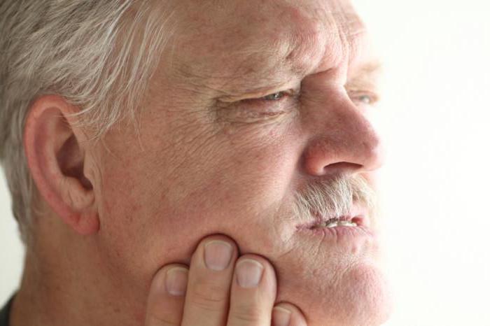
- The presence of shooting pain, localized in the gland that has become inflamed. Pain may spread to the ear, neck or mouth. There may also be pain caused by chewing food or minimal opening of the mouth.
- Swelling and noticeable hyperemia of the skin in direct projection to the salivary gland that has become inflamed.
- The presence of an unpleasant taste and odor in the mouth, which is caused by suppuration of the salivary glands.
Signs of salivary gland disease are varied. Sometimes patients complain of a feeling of pressure on the affected area, which is evidence that purulent contents have accumulated at the site of inflammation.
As a rule, in the presence of the disease, body temperature rises to 40 degrees. In this case, asthenia and a feverish state are noted.

The most dangerous form of sialadenitis
Sialadenitis, the symptoms of which are varied, occurs in different forms. The most dangerous of the salivary glands is considered to be which is also called mumps. This virus is fraught with serious complications, since in addition to the salivary glands, it can also infect other glands, for example, mammary or reproductive glands. Sometimes the pathology even extends to the pancreas.
Mumps belongs to the category of highly contagious diseases, therefore, if standard symptoms appear, indicating the onset of an inflammatory process in the salivary glands, the patient should stop communicating with healthy people and urgently seek help from a specialist to clarify the diagnosis.

In the absence of timely treatment for diseases of the salivary glands, purulent complications may develop in the human body. If an acute abscess occurs in one of the salivary glands, the patient’s body temperature will certainly rise sharply.
As a rule, the general condition of a person is serious. Sometimes pus is released directly into the mouth. A fistula may also form, from which pus oozes onto the skin.
Carrying out diagnostics
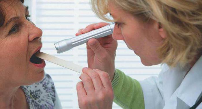
With a disease such as sialadenitis, the symptoms of which are varied, diagnostics is required. Typically, an increase in the size and change in the shape of the salivary glands may be noted during a series of standard examinations performed by a physician or dentist. In addition, the patient may complain of pain. This happens if the disease has a bacterial basis. Often, with viral infections, for example mumps, pain may not bother you at all.
If a purulent process is suspected, the therapist may prescribe a CT scan or ultrasound.
Below is a list of standard diagnostic methods for mumps:
- The use of computed tomography is a modern method that allows you to obtain clear images.
- X-ray.
- MRI (Magnetic Resonance Imaging) provides high-quality images of the affected area using nuclear magnetic resonance.
- Ultrasound examination. This diagnosis is the most common way to detect damage to the salivary glands. It is carried out using ultrasonic waves and has minimal negative effects on the human body.
Preventive measures
To fully prevent the occurrence and subsequent spread of the inflammatory process to other salivary glands, the patient must observe basic hygiene, monitor the condition of the oral cavity, tonsils, gums and teeth.
If elementary diseases of a viral or cold nature occur, timely therapy should be carried out.
At the first signs of disruption of the salivary glands, you should irrigate the oral cavity with a solution of citric acid. This method makes it possible to free the salivary ducts in the most common and harmless way by provoking intense salivation.
Therapy methods
Inflammation should be treated by a specialist, since incorrectly chosen treatment tactics can complicate the course of the disease and provoke its transition to a chronic form. The chronic course is dangerous due to its periodic exacerbations and resistance to the effects of drugs.
If treatment is started in a timely manner, it is usually sufficient for patients to undergo conservative therapy. In some cases, therapy is performed on an outpatient basis. Sometimes the patient requires bed rest and a balanced diet.
In some cases, patients complain of acute pain in the mouth and difficulty chewing. They need to eat crushed food to relieve discomfort.

To reduce the manifestations of a process such as inflammation of the parotid salivary gland, doctors advise drinking plenty of fluids. You can consume compotes, juices, herbal fruit drinks, rosehip decoction and even milk. Local treatment is highly effective.
Sometimes patients are indicated for certain physical procedures. For example, a UHF or Sollux lamp will be used.
To ensure the outflow of saliva, it is recommended to follow a diet that promotes the outflow of saliva. In this case, before eating, you should hold a thin slice of lemon in your mouth.
Before meals, you can eat crackers and sauerkraut. Sometimes cranberries or other acidic foods are used. This makes it possible to avoid stagnation in the salivary glands and promotes the rapid removal of dead cells and bacterial breakdown products.
Depending on the progression of the disease, the doctor can decide when to begin active stimulation of salivation. To reduce body temperature and reduce pain, patients are advised to take non-steroidal anti-inflammatory drugs. For example, Baralgin, Ibuprofen or Pentalgin are used.
If the patient’s condition continues to worsen and specific signs of purulent lesions appear, then in this case they resort to the use of antibiotics.
Surgical intervention
Inflammation of the salivary glands, the symptoms of which we are currently studying the treatment, in some cases can be eliminated surgically. Surgery involves opening and subsequent drainage of the affected gland. This method is especially used for severe purulent processes. In such cases, medications are injected directly into the salivary gland.
Treatment of a disease that has taken a chronic form is considered a very long and complex process.
It should be noted that the chronic form can be either the result of an acute process or a primary manifestation. Often a protracted course is observed with rheumatoid arthritis, Sjögren's syndrome and other pathologies.
The main forms of chronic nonspecific sialadenitis
The chronic nonspecific form is divided into the following types:
- parenchymal;
- interstitial, expressed in damage to the ducts (chronic sialodochitis);
- calculous, characterized by the appearance of stones.
In most cases, the patient does not complain of pain
Chronic disease of the salivary gland in the acute period is characterized by retention of saliva (colic). A thick secretion resembling mucus is released from the mouth of the duct. It tastes salty.

Diseases contributing to the development of sialadenitis
With various pathological processes in the body (diffuse damage to connective tissue, damage to the digestive organs, disruption of the endocrine system, malfunction of the central nervous system), dystrophic diseases of the salivary glands can develop, which are expressed in an increase and disruption of their functionality.
As a rule, a reactive proliferation of intermediate connective tissue occurs, which provokes the development of interstitial sialadenitis. This condition can occur with botulism, diabetes mellitus, thyrotoxicosis, scleroderma, Sjögren's syndrome.
Conclusion
Sialadenitis, the symptoms, diagnosis and treatment of which you already know, is an inflammatory process in the salivary glands. It can be triggered by certain diseases, as well as lack of oral hygiene.
An important condition is the timely implementation of therapy. Otherwise, the disease can take a purulent form and even become chronic. In advanced forms, surgical intervention is indicated.
In addition to the minor salivary glands (labial, buccal, palatine, lingual), the excretory ducts of 3 paired major salivary glands open into the oral cavity: 1) parotid; 2) submandibular and 3) sublingual.
General plan of the building. Each large salivary gland is covered with a connective tissue capsule, from which septa (trabeculae) extend, dividing the gland into lobules. The lobules include terminal sections and intralobular excretory ducts. The intralobular excretory ducts include intercalary and striated ducts.
The terminal sections of the lobules are not the same in each gland. The parotid gland has only protein (serous) terminal sections; in the submandibular region - proteinaceous and proteinaceous mucous membranes; in the sublingual gland - protein, mixed and mucous.
The interlobular trabeculae contain blood and lymphatic vessels, nerves and interlobular excretory ducts, into which the striated intralobular ducts flow. The interlobular ducts empty into the duct of the gland, which opens either into the vestibule of the oral cavity (duct of the parotid gland) or into the oral cavity (duct of the submandibular and sublingual glands).
Parotid salivary glands. These are the largest glands of all the salivary glands, covered with a connective tissue capsule, from which trabeculae extend, dividing it into lobules. The lobules include protein terminal sections, intercalary and striated ducts. These glands belong to the complex branched alveolar glands and produce protein (serous) secretion.
Protein tails They have a round or oval shape, and consist of 2 types of cells: I) glandular cells called serocytes, and 2) myoepithelial. Between the terminal sections there are thin layers of connective tissue that form the stroma of the gland.
Interlobular excretory ducts- the smallest, starting from the terminal sections, consist of an inner layer of cubic or flattened epithelial cells and myoepithelial cells. In the parotid gland, these ducts are well developed and branch. These ducts empty into intralobular striated ducts.
Striated intralobular excretory ducts well developed consist of one layer of prismatic epithelial cells and a layer of myoepithelial cells. The striated ducts flow into the interlobular excretory ducts.
located in interlobular connective tissue. At the sources, these ducts are lined with a two-layer, at the mouth - with multilayer cubic epithelium. The interlobular excretory ducts flow into the common duct of the gland.
Common duct of the gland at the sources it is lined with multilayered cubic, at the mouth - with multilayered squamous non-keratinizing epithelium. The duct pierces the masticatory muscle and opens into the vestibule of the oral cavity at the level of the upper 2nd molar.
Submandibular salivary glands. These are complex, branched, alveolar-tubular glands, located under the lower jaw and also covered with a connective tissue capsule, from which connective tissue trabeculae extend, dividing it into lobules. The lobule of these glands consists of proteinaceous and proteinaceous-mucous terminal sections, intercalary and striated ducts. The structure of the protein terminal sections of the submandibular salivary gland is similar to their structure in the parotid gland.
Protein-mucous (mixed) end sections consist of mucous cells - mucocytes (mucoctus), serocytes and myoepitheliocytes. Serocytes are located along the periphery in the form of serous (protein) crescents of Gianuzzi.
Protein crescents consist of cubic-shaped serocytes, between them there are intercellular microtubules. Mixed end mucocytes located in their central part, have a conical shape, light color, and there are microtubules between them. Myoepitheliocytes of mixed end sections located between the basal ends of serocytes of the protein crescents and the basement membrane. Their function is to participate in the secretion of secretions from glandular cells and terminal sections.
Intercalated intralobular ducts in the submandibular gland they are poorly developed, they are short and do not branch.
Striated intralobular spaces well developed, branching, have extensions. The wall of these ducts includes tall light cells, wide dark cells, goblet-shaped cells and poorly differentiated conical-shaped cells. These cells produce some hormonal products: growth factors, insulin-like factor, etc. The striated ducts flow into the interlobular ones.
Interlobular ducts at the sources they are lined with two-layer, at the mouth - with multilayer cubic epithelium. They flow into the gland duct.
gland duct, lined at the origins with multilayer cubic epithelium, at the mouth with multilayered squamous epithelium, opens under the tongue, next to its frenulum.
Sublingual salivary glands. These are the smallest glands among the major salivary glands. They are also covered with a connective tissue capsule and are also divided into lobules by trabeculae extending from the capsule. In the lobules of these glands there are 3 types of terminal sections: 1) protein: 2) protein-mucosal and 3) mucous. The proteinaceous and proteinaceous-mucosal terminal sections are similar in structure to the previously described proteinaceous ones in the parotid gland and the proteinaceous mucous membranes in the submandibular gland.
Mucous end sections consist of conical mucocytes and myoepitheliocytes. Mucocytes are light in color and have intercellular microtubules between them. The functional significance of these cells is the synthesis and secretion of mucous secretions. Myoepitheliocytes are located between the base of mucocytes and the basement membrane.
Intercalated excretory ducts poorly developed.
Striated excretory ducts in the sublingual salivary glands are poorly developed. They flow into the interlobular excretory ducts.
Interlobular excretory ducts at the sources they are lined with two-layer, at the mouth - with multilayer cubic epithelium; flow into the gland duct.
gland duct, initially lined with stratified cubic epithelium, at the mouth with stratified squamous epithelium, it opens next to the duct of the submandibular salivary gland.
