Asymmetry of v4 segments of vertebrates. Hypoplasia of the intracranial v4 segment of the right vertebral artery: MRI signs, consequences
Post date: 07.08.2011 16:16
shift
My mother did an MRI: MRA of the cerebral arteries + venous sinuses.
This is what showed
A series of MR angiograms performed in TOF mode in an axial projection with subsequent processing using the MIP algorithm and three-dimensional reconstruction in the coronal and axial planes visualized the internal carotid, main, and intracranial segments of the vertebral arteries and their branches. A variant of the development of the circle of Willis in the form of a lack of blood flow through the left posterior communicating artery. The median artery of the corpus callosum is visualized, equal in diameter to the anterior cerebral arteries.
There is a moderate narrowing of the lumen of the intracranial segment of the right vertebral artery, along its entire length in the study area.
A pronounced asymmetry of the lateral ventricles (D>S) is determined.
In a series of MR venograms of the brain performed in TOF mode, followed by processing using the MIP algorithm and three-dimensional reconstruction in the coronal projection, the internal and external jugular veins and their branches, sinuses (superior longitudinal, straight, sigmoid and transverse sinuses).
There is a pronounced asymmetry of blood flow along the transverse and sigmoid sinuses (D>S). The remaining sinuses are without features.
No additional venous network was identified.
Conclusion: MR picture of the development variant of the Circle of Willis. Moderate narrowing of the lumen of the intracranial segment of the right vertebral artery. Asymmetry of blood flow along the transverse and sigmoid sinuses (D>S). Lateroventriculoasymmetry.
Please decipher what is here and, if so, how to treat it. she is very worried because... nothing is clear.
Post date: 07.08.2011 20:43
Papkina E.F.
shift, your mother’s MRI angiography reveals impaired blood flow in the area of the right vertebral artery and uneven blood flow through the sinuses, where venous blood flows. This is most likely a developmental option, that is, it has been like this since birth. Under conditions of stress or physical exertion, with age-related changes vessels, a previously silent anomaly of vascular development can manifest itself as headaches, dizziness, loss of memory, and loss of coordination. You need to consult a neurologist to carry out adequate treatment and eliminate neurological symptoms. The prognosis is favorable, since in most cases the surrounding vessels take over their functions narrowed, altered vessel.
Post date: 08.08.2011 19:40
Guest
Commenting on MRI without a clinic is not grateful. I think your doctor sent you for research? So you need to ask him if he found what he was looking for.
Post date: 05.10.2011 19:48
Guest
consultations of this kind can be given by an MRI doctor (he was specially trained for this purpose!))) and treatment is prescribed by the attending physician who referred.
A full examination is necessary - this also includes an MRI of the brain (the cause of lateroventriculoasymmetry) and cervical spine spine (a decrease in blood flow through one of the vertebral arteries may not be congenital, but acquired as a result of osteochondrosis!)
Post date: 25.06.2012 12:28
Guest
My son had a draft board, they did an MRI (complaints of headaches, pain in right temple, B in the conclusion they wrote: MRA picture of the variant development of the Circle of Willis in the form of decreased blood flow in both posterior communicating arteries. Please explain what kind of circle of Willis this is, and how dangerous it is for health, what to do with the army?
Post date: 27.06.2012 19:17
Papkina E.F.
In practice, very often there are variants of the development of the so-called circle of Willis (this system of arterial supply to the base of the brain). These changes are not life-threatening. The issue of conscription is decided individually after an examination by a neurologist.
Post date: 24.04.2013 22:46
Olga
The woman underwent an MRI, the conclusion: the MR picture is moderately pronounced external replacement hydrocephalus. Option for the development of the Circle of Willis. Decreased blood flow in the intracranial segment of the right VA. HELP DECORD.
Post date: 14.11.2013 23:50
Angela
My daughter gave an MRI report...please explain how serious this is and whether it can be treated. She has a 9 month old baby and we are very worried. Conclusion: MR picture of AVM in the left MCA and PCA basin. The circle of Willis is closed. Decreased blood flow along the A1 segment of the left ACA (hypoplasia). Decreased blood flow in the intracranial segment of the right VA (hypoplasia).
Post date: 30.11.2013 17:40
Galina
I had an MRI today. I have pain in my neck when I turn my head.. the noise in the left one is already... terrible, for about 10 years.. the right eye can’t listen.. double vision when looking straight.. I tilt my head to the left.. the double vision goes away... so I’ve been adapting for more than 10 years... tilting my head to the left. Here is the result -
In a series of MR tomograms (T2 TSE sag+cor+tra, T1 SE sag), the lordosis is straightened. The height of the intervertebral discs C4-C7 is reduced. In the segments C4-C5, C5-C6, C6-C7, marginal bone sharpenings are determined along the posterior and posterolateral sides surfaces of the vertebral bodies, covering old disc protrusions, smooth out the anterior contour of the dural sac. A small perineural cyst in the right intervertebral foramen C6-C7, with a cross-section of 0.5 x 0.4 cm. The lumen of the spinal canal at the level of C4-C7 is 1.1 cm. Spinal the brain is structural, the signal from it (according to T1 and T2) is not changed. The shape and size of the vertebral bodies are normal, without signs of bone marrow edema. The cranio-vertebral region is without features. Asymmetry in the width of the vertebral arteries S>D. Doctor, this is indicated for me manual therapy? And what else? Thank you.
Post date: 16.12.2013 19:46
Catherine
What is the asymmetry of the transverse and sigmoid sinuses D>S, moderately pronounced dilation of collateral veins in the left PCF and what does this mean?
Post date: 04.02.2014 13:31
Ramilla
Good afternoon Tell me please! I did an MRI, but I don’t understand everything. A variant of the development of the circle of Willis is the absence of blood flow in both posterior communicating arteries (open). The vertebral arteries in the v5 segment are asymmetrical: d up to 2.2 mm on the right, up to 3 on the left. Transverse sinuses are symmetrical: d up to 7 mm. In the white matter of the left frontal and parietal lobe subcortically, single foci are identified, max sizes up to 4 mm, hyperintense on T2 VI - probably of vascular origin. The subarachnoid space is locally expanded in the frontoparietal regions. The right vertebral artery is of small diameter. How dangerous is this? I am worried about severe headaches, but not constantly, as well as blood pressure up to 150 over 100, all these symptoms appeared during pregnancy, after that they seemed to go away, but now they are bothering me again. I am 24 years old, I gave birth 3 months ago. I would like to know what this means and what a diagnosis! Thanks in advance!
Post date: 11.06.2014 12:45
Guest
MRI: MR signs of decreased blood flow in segments A1 of both ACAs, segments M3 M4 of both MCAs, in the distal segments of both PCAs, segment V5 of the ACA, most likely due to atherosclerotic changes. What does this mean and what therapy is needed. What are your recommendations?
Post date: 15.06.2014 18:00
Irishka
Please tell me whether it is worth doing an MRI on the machine open type, like this http://radio-med.ru/makers/mrt/open-mri-10/hitachi-airis-ii-0.3t
Or should I still find a clinic with the usual one?
What do you think about mobile MRI scanners?
Post date: 21.08.2014 19:19
Natalya *d*
help me figure it out! MRA signs of lack of blood flow in the right vertebral artery, pathological tortuosity of the internal carotid arteries,development option venous system with decreased blood flow in the transverse sinus on the left. Thank you.
Post date: 25.08.2014 14:17
Christina
Hello! Please consult the results of MRI of the brain vessels. I went for examinations due to frequent headaches from an early age, now I am 23. Here is the conclusion: there is a variant of the development of the circle of Willis (aplasia A1 of the ACA on the right, aplasia of the posterior communicating artery on the right and hypoplasia on the left ).MR signs of lack of blood flow in the intracranial segment of the right vertebral artery (aplasia). Please tell me how serious this is and what treatment to choose? Thank you in advance for your consultation
Vertebral artery syndrome with cervical osteochondrosis- this is one of the most insidious and severe ailments that can strike different people. Cervical spinal stenosis affects both old and young patients. It is caused by disturbances in the supply of blood to brain cells due to lesions in one or both arteries that carry blood components to the hemispheres of the brain.
Causes of vertebral artery syndrome
This phenomenon occurs in several cases:
- Influenced by various unfavorable factors in a person, the main vessels that supply the brain cells with blood are compressed. Usually, narrowing occurs in one of the arteries, less often in both.
- Nutrients and oxygen stop getting into the right places in the required quantity.
- Show themselves various signs illness: dizzy, dark vision.
- At untimely treatment A stroke of an ischemic nature may develop.
- A risk factor may be atherosclerosis or hypoplasia of the spinal artery.
 Blood enters the brain mainly through the carotid arteries (up to 70%), and the rest is nutrient liquid is coming along 2 lateral vessels. When the main blood flow channels are damaged, lesions occur that are usually incompatible with life, and if there are problems in the other 2 arteries, a person’s health may worsen, then vision problems and damage will begin hearing aid, which can lead to disability.
Sometimes the disease is caused by asymmetry of blood flow along blood vessels spine - it cannot be treated, but can develop into other diseases. Another risk factor is instability in the cervical spine, which leads to prolapsed discs. spinal column. This can also happen after receiving an injury (both ordinary and birth), when sedentary life.
The appearance of the syndrome may great influence cause osteochondrosis or a pathology known as tortuosity of the arteries in the spine.
Symptoms of the disease
It is quite difficult to immediately identify signs of vertebral vein syndrome when a person is diagnosed with spinal canal stenosis in the cervical region. This is because the manifestations of this disease strongly resemble osteochondrosis or diseases that usually cannot be associated with problems in the spine. Therefore, if at least one symptom of those listed below is detected, it is necessary to urgently take the patient for examination to a medical institution.
 The most common symptom of this disease is headaches. They can manifest themselves in the form of attacks that come over the patient with some frequency, or as constant painful sensations. The main area of distribution of such pain is the back of the head, but they can move to the temporal lobes and to the frontal part.
Over time, such pain intensifies and manifests itself when bending or turning the head. Then the pain moves to the skin under the hair. It appears when you touch your hair with your hands. This action may cause a burning sensation. The vertebrae of the neck begin to crunch when turning the head.
All of the above troubles are complemented by the following symptoms:
- The patient's blood pressure increases.
- There is noise and ringing in the ears.
- The person may vomit.
- Pain begins in the area of the heart muscle.
- The patient gets tired quickly.
- With frequent dizziness, the patient may faint or lose consciousness.
- Spinal stenosis causes sharp pain in the cervical region.
- arise various disorders in the eyes and pain in the ears. Usually they are one-sided.
![]() If the disease lasts for a long time, it can cause increased pressure inside the skull and vegetative-vascular dystonia. The patient's fingers become numb. A person develops irritability, mood swings, and unreasonable fear.
If the disease lasts for a long time, it can cause increased pressure inside the skull and vegetative-vascular dystonia. The patient's fingers become numb. A person develops irritability, mood swings, and unreasonable fear.
Diagnosis of arterial syndrome in cervical osteochondrosis
The examination of the patient begins with an external examination. At the same time, doctors pay special attention factors such as: painful sensations on skin the patient has muscle tension in the occipital region, pain on the vertebrae of the neck when pressing.
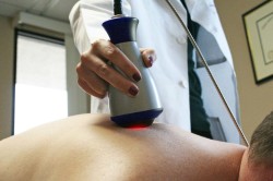
Diagnosis is carried out using Doppler ultrasound. Using this equipment, you can examine all the vessels that supply the brain with blood, determine their condition at the time of testing and identify various disorders and deviations.
Another way to diagnose the disease is by using radiographic equipment. If during the examination a person becomes worse, then to clarify the reasons, the patient is sent for magnetic resonance imaging to check areas of the brain. Based on the results of such a study, the patient may be urgently hospitalized.
Errors in diagnosing the disease (they are possible, since the disease's symptoms coincide with other diseases) can lead to irreparable consequences. Therefore, when making a primary diagnosis, it is advisable to repeat the examination in order to identify signs of the disease that were missed during the first diagnosis.
What methods are used to treat the disease?
If the cause of the disease is precisely established and it is pinched arteries, then doctors prescribe a course of therapy that should help completely cure the person. Treatment should be carried out under the strict supervision of the attending physician, even when the patient is at home.
Self-medication in this case is strictly prohibited, as it can lead to irreversible consequences, even death.
 The treatment process must be complex. Below is a list existing methods fight the syndrome. Doctors can use all of these methods, or choose the most appropriate for a particular case. The disease is treated in the following ways:
- A course of vascular therapy is prescribed.
- The patient is prescribed therapeutic exercises.
- The patient is discharged medications to improve blood flow.
- To get rid of fainting, it is recommended to use special stabilizing medicines. They are also needed to relieve dizziness, vomiting, nausea and eliminate problems with the vestibular system.
- Sometimes acupuncture and acupuncture methods are used.
- The patient is prescribed a massage, which must be performed by a licensed specialist.
- In some cases, manual therapy may be used.
- Reflexology may be used.
- IN curative therapy include autogravitational methods of curing the disease.
Non-drug types of therapy can also be used, which are prescribed only by the attending physician. This is done depending on the severity of the disease, its stage and the main reasons that led to the appearance of the syndrome. The main thing is complex use various methods for the most effective fight with illness.
If a patient has a congenital pathology of asymmetry of the spinal arteries, then doctors will only cure the secondary syndrome, and the main cause is incurable. This can lead to relapse of the disease if the person does not follow the recommendations of the attending physician.
According to the WHO definition (1970), vertebrobasilar insufficiency is “a reversible disorder of brain function caused by a decrease in the blood supply to the area supplied by the vertebral and basilar arteries”
Both vertebral and basilar arteries form vertebrobalibular system (VBS), which has a number of features.
It supplies blood to various and functionally heterogeneous formations: the posterior sections cerebral hemispheres brain (occipital lobe and mediobasal parts of the temporal lobe), thalamus optic, most of the hypothalamic region, cerebral peduncles with quadrigeminal, pons, medulla oblongata, reticular formation of the trunk - reticular formation (RF), upper sections spinal cord.
The same sections often have several sources of blood supply, which determines the presence of zones of adjacent blood circulation, which are more vulnerable in case of circulatory failure.
Trunk is supplied by intracranial sections of the vertebral arteries and their branches, the main artery and its branches. The area of adjacent blood supply is the reticular formation.
Cerebellum receives blood supply from three pairs of cerebellar arteries: superior and anterior inferior(branches of the basilar artery) and posterior inferior cerebellar artery (terminal branch of the vertebral artery).
Especially significant area adjacent blood supply- area of the worm.
Posterior parts of the cerebral hemispheres receive blood supply from front, middle(branches of the internal carotid artery) and s posterior cerebral artery(terminal branch of the basilar artery).
The most important area of adjacent blood supply: posterior third of the interparietal sulcus(the junction zone of the branches of all three cerebral arteries); cuneus and precuneus, posterior part of the corpus callosum and pole of the temporal lobe(junction area between PMA and PMA); superior occipital, inferior and middle temporal and fusiform gyri(junction area of the PCA and SMA).
Fusion of the vertebral arteries into the main - unique feature all arterial system , because the main artery represents a pre-prepared path collateral circulation without spending time on its formation. This has a positive meaning - the rapid inclusion of collateral circulation leads to the restoration of blood flow in the vertebral artery during its compression, and a negative one, because creates conditions for the development of the syndrome "subclavian steal", i.e. with proximal obstruction subclavian artery before the vertebral body leaves it, blood is redistributed to the arm, sometimes to the detriment of the VBS, which can, if hard work hand lead to the development of transient ischemia in VBS.
IN normal conditions blood flows from the vertebral arteries continue their movement in the main artery, maintaining the same volumes of blood flow and without mixing with each other. Zones of “mobile” (dynamic) equilibrium are created between these flows. Occlusion or stenosis of one of the vertebral arteries disrupts it, mixing of flows occurs, displacement of zones of “moving” equilibrium and blood flow from another vertebral artery through the main artery. This can lead to the development of thrombosis even without pronounced atherosclerosis - “stagnant” blood clots at points of “moving” equilibrium.
Small penetrating arteries They depart from the large arteries (basilar, posterior cerebral) at a right angle, have a straight course and the absence of lateral branches.
Blood circulation in the VBS (according to angiography) is two times slower than in the carotid system. Cerebral blood flow in the cerebral hemispheres (internal carotid artery system) is 55-60 ml per 100 g of brain tissue per minute, and in the cerebellum - 33. This enhances the influence of the hemodynamic factor in the development of reversible cerebral ischemia in VBS. Transient ischemic attacks in the VBS are much more common, accounting for 70% of all TIAs. Collateral circulation, improving or restoring cerebral perfusion, develops and is created in case of stenosis or occlusion of the artery based on existing anastomoses. Of the intracranial anastomoses, the circle of Willis is extremely important. A decrease in blood flow in the VBS leads to retrograde blood flow through the posterior communicating arteries, sometimes to the detriment of the carotid system - “internal theft”. Extracranial retromastoid anastomosis provides two additional sources of blood supply for the VBS. Large branches arising from the vertebral artery at the level of the atlas anastomose with the branches of the occipital artery from the external carotid artery system and the ascending and deep cervical arteries from the subclavian artery system. Anastomoses between cerebellar arteries: back lower ( final branch vertebral artery) and the superior and anterior inferior cerebellar arteries (branches of the basilar artery). Good development anastomosis ensures sufficient functioning of collaterals and, in the event of a decrease in blood flow in the VBL, prevents the development of neurological disorders.
In 70% of cases, the left vertebral artery is 1.5-2 times wider than the right
, which predetermines its importance as the main source of blood supply posterior sections brain The asymmetry of the caliber of the vertebral arteries creates the possibility of thrombus formation in the main artery.
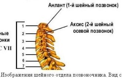
The uniqueness of the course of the vertebral artery: at the level of the CVI–CII cervical vertebrae it runs in its own bone canal, then, emerging from it, bends around the CI, describing an outwardly convex arc around it, then rises upward and, perforating the hard meninges, through the foramen magnum enters the cranial cavity.
Anomalies of vascular development are common in VBS. 20% In patients with VHD pathology, anomalies in the development of the vertebral arteries are detected. According to Powers et al, (1963) hypoplasia occurs in 5-10%
cases, aplasia - 3%
, lateral displacement of the mouth of the vertebral artery – in 3-4%
, origin of the vertebral artery from the posterior surface of the subclavian artery – 2%
, entry of the vertebral artery into spinal canal at the level of CV, CIV, sometimes CIII – at 10,5%
cases, there are also other anomalies: the origin of the vertebral artery from the aortic arch, from the subclavian artery in the form of two roots, etc.
A decrease in blood supply with insufficient compensation by collateral circulation leads to the development of ischemia of brain tissue fed from the VBS.
Pathogenesis of ischemia.
Thanks to research recent years it has been shown that cerebral ischemia, or circulatory hypoxia of the brain, is a dynamic process and involves the potential reversibility of functional and morphological changes in brain tissue, not being identical to the concept “ cerebral infarction", reflecting the formation of an irreversible morphological defect - structural destruction and disappearance of neuronal function. The stages of hemodynamic and metabolic changes occurring in brain tissue at various stages of circulatory failure have been identified. A scheme of successive stages is proposed "ischemic cascade" based on their cause-and-effect relationships (Gusev E.I. et al., 1997,1999):
>decrease cerebral blood flow;
> glutamate “excitotoxicity”;
> intracellular accumulation of calcium ions;
> activation of intracellular enzymes;
> increased synthesis of nitric oxide NO and development of oxidative stress;
>expression of early response genes;
> “long-term” consequences of ischemia (reaction local inflammation, microvascular disorders, damage to the blood-brain barrier;
> apoptosis.
For normal course metabolism brain tissue requires constant cerebral blood flow to ensure sufficient supply to the brain nutrients: proteins, lipids, carbohydrates (glucose) and oxygen. Stable maintenance of cerebral blood flow at a level of 50-55 ml/100 g of brain tissue per minute. at the level of the hemispheres and 33 ml/100 g of brain tissue per 1 min. at the level of the cerebellum is supported by autoregulation of cerebral blood flow, which at the level of large vessels is carried out reflexively due to adrenergic and cholinergic receptors of their walls with the help of the regulatory mechanism of the carotid sinus and chemical regulation in the vessels of the microvasculature (with excess supply of O2, i.e. hypocapnia, the tone of the precapillary arterioles increases; with insufficient supply of O2 to the brain, hypercapnia, the tone decreases; under conditions of increasing the amount of carbon dioxide, the sensitivity of microvessels to it increases). The rheological properties of blood (viscosity, aggregation ability) matter shaped elements blood, etc.) and the value of perfusion pressure, which is defined as the difference between mean blood pressure and mean intracranial pressure. Critical level cerebral perfusion pressure – 40 mmHg, below this level cerebral circulation decreases and then stops.
In case of acute circulatory failure In a specific area of the brain, the latter is capable of temporarily compensating for local ischemia through mechanisms of autoregulation and enhancing collateral blood flow. However, a further decrease in cerebral blood flow leads to a breakdown of autoregulation and the development of metabolic disorders. It has been established that the processes of consumption of O2 and glucose by the brain occur in parallel. Glucose is the only supplier of energy necessary for the normal course of metabolic processes, because most of them are energy-dependent: synthesis of proteins, many neurotransmitters, binding of a neurotransmitter to a receptor, impulse transmission, ion exchange through plasma membrane etc. The first reaction to brain hypoxia occurs in the form of inhibition of protein synthesis. Protein and RNA synthesis occurs more actively in the cerebral cortex and cerebellum. Glucose metabolism usually occurs with a predominance of the aerobic pathway, giving more high-energy compounds (36 ATP molecules from 1 glucose molecule). Increasing hypoxia leads to the predominance of anaerobic glycolysis, which is more energetically disadvantageous (2 ATP molecules from 1 glucose molecule). Due to energy deficiency in mitochondria, oxidative phosphorylation is inhibited, and lactic acid accumulates in the cell. At the same time, the carbon dioxide content in the brain tissue increases and the pH shifts to the acidic side. Lactic acidosis occurs. As a result, in the focus of ischemia there is a decrease in cerebral blood flow, while in its surroundings there is an increase in blood flow to the detriment of the ischemic zone - the phenomenon of “luxurious perfusion” (according to Lassen). The growing energy deficit under these conditions leads to further disruption of energy-dependent processes. The transition to anaerobic glycolysis leads to an increase in alpha-ketoglutaric acid, unused in the Krebs cycle, into the amino acid glutamate, which also has the properties of an excitatory mediator (Swanson et al., 1994). In addition, increasing lactic acidosis blocks the reuptake of glutamate. Thus, the excitatory neurotransmitter accumulates in the intercellular space, which leads to the development of “glutamate excitotoxicity,” i.e. stimulation of cells by glutamate. Lactic acidosis in combination with increasing hypoxia causes disorder electrolyte balance nerve and glial cells: the release of K+ ions from the cell into the extracellular space and the movement of Na+ and Ca++ ions into the cell, which suppresses the excitability of neurons and reduces their ability to conduct nerve impulses.
Exciting amino acids(glutamate, aspartate) act on neuronal receptors for N-methyl – D-aspartate (NMDA receptors), which control calcium channels. Their overexcitation leads to a “shock” opening of ionic calcium channels and additional excess influx of Ca++ ions from the intercellular space into neurons and its accumulation in them.
Norepinephrine, the release of which initially increases sharply during hypoxia, activates the adenylate kylase system, which stimulates the formation of AMP, which causes an increase in energy deficiency and leads to an increase in Ca++ ions in nerve cells.
Excessive intracellular accumulation of Ca++ ions leads to the activation of intracellular enzymes: lipase, protease, endonuclease, phospholipase and the prevalence of catabolic processes in the nerve cell. Under the influence of phospholipases, phospholipid complexes disintegrate in the membranes of mitochondria (phospholipase A2), intracellular organelles (lysosomes) and in the outer membrane. Their breakdown enhances lipid peroxidation (LPO). The end products of LPO are: malondialdehyde, unsaturated fatty acids(especially arachidonic acid) and O2 free radicals. End products of decomposition arachidonic acid: thromboxane A2 and others, hydroperoxides, leukotrienes. Thromboxane A2 and others cause spasm cerebral vessels, enhance platelet aggregation and coagulation changes in hemostasis. Leukotrienes have vasoactive properties. Microvascular disorders lead to an increase in ischemia in the ischemic area. Free radical O2 is a molecule or atom that has an unpaired electron in its outer orbit, which makes it aggressive in converting cell membrane molecules into free radicals, i.e. provide a self-sustaining avalanche-like reaction. Activation of lipid peroxidation processes is also facilitated by the rapid depletion of the antioxidant system, the enzymes of which inhibit the formation of peroxides and free radicals and ensure their destruction. In addition, in the ischemic focus the content of substances decreases: alpha-tocopherol, ascorbic acid, reduced glutamate, which bind the end products of LPO. The accumulation of hydroperoxides leads to the formation of hydroxy acids and the development of oxidative stress.
Activated by increasing hypoxia, microglial cells synthesize potentially neurotoxic factors: proinflammatory cytokines (interleukins 1,6,8), tumor necrosis factors, ligands for the glutamate NMDA receptor complex, proteases, superoxide anion, etc. Excitation of NMDA receptors leads to activation of the NO- enzyme synthetase involved in the formation of nitric oxide from arginine. The complex of nitric oxide with superoxide anion helps reduce the production of neutrophins. Neutrophins are regulatory proteins nerve tissue, synthesized in its cells (neurons and glia), acting locally - at the site of release and inducing dendritic branching and axonal growth. These include: nerve growth factor, cerebral growth factor, neutrophin-3, etc. Anti-inflammatory factors (interleukins 4,10) and neutrophins prevent the damaging effects of neurotoxic factors, the end products of LPO, on the ultrastructures of nerve and glial cells. Destruction of the phospholipid complex nerve cells leads to the production of antibodies to them. The release of anti-inflammatory and vasoactive substances from ischemic brain tissue leads to the penetration of neurospecific proteins into the blood, which leads to the development of an autoimmune reaction and the production of antibodies to the nervous tissue.
Under conditions of increasing energy deficiency, further inhibition of the synthesis of RNA, proteins, phospholipids, and neurotransmitters occurs. Inhibition of the synthesis of neurotransmitters disrupts connections between neurons and deepens metabolic disorders in them. A decrease in protein synthesis in the ischemic focus leads to the expression of cell death genes and triggers a genetically programmed mechanism of cell death - apoptosis, in which the cell disintegrates into parts in the form of apoptotic bodies, separated in membrane vesicles that are absorbed by neighboring cells and/or macrophages. IN pathological process glial cells are involved faster and to a greater extent, brain neurons are involved more slowly and less significantly (Pulsinelli, 1995). At this stage of ischemia, metabolic disorders are reversible.
The volume of blood flow is 10-15 ml per 100 g of brain tissue per minute. - critical threshold beyond which irreversible changes– necrosis (Hossman, 1994), which is accompanied by the release of cell contents into the intercellular space and the development of an inflammatory reaction.
Acute failure cerebral circulation in VBS is considered as transient disorder cerebrovascular accident or transient ischemic attack (TIA) in VBS. It is characterized acute occurrence focal neurological symptoms are usually without cerebral symptoms(less often against the background of their weak severity) due to short-term local cerebral ischemia. Focal neurological symptoms last from several minutes (usually 5-20 minutes) to several hours (less often up to 24) and end full restoration impaired functions within 24 hours. Chronic cerebral circulatory failure in VBS is considered discirculatory encephalopathy. However, the ischemic process of brain tissue in dyscirculatory encephalopathy is irreversible and is accompanied by the development of necrosis (i.e. infarctions in the white matter and basal ganglia, less often in the optic thalamus, pons) and an inflammatory reaction (spongiosis, proliferation of astrocytes, myelin disintegration with partial disintegration of the axial cylinders), occurring mainly perivascularly. In this case, CT and MRI reveal infarcts in the white matter and subcortical nodes and signs intracranial hypertension: expansion of the ventricles of the brain (mostly the anterior, less often the posterior horns of the lateral ventricles with the phenomenon of “leukoaraiosis” around them due to a decrease in density white matter) or cortical atrophy with expansion of the subarachnoid spaces of the cerebral hemispheres. For the same reason, chronic cerebral vascular insufficiency in the arteries of the VBS cannot be considered as a minor ischemic stroke, i.e. lacunar infarction. This makes it possible to distinguish TIA and CNMK in VBS in special shape
vascular pathology brain - vertebrobasilar insufficiency (VBI). The ischemic process in VBI is reversible and CT and MRI, as a rule, are not detected morphological changes.
In the mechanism of origin of VBN more significant are the atherothrombotic and hemodynamic factors, “subclavian steal”, less significant: the embolic factor, vasospasm and changes rheological properties blood (hyperlipidemia, hyperfibrinemia, polycythemia, etc.).
The following lead to the development of VBN (in order of importance):
occlusive and stenotic lesions of the VBS arteries (especially stenosis of the vertebral arteries and main thrombosis);
1) deformation of the vertebral arteries;
2) extravasal compression of the extracranial parts of the vertebral arteries.
Occlusions more often develop as a thrombosis, less often as an embolism. The main cause of occlusion is atherosclerosis. Atherosclerotic plaques in atherosclerosis are often localized at the mouths of the vertebral arteries, in the area of bifurcation of the basilar artery and at the mouths of their branches. As atherosclerotic plaques disintegrate, they become the cause of thrombosis. A blood clot that has broken away from atherosclerotic plaque, clogs the distal branches of these arteries. The second most important is arterial hypertension. It plays a dual role: firstly, it promotes the formation and development of atherosclerotic plaques at the mouths of small penetrating arteries and their embolism (which is facilitated by the characteristics of these arteries) and, secondly, they cause pathological tortuosity of these vessels, changing vascular wall. Parietal thrombi are of less importance in vasculitis: nonspecific aortoarthritis (or pulseless disease or Takayasu disease) and secondary manifestations in tuberculosis, SLE, syphilis, AIDS, etc. Rarely, emboli in the vertebral arteries can come from atheromatous plaques or vegetations on the heart valves in diseases hearts, even less often from veins lower limbs And internal organs at congenital pathology heart (non-closure of the foramen ovale).
When you suddenly throw your head back the vertebral artery may be strangulated by the posterior edge of the foramen magnum. With lateral displacement of the mouth of the vertebral artery, turning the head can lead to compression of the vertebral artery, often together with the subclavian artery.
The vertebral artery can be elongated and have a “C” and “S” shaped course, go in the form of a loop, or have kinks and tortuosities. The pathology can be congenital or acquired (with hypertension, age-related changes).
Cervical osteochondrosis can cause compression of the vertebral artery due to the penetration of lateral and posterolateral osteophytes of the uncovertebral joints into its canal, as well as by the spasmodic scalene muscle (scalenus syndrome) in the section before it enters the bone canal. Paired intervertebral joints forming back wall canal of the vertebral artery are introduced into the canal during subluxations according to Kovacs (slipping in front of the vertebral body) due to inferiority of the muscular and ligamentous-articular apparatus or injury to the cervical spine, due to arthrosis with deforming spondyloarthrosis, rheumatoid arthritis, ankylosing spondylitis. The vertebral artery is especially often subject to compression due to anomalies of the craniovertebral junction. This is the Kimmerle anomaly, i.e. an abnormal bony canal instead of a wide and shallow groove of the vertebral artery on the dorsal side of the lateral mass of the atlas; assimilation of Atlas (CI), i.e. its fusion with the base occipital bone; asymmetry or high location of the odontoid process of the epistrophy (CII), basilar impression, i.e. funnel-shaped depression of the hypoplastic edges of the foramen magnum, atlanto-occipital joints and distal parts of the Blumenbach clivus into the cranial cavity and the frequent combination of the latter with an anomaly of the CI and CII vertebrae; Arnold-Chiari malformation, i.e. descent of the cerebellar tonsils through the foramen magnum into the upper parts of the spinal canal. Long-term compression of the vertebral arteries contributes to the formation and growth of atherosclerotic plaques.
Clinical picture
The clinical picture is determined location and degree of damage to the arteries of the vertebrobasilar region, general condition hemodynamics, blood pressure level, state of collateral circulation and is manifested by transient focal neurological disorders in several (at least two) different departments brain powered by VBS. Particularly significant are dizziness, ataxia and visual disturbances (according to Hutschinson, 1968). An attack of dizziness is often the first symptom of VBI, often accompanied by nausea and vomiting. The cause of dizziness is: ischemia of the labyrinth, vestibular nerve and/or trunk. In the first two cases, the dizziness is systemic: by the type of rotation of an object with the appearance of horizontal or rotatory nystagmus, often accompanied by hearing impairment; in the second - non-systemic, increasing when turning the head with small-scale horizontal nystagmus, accompanied by dysphonia and dysarthria. With ischemia of the occipital cortex, visual disturbances occur: simple photopsia (flickering of sparkles, stars, etc.), visual hallucinations, visual field defects in the form of homonymous hemianopsia, often of the upper quadrant type. Transient ischemia of the mesencephalic part of the brain stem is manifested by oculomotor disorders in the form of diplopia, paresis oculomotor muscles, short-term gaze paresis (vertical or horizontal) with convergence paresis, mild strabismus, eyelid ptosis. Ischemia in the area of the ascending systems of the Russian Federation can cause loss of consciousness. It is usually accompanied by brainstem symptoms: double vision, dizziness, nystagmus, dysarthria, facial numbness, ataxia or hemianopsia. Transient ischemia inferior olive and RF may cause a sudden fall attack due to bilateral leg weakness and immobility. An attack of a sudden drop in postural tone without loss of consciousness is called a drop attack. With VBI, after an attack the patient cannot get up immediately, although he was not injured. Transient ischemia of the mediobasal areas temporal lobes accompanied by the development of global amnesia - short-term loss RAM. During this period, patients are not completely adequate, lose their plan of behavior, and do not express their thoughts clearly. After a few hours, they develop transient amnesia for a certain period of time. Transient cerebellar ischemia causes ataxia, most often in standing and walking. Paresis of facial muscles is often observed peripheral type(the entire half of the face). Sensitivity disorders: paresthesia, hyper- and hypoesthesia, more often around the mouth, less often on both halves of the face or body, in the limbs in any combination, including all four. Movement disorders manifest themselves in the form of increased tendon reflexes, weakness, awkwardness of movements in the limbs in any combination. During various attacks, the side of motor and sensory disturbances changes. An inverted type of transient paresis is noted - muscle weakness, mainly in the proximal parts of the arm and/or leg, and a more persistent nature of changes in tendon reflexes. Unlike strokes, VBS is not characterized by alternating trunk symptoms. The permanent form is also characterized by headaches in the occipital region, which sometimes appear in the paroxysmal form.
Currently in the diagnosis of VBI The leading place is occupied by ultrasound research methods. Ultrasound Doppler ultrasonography (USDG) is based on the Doppler effect: when a sound source moves relative to the receiver, the frequency of the sound perceived by it differs from the frequency of the source sound by an amount directly proportional to the speed of the relative (linear) movement. Ultra beep from moving blood (erythrocytes) is perceived by a device that records LSV (linear velocity of erythrocytes). For analysis ultrasound results the LSC asymmetry coefficient is displayed. It is defined as the ratio of the difference in BFB in both vertebral arteries to the lesser BFB in one of them and is expressed as a percentage. Normally it does not exceed 20%. The method allows you to detect stenosis with a degree greater than 50%. Duplex scanning allows you to obtain an image of the vascular bed with the characteristics of blood flow in it. In this case, the ultrasonic signal sent in the direction of the vessel being examined is reflected from moving red blood cells. The difference between the frequency of the sent and reflected ultrasound wave is the linear speed of blood flow. By sequentially scanning the area above the vessels with a sensor mounted on the sensor arm, data on the spatial location of the sensor is obtained, synchronous with the Doppler signal, which is subjected to computer analysis and on the basis of which a map of the studied vascular zone is drawn - a Doppler ultrasonogram. It can be either in gray or color, if the device has a color Doppler coding program.
Normal spectrogram cerebral vessels has the form of a pulse half-wave lying above the isoline, with a systolic peak and a diastolic incisura. Changes in blood flow lead to changes in Doppler spectra. With arterial stenosis, the speed of movement in the stenotic area increases in proportion to the degree of stenosis, and upon exiting it there is an expansion of the speed range and a partially reverse movement of blood. Accordingly, on the spectrogram this looks like a sharp increase in the amplitude of the systolic peak, an expansion of the speed range, and with a stenosis of 75% or more, the appearance of spectral components below the isoline. A complete ultrasound examination includes recording spectrograms of the main great vessels head: common (CCA), external (ECA), internal (ICA) carotid arteries, right and left vertebral arteries (VA), branches of the ophthalmic artery (OA) and facial artery. Performing functional compressor tests allows us to judge the safety of the circle of Willis. The test is considered positive (i.e. the anastomosis is functioning) if there is an increase (or registration) of blood flow. Doppler ultrasound allows you to examine the extracranial sections of the carotid and vertebral arteries and identify their occlusive lesions and deformation of the vertebral arteries.
Transcranial Doppler (TCD or TCD) is based on the use of lower frequency ultrasound waves that can penetrate the thin part of the skull bones. The method allows you to study blood flow in the anterior, middle and posterior cerebral and basilar arteries and evaluate intracranial hemodynamics in occlusive lesions of extracranial arteries, identify vasospasm in SAH, occlusive lesions of intracranial arteries, arterial aneurysms, arteriovenous malformations, and disorders of the venous circulation of the brain. TKD in combination with functional tests(compression, orthostatic, with nitroglycerin, with CO2, etc.) allow you to assess the state of cerebral hemodynamics and vascular reserves of the brain. Ultrasound Doppler and TCD examination with duplex scanning(and especially enhanced by color duplex coding) make it possible to isolate the vascular wall from the bloodstream, which makes it possible to study the intima-media complex, the nature of the atherosclerotic plaque, and resolve the issue of its embologenicity (homogeneous with low density and heterogeneous with a predominance of structures of low ultrasound density). TCD monitoring by fixing sensors on the head in a special helmet makes it possible to detect microemboli in the vessels of the brain, in which both the analysis of spectrograms and the sound signal from the emboli (“chirping”, “whistle”, “pop” or constant sound) are important.
Thus, the diagnosis of VBI should include an ultrasound examination to identify lesions of extracranial vessels and be supplemented by TCD to study the state of vascular reserves of the brain. Doppler ultrasound and TCD also allow for an objective assessment of VBI treatment.
References
1. Vilensky B.S. Stroke. - St. Petersburg, 1990.
2. Differential diagnosis nervous diseases./ Ed. G.A. Akimova and M.M. Same Guide for Doctors. - St. Petersburg: Hippocrates, 2001.
3. Neurological journal No. 3, 2001. V.I. Skvortsova. Ischemic stroke: pathogenesis of ischemia, therapeutic approaches.
4. Neurology./ Ed. M. Samuels. Per. from English - M.: Praktika, 1997.
5. Nervous diseases./ Ed. M.N. Puzina. Tutorial for students of the postgraduate education system - M.: Medicine, 2002.
6. Troshin V.D. Vascular diseases nervous system. Early diagnosis, treatment, prevention. Guide for doctors. - N. Novgorod, 1992.
7. Troshin V.M., Kravtsov Yu.I. Diseases of the nervous system in children. Guide for doctors and students, vol. 2 - N. Novgorod: Sarpi, 1993.
8. Shtulman D.R., Levin O.S.. Neurology. Handbook of a practical doctor - M.: MED press-inform, 2002.
9. Yakovlev N.A. Vertebro-basilar insufficiency. Syndrome of the vertebrobasilar arterial system - Moscow, 2001.
10. Yakhno N.N., Shtulman D.R. Diseases of the nervous system. Guide for doctors, vol. 1 - M.: Medicine, 2001.
