Normal thyroid isthmus in women. Dimensions of thyroid nodules: normal, characteristics and types. Ultrasound can help determine the size of the organ, its lobes, structure and tissue heterogeneity, and thereby identify various problems of the thyroid gland -
When visiting an endocrinologist, most patients do not even have basic knowledge about the design and operating features thyroid gland.
What should be the normal size of the thyroid gland? What sizes are considered borderline?
How to correctly interpret the ultrasound result?
Unfortunately, the endocrinologist does not always have the time and opportunity to talk in detail about everything that worries the patient.
What you need to know about thyroid nodules? What are the methods of treating them?
Knowing the answers to these questions will help in your recovery and save you from a lot of trouble.
Pathologies and normal indicators of the thyroid gland
Normally, the thyroid gland should not bother a person. If there are grounds for concern, you should visit an endocrinologist.
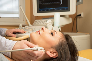
The doctor must analyze in detail clinical picture, so the patient is asked to undergo an ultrasound.
Due to lack of medical knowledge, the description of the image may not be clear to the patient.
It is better to familiarize yourself with the basic concepts of thyroid examination in advance.
What is thyroid volume?
There is a concept of norm and deviation from it. The size of the thyroid gland varies between men and women.
Size standards also depend on a person’s body weight and age.
Ultrasound is used to determine the size.
In women, the thyroid gland should not exceed the size of 18 cm³, and in men 25 cm³.
If the sizes differ by 1-5 mm, these are borderline sizes and are more likely to be normal.
Depending on body weight is given in the table.
In adolescents during puberty and in pregnant women, the volume of the gland increases, and this is considered normal.
The size of the gland in children under 15 years of age differs depending on age as shown in the table.
Left and right lobe, isthmus
The thyroid gland has two lobes: right and left.
Between the lobes there is an isthmus.
Normal on ultrasound is the right and left lobe should not differ significantly from each other in volume.
The normal share size is:
- height 4 cm;
- width 2 cm;
- thickness 2 cm.
The size of the isthmus should not exceed 5 mm.
If the deviation from these indicators is 1-5 mm and the patient has no complaints, most likely we're talking about about individual characteristics.
An endocrinologist must understand each specific case and identify the pathology.
Outlines of the thyroid gland
By contours we mean the outlines of the organ, which are displayed on the monitor at the time of the ultrasound.
Normally, the gland has clear and continuous parenchyma contours.
The word “parenchyma” is used in medicine to designate the entire collection of tissues of an organ, for example, the thyroid gland.
If the contours of the parenchyma are blurry or unclear, then inflammation, for example, of an autoimmune nature can be suspected.
In the picture, this process is displayed by the non-uniform color of the tissues, which indicates their different densities.
Echogenicity
Echogenicity is the level of reflection sound signal ultrasound machine from thyroid tissue.
It could be:
- Normal. This means that there are no formations in the organ: cysts, nodes, tumors.
But if the size of the gland is changed, even with normal echogenicity, this indicates a disease.
If the gland contains formations of the rest of the parenchyma, then the echogenicity always differs at the edges of the pathological element.
- Reduced. This indicator means that there are nodes or cystic formations which are visible on ultrasound.
- Elevated. Observed if thyroid gland prevails connective tissue.
Accordingly, its functions in this case are reduced, because connective tissue cannot synthesize hormones the way glandular tissue does.
- Zero. That is, there is no reflection of the sound signal of the ultrasound machine from the structures of the gland.
The conclusion about what echogenicity the ultrasound machine detected can be read in your chart.
Fabric structure
Normally, the tissue structure has a uniform characteristic granularity, the cell size does not exceed 1 mm in diameter.
This granularity is due to the diameter of the follicles.
At various pathologies the structure will be heterogeneous, for example, with macrofollicles the cell diameter will increase.
Regional lymph nodes
Examination of surrounding tissues also provides the endocrinologist with a lot of useful information.
Normal regional lymph nodes not enlarged.
An increase in them usually indicates cancer or inflammation of the thyroid gland.
The diagnosis is confirmed with a biopsy.
Thyroid nodules
A thyroid nodule is an area that is morphologically distinct from the main gland tissue.
Differences can be identified using ultrasound.
Thyroid nodules
a common phenomenon. They are diagnosed in 11% of the world's population.
These formations can be benign or malignant.
For differentiation, a fine-needle biopsy method is used.
The biopsy is performed under ultrasound guidance. Typically, a biopsy examination is prescribed when the size of the node is more than 5-7 mm.

On an ultrasound of the thyroid, it is possible to see formations even less than 2-3 mm in diameter.
If their structure is homogeneous and they do not have too low or too high echogenicity, then doctors call such elements follicles and classify them as normal.
If the formation is more than 4 mm, then it is called a node.
Depending on their echogenicity, nodes are:
- Isoechogenic, that is, the echogenicity of the nodes is the same as that of healthy tissue.
- Hyperechoic, that is, very dense. On ultrasound, such structures are displayed as light spots.
- Hypoechoic, that is, not dense. On an ultrasound, such nodes will appear dark.
Ultrasound also shows the number of nodes, but does not show their impact on hormonal background.
To find out whether the node is toxic or euthyroid, the endocrinologist gives a referral to biochemical analysis blood.
Methods of nodule therapy
After a biopsy and blood test, the doctor can make an accurate diagnosis.
The node may be:
- Benign tumor.
This may be a colloid formation that has formed as a result of autoimmune thyroiditis.
Among all nodes, these are the ones that occur in 93% of patients.
Treatment is not required if the formation does not interfere with the person, does not create cosmetic defects, does not compress neighboring tissues or blood vessels, and does not produce excessive amounts of hormones.
Otherwise, surgical removal of the nodes is prescribed.
- Follicular tumor.
According to rough estimates, in 86% of cases the tumor is benign, and in the remaining cases it is malignant.
In case of malignancy, the tumor grows into the capsule of the node, but in case of benign it does not.
- Malignant tumor.
In this case, surgery to remove the tumor is always indicated.
During the operation, the surgeon looks at how far the tumor has spread and decides how much of the gland needs to be removed.
After this, radiation and chemotherapy courses are prescribed.
On initial stages percent complete cure high.
The prognosis for recovery, according to endocrinologists, directly depends on the timeliness of diagnosis.
That is why patients over 45 years of age should undergo preventive ultrasound at least once a year.
It is necessary to distinguish the norm from the pathology in time
The thyroid gland is an integral part of the endocrine system.
Regular examinations and ultrasound monitoring, compliance with all doctor’s prescriptions will help maintain her health.
In the treatment of thyroid diseases Self-medication is unacceptable!
At the first symptoms, you should consult an endocrinologist.
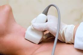
Ultrasound examination helps to thoroughly examine the thyroid gland and adjacent structures and tissues of the cervical region.
The standard results depend on the age and gender of the patient. After the examination, the doctor does the decoding, but you can understand some things on your own.
For more information on deciphering the results of an ultrasound examination of the thyroid gland: the norm in women and men depending on age, as well as tables of values, see below.
Ultrasound is performed to:
- Determine the size and volume of the thyroid gland, its structure and homogeneity.
- Determine whether inflammatory processes are present in it.
- Diagnose hypo-/hyperthyroidism.
- Determine whether the tumor is a mass of the thyroid gland or a mass of an adjacent structure (for example, the sternum).
- Assess whether there are manifestations of (toxic, nodular, diffuse) goiter.
- Estimate size parathyroid gland. A normal parathyroid gland is difficult to distinguish on ultrasound and is not palpable.
- Assess the growth of previously diagnosed formations and the effectiveness of the chosen treatment method.
What indicators are considered normal: tables of values
Taking into account the patient’s weight, the optimal sizes of the thyroid gland in adults are as follows:
Formula for calculating volume:
V lobes = height*length*width*0.479,
where 0.479 is the elliptic coefficient.
V thyroid gland = V right lobe+ V left lobe.
Normal size of the thyroid gland and signs of pathology
Normally, in men its size is 23-25 cm^3, in women it varies from 16-18 cm^3. The shares must be equal in size. The size of normal lobes is usually 4*2*2 cm. Normal thickness isthmus - 4-5 mm (in 7% of people there is no isthmus). Optimal size The parathyroid glands are 4*5*5 cm. The weight of a healthy thyroid gland in a woman is 17-19 grams, in men - 18-20 grams.
In children under 16 years of age, the volumes are correspondingly smaller. The size of the thyroid gland in girls is always smaller and varies by 1-2 cm^3.
Indicators for pathology
They are divided into two types: those associated with abnormal hormone production and those associated with an increase in the size of the thyroid gland.
Each of the deviations causes a change in the shape and structure of the gland. An ultrasound will detect any of the following diseases.
Assessment of the echogenicity (color/tone on the screen in gray scale) of the gland is used to diagnose the presence of compactions, which indicates the possible occurrence of a cyst (usually filled with fluid) or single nodules.
More often they are benign. Very rarely they pose a risk to the patient and require surgical excision.
When assessing echogenicity, the following diseases can also be diagnosed:
- Colloid nodular goiter.
- Hyperparathyroidism (osteitis fibrocystis - inflammation of the endocrine system).
- Multiple endocrine neoplasia syndrome (tumor/increase in the number of several endocrine glands).
- Thyroid cancer gives a good survival rate if you contact an oncologist-endocrinologist early.
Echogenicity shows a change in the tone of the thyroid gland on the screen (from light gray to black). Normally, all areas should be gray (isoechoic).
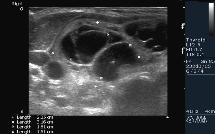
Multiple nodes and cystic formations in enlarged right lobe thyroid gland
Pathological manifestations may indicate the presence of certain diseases.
- Enlargement of the thyroid gland/change in its contours and shape is often caused by the occurrence of goiter (nodular, toxic, diffuse, diffuse-nodular mixed goiter, Hashimoto's goiter). It's being removed surgical method, based on cosmetic reasons. A goiter also compresses the esophagus and trachea, causing difficulty breathing and pain when swallowing. Sometimes it can grow into the sternum.
- Swelling and aberrant location indicate thyroiditis, a severe inflammatory process often caused by bacteria, fungi or viruses. Symptoms characteristic of thyroiditis: high fever and pain; during inflammation, manifestations of hypo-/hyperthyroidism are also observed.
- Enlargement of the gland and inflammation of the lymph nodes may be associated with hyperthyroidism - the production is extremely large quantity hormones T3 and T4. Treatment methods include treatment with iodine and antithyroid drugs (inhibit hormone biosynthesis). Rarely: surgery.
- A decrease in thyroid gland and isthmus thickness is usually caused by hypothyroidism - insufficient production of hormones. It may develop for several months before being recognized. The disease is often associated with pregnancy.
 An examination of the thyroid gland is first done visually by a doctor and by palpating the organ with your hands. What kind of failures does it mean and what? additional methods diagnostics are necessary in this case, read carefully.
Why thyroid inflammation is difficult to diagnose immediately and how to treat it pathological process, you will find out.
Up to 40% of the world's population have small nodules in the thyroid gland that do not manifest themselves in any way. But sometimes the nodes reach large sizes, are easily palpable and can even deform the neck. Follow this link to learn about folk methods for treatment and prevention nodular goiter, which can be used along with the main therapy.
What should be in the conclusion
The conclusion must indicate:
- Lymphatic condition cervical nodes(whether they are inflamed or not).
- The condition of the parathyroid glands (can be seen on ultrasound), as well as their size and volume.
- Echostructure (indicates the presence of inflammatory processes; hyper-, hypo- and anechogenic areas should be absent).
- The shape of the thyroid gland (classical, not visible), as well as its size and volume.
- Location (presence of shifts, below/above the pole, size and location of the isthmus, its thickness).
- Description of contours (clear, unclear, blurry).
- The presence of formations (absent, single or multiple) and their characteristics (less than 1 cm - focal, diffuse formation; more than 1 cm - a knot, the permissible size of a knot is up to 3-4 cm).
- Blood flow (increased blood flow may be a symptom of malignancy).
The conclusion is drawn up in the form of a protocol.
The norm is considered to be non-inflamed lymph nodes in the neck with regular outlines, the location is typical, normal, the structure of the gland is homogeneous (sometimes granular or moderately heterogeneous), there are no changes in the size, volume and shape of the organ, there are no compactions, the contours are smooth, well distinguishable, clear, the echostructure is homogeneous, blood flow is not increased, there are no signs of compaction or relapses.
If one or more seals are identified during the inspection, the specialist will study their features in detail. If the endocrinologist suspects that the formation is malignant tumor, a biopsy will be scheduled. Ultrasound results do not indicate with 100% accuracy the malignancy of the tumor. To do this, the specialist will definitely prescribe a “Blood Biochemistry” test (minimal or extended profile) and an absorption test radioactive iodine.
 Heredity and iodine deficiency are risk factors for the development of thyroid pathologies in children. difficult to recognize on your own. What parents should be wary of and how to suspect a pathology in a child, read on our website.
What is thyrotropin and what does increased production of this hormone lead to, we will tell you in the topic.
Video on the topic
The normal size of the thyroid gland is a relative concept, since there is a difference between the anatomical, ultrasound and x-ray volumes of the organ. Doctors rely on ultrasound for the distance between the lobes and the isthmus of the thyroid gland. If they deviate from the norm, the likelihood of disease is high.
However, thyroid enlargement on ultrasound scanning may not extend beyond normal anatomical boundaries.
The anatomical size of the thyroid gland is normally impossible to estimate. When palpating (finger palpating) the neck area, it is impossible to accurately determine the boundaries. The thyroid gland of an adult is close to the larynx, so it is problematic to feel it with your fingers.
To determine the volume of the thyroid gland, you need to distinguish the following anatomical components of the organ:
- Left lobe;
- Right lobe;
- Isthmus.
Finger identification of parts is hampered by the subcutaneous fatty tissue, muscles, carotid arteries and cervical tendon fascia.
Features of thyroid size in women and men
In women, the thyroid gland has large sizes and it is located more to the side of the cricoid and thyroid cartilage. In men, the organ is located below the upper edge of the sternum. For representatives of the fair half, the weight of the organ is 17-40 grams, vertical size– 60-80 mm, and the size of the isthmus – from 7 to 15 mm. The diameter of the thyroid gland in the anteroposterior direction is approximately 20 mm. The volume of the organ in the fair half is slightly larger than in men.
Throughout life, the thickness and length of the thyroid gland changes. In a newborn, the weight of the organ is about 2 grams, and at the end of the first year of life it reaches 14 grams. The most active increase in its thickness is observed in the interval of 5-10 years.
Involution of the thyroid gland begins at age 55. The reduction in size occurs due to follicle atrophy. In this case, the functionality of the organ is not impaired. Reserve reserves of hormonal tissue are maintained until the onset of menopause.
A fifth of people have an additional thyroid gland located in the front hyoid bone. It can be large in size and, in the absence or decrease in the activity of the main analogue, partially compensate for its lost functions.
The right lobe in humans is more developed than the left. The isthmus is normally fixed by a ligament to the cricoid cartilage. In 5% of people, the isthmus may be physiologically absent. In less than 1% of people, the thyroid gland may have abnormalities: bifurcation or the presence of additional lobes.
To correctly determine the size of the thyroid gland, you should consider anatomical features organ structure.
Thyroid volume upon palpation
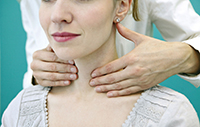
The total volume of the gland upon palpation is determined by the sum of the right and left lobes. The isthmus under the fingers is difficult to determine. To determine the size, the endocrinologist feels the gland from back to front. The doctor stands behind the patient and wraps his hands around the neck. By applying gentle pressure with the fingertips, the specialist determines its structure, density and the presence of pathological nodes.
The volume of the thyroid gland is normally comparable to the size nail phalanx thumb. Normally, the organ has a homogeneous structure and moderate density. Normally, the organ moves well without painful sensations. If the thyroid gland cannot be felt under the fingers, this does not mean the presence of pathology. Reducing volume and size – physiological norm in older people and children.
Ultrasound dimensions of the thyroid gland
Ultrasound dimensions of the thyroid gland are assessed in medicine to determine pathological nodes and malignant tumors in organ tissue. Using ultrasound, you can determine the following characteristics of an organ:
For the prevention and treatment of thyroid diseases, our readers recommend Monastic Tea. It consists of 16 most useful medicinal herbs, which have extremely high efficiency in the prevention and treatment of the thyroid gland, as well as in cleansing the body as a whole. The effectiveness and safety of Monastic tea has been repeatedly proven clinical studies and many years of therapeutic experience. Doctors' opinion..."
- Structure;
- Height;
- length;
- Width;
- The size of the nodes.
In medicine after definition pathological changes Hormonal levels are determined using an ultrasound. Every doctor knows the clinical standards of the thyroid gland.
To calculate the indicators, the formula is used: multiplying 0.479 by the height, length and width of the organ. This formula calculates the total volume of the share. The value of 0.479 reflects the ellipsoidality of the thyroid gland. The resulting volume is calculated in cubic millimeters and centimeters.
To determine the total volume of an organ, the sum of both shares is necessary.
Normal thyroid volume:
- For women – 25 ml;
- For men - 18 ml.
In children, physiological dimensions differ depending on age.
You need to understand that a reduced isthmus or lobe of the thyroid gland in a child does not mean the presence of pathology. If the size of an organ is reduced by 1 cubic cm, the functionality of the organ is not impaired. Detection of an enlarged thyroid gland on ultrasound requires additional examination to identify the causes of pathology.
In adults, there are the following ultrasound standards for the thyroid gland depending on body weight:
- 12.3 cubic centimeters – weight up to 40 kilograms;
- 15.5 – up to 50 kg;
- 18.7 – up to 60 kg;
- 22 – 70 kg;
- 25 – 80 kg;
- 28.4 – 90 kg;
- 32 – 100 kg;
- 35 – 110 kg.
When assessing the size of the thyroid gland, the doctor determines the following tasks:
- Assessment of thyroid localization: low, typical, aberrant. At ultrasound examination ectopic areas can be detected. Pathological tissues may be located near the main body of the organ;
- The structure of the thyroid gland: two lobes and an isthmus should be located in the midline. The pyramidal lobe is located near them. Outgrowths of thyroid tissue extend from the lower poles to the tongue. The small projections of thyroid tissue are called “antipyramidal.” They arise as a result of decreased functionality and tissue atrophy;
- Contours of the gland: unclear or clear. Fuzzy boundaries are observed against the background of inflammatory diseases;
- Dimensions: isthmus - in the anteroposterior direction consists of 3 dimensions, determined in perpendicular planes;
- Structure: homogeneous, granular, heterogeneous;
- Echogenicity: increased, decreased, normal;
- Availability focal formations: calcifications, cysts, nodes;
- Condition of the cervical lymph nodes: increase, internal structure, soft tissue changes, cystic transformation of the organ.
It is not enough to determine the volume of the thyroid gland using ultrasound. To form a conclusion, you need to study the above-described properties of the organ.
Ultrasound norm and pathology during ultrasound scanning of the thyroid gland
Ultrasound normal does not mean that a person is healthy. The acoustic wave does not reflect uniformly from all anatomical structures, so some formations are not detected on the monitor. If pathological changes or suspected diseases are detected, laboratory tests are prescribed.
Even the most qualified ultrasound specialist will not be able to reliably establish a diagnosis. Medical criteria do not allow the use of ultrasound as the main method for diagnosing endocrine pathology.
When implementing ultrasound scanning the doctor will not be able to determine the internal and external structure of the organ. Only an objective and comprehensive study will determine the diagnosis of the disease.
The norm on ultrasound and physiological laboratory criteria allows the endocrinologist to assume the absence of anatomical growths of the thyroid gland. To study the functional properties of the organ, tests for thyroid hormones are required.
In conclusion, we will formulate a definition of what a diagnostic norm is:
- Physiological dimensions;
- Absence of echoacoustic changes in the isthmus and lobes;
- No soft tissue pathology;
- Lymph nodes are not enlarged.
In medicine, the normal size of the thyroid gland is somewhat distorted and is equated to ultrasound values. This approach is formed for adequate and high-quality diagnosis of the disease.
It still seems like it’s not easy to cure your thyroid?
Considering that you are now reading this article, we can conclude that this illness still haunts you.
You've probably also had thoughts about surgical intervention. This is clear, because the thyroid gland is one of the most important organs, on which your wellness and health. And shortness of breath, constant fatigue, irritability and other symptoms clearly interfere with your enjoyment of life...
But, you see, it is more correct to treat the cause, not the effect. We recommend reading the story of Irina Savenkova about how she managed to cure her thyroid gland...
Ultrasound examination of the thyroid gland is included in the mandatory examination of the endocrine system, since this method allows us to identify structural changes and assess the size of the gland. It is also necessary to take hormones to clarify the thyroid status - status functional state systems. In this case, it is necessary to examine the blood for TSH (thyrotropin), T 4 (tetraiodothyronine) and antibodies to TPO (thyroid peroxidase), since without them the results will not be complete. These are the main indicators of the gland.
Thyrotropin is a hormone of the pituitary gland that acts on the receptors of the organ described in the article and stimulates the production of thyroid hormone. The fact is that if there is insufficient production of thyrotropin, the synthesis of thyroid hormones is not possible, which leads to its hypertrophy.
Tetraiodothyronine – main hormone The thyroid gland produces thyrotropin. It is considered the primary hormone that is transformed into triiodothyronine. When there is an excess of it, it happens Graves' disease, characterized by excessive growth of the goiter. With a deficiency - cretinism, and over the years - myxedema - swelling of the limbs.
Thyroid peroxidase is an important enzyme of the gland. The volume of the thyroid gland can be guessed by an endocrinologist during an examination, but it can only be accurately determined by ultrasound. The normal size of the thyroid gland differs among women. The size of the lobes can vary between 2.5-4 cm (length), 1.5-2 cm (width) and 1-1.5 cm (thickness). For diagnostics important has a gland volume:
- For men it should be up to 25 cm 3
- In women up to 18 cm 3
Most often, in the presence of the disease, a change in the volume and size of the thyroid gland occurs. This sign is especially characteristic of endemic goiter(a disease caused by a lack of iodine in food). Also, with thyrotoxic goiter, an increase in all parameters may also be observed. But the presence of normal size of the thyroid gland does not mean the absence of the disease. The main indicator of normal functioning of the thyroid gland is hormonal status. If the volume of the gland is normal, but there is an increase in the level of T4, then this indicates the development of thyrotoxicosis. Therefore, it is imperative to take hormones, especially if you have health problems.
Vivid deviations and diseases
Any hyposecretion or hypersecretion leads to disorders and, as a consequence, diseases. Let's look at the most striking pictures of deficiency and excess of thyroid hormones. Health problems in this regard can result in problems with childbearing.
Thyroiditis - the ending of the word “itis” indicates an inflammatory process of the gland, in which there is significant pain in the area of the thyroid gland, enlargement of the lymph nodes in the neck (another sign of any inflammatory process). People get sick with CVD weak immunity for poisoning and infections. Treatment is both outpatient and surgical, depending on the progression of thyroiditis.
Cancer and other gland tumors. Unfortunately, this disease can affect any organ and any gland.
Graves' disease or goiter in the common people - when from increased output hormones, a bright external picture is observed: characteristic bulging eyes, enlargement of the gland visible from the side, excessive activity nervous system and toxic state of the body. The disease is memorable and is best treated surgically, but more conservative treatment is also possible.
Cretinism - most often congenital disease, when the mother’s body lacked iodine during pregnancy. Most often, those born with this diagnosis are significantly delayed in development. In 90 percent it is not curable.
Myxedema occurs when there is insufficient supply of organs and limbs with thyroid hormones. It occurs due to hormone deficiency, just like cretinism, but over time (in adults). The patient's tissues become swollen and purple-blue. Treatment is hormonal, conservative.
Endocrine diseases affect all types of metabolism, any organs and systems. Therefore, complaints for such diseases are comprehensive. Timely detection will help carry out full treatment and stay healthy. Come on in and be attentive to your health. You have only one.
You can learn about indicators of thyroid disease from the following video:
Noticed a mistake? Select it and click Ctrl+Enter to let us know.
The thyroid gland plays a huge role in providing normal functioning endocrine and reproductive system female body.
Structure of the thyroid gland
The thyroid gland is an unpaired organ that belongs to the endocrine system. Functional work glands obey the central nervous system and hormones produced by the pituitary gland. The thyroid gland is the largest gland in the body, which performs an intrasecretory function.
The gland gets its name from the adjacent thyroid cartilage, which is part of the larynx. The location of the gland allows for physical diagnostic methods. The thyroid gland is able to store hormones in the form of thyroglobulin.
Secretion of hormones can occur in the presence of iodine. The thyroid gland is similar in appearance with a butterfly with open wings. It is located on the front of the neck at the level of the trachea and the lateral walls of the larynx.
The thyroid gland consists of 2 lobes: right and left. The isthmus connects the two lobes, forming a process. The gland receives iodine through circulatory system, which supplies it with the carotid and subclavian arteries.
The endocrine gland is represented by two types of cells:
- Follicular cells participate in the structure of the parenchyma of the gland and form follicles consisting of a cavity. This cavity filled with colloid.
- Parafollicular cells distributed between follicles. And the follicles are united into conglomerates that surround capillary network, mast cells and connective tissue.
These conglomerates form lobules that produce thyroid hormones. Thyroid hormones affect the functioning of organs and systems of the body. Hyposecretion of thyroid hormones can disrupt the functioning of the nervous system and lead to delayed mental and physical development.
Main Dimensions
There is a value that is used to differentiate normal from pathology of the thyroid gland - this is volume. If given value changed, then it indicates the presence of changes in the structure of the thyroid gland. The second important value is the weight of the thyroid gland; in the female population it is 17-19 grams.
Thyroid volume in women
The size of the thyroid gland in women varies between 16 and 18 cm3. The lobes of the gland should be equal in size, which is normally 4*2*2 cm. At the same time, the thickness of the thyroid isthmus is 4-5 cm, and in some people it is absent.
Formula for calculating volume
There is a formula for determining the volume of the thyroid gland, thanks to which you can accurately determine its size:
- V lobes = height*length*width*0.479, where 0.479 is the elliptical coefficient.
- V of the thyroid gland = V of the right lobe + V of the left lobe.
Standard table
In order to monitor the condition women's health, you need to know the size of the thyroid gland. The table of norms will help you navigate normal sizes thyroid gland, determine pathological condition and consult a specialist.
| Weight, kg | 50 | 60 | 70 | 80 | 90 | >90 |
| Volume, cm3 | 15, 5 | 18,7 | 22 | 25 | 28,4 | 32 |
The size of the thyroid gland in women changes during hormonal surges, such as pregnancy, puberty and menopause.
Reasons for changes in thyroid volume in women
Like all glands internal secretion, the thyroid gland needs a catalyst. The catalyst for this gland is the trace element iodine. In the absence required quantity iodine, changes occur in the volume of the thyroid gland, it increases in size irreversibly.
In addition, there are other reasons that provoke the growth of goiter:

- Nervous exhaustion and stress. Exhaustion and overstrain of the nervous system undermines the functioning of the entire system as a whole. Due to disruption of tissue innervation, it leads to spasm of certain muscle groups, which impairs blood circulation.
- Decreased immunity. Viral and infectious diseases reduce immunity, depleting it. Inflammatory processes pharynx and neck area can provoke activation protective forces, thanks to which cells actively grow.
- Hormonal dysfunction. During periods of hormonal surges endocrine system forced to work at the limit, which may disrupt normal work thyroid glands
- Stagnation of blood and lymph. Atherosclerotic changes in blood vessels can disrupt the outflow of blood and lymph from the gland. Stagnation of thyroid metabolic products can cause swelling and tissue enlargement due to intensive cell growth.
- Hereditary factors. Features of the body are inherited, in which the thyroid gland works more intensely to produce more endocrine hormones.
- Bad environment. The danger is posed by areas with a radioactive background, where grown food products are poor in microelements. These changes lead to mutation of thyroid cells.
- Chronic diseases. Chronic diseases can provoke changes in the structure of the thyroid gland due to compensatory and decompensatory processes.
How is the size of the thyroid gland diagnosed?
You can diagnose the size of the thyroid gland in the endocrinologist’s office, thanks to visual inspection and palpation. If an examination reveals an enlarged thyroid gland, the endocrinologist may refer you for diagnostic tests.
Basic diagnostic methods:
- Blood test. Laboratory tests involve determination of concentration TSH hormones, T4 and T3. Based on laboratory data, you can learn about the functioning of the thyroid gland and its size.
- Nuclear scan or radioisotope study. This method research involves taking radioactive iodine or introducing it parenterally into the body. The doctor receives images in three projections, from which one can learn about the size, shape, and the presence of areas that are not working correctly.
- Ultrasound examination. Thanks to the study, the size of the thyroid gland is determined and its lobes are examined. Ultrasound can detect the presence of nodes and their number.
- Fine needle biopsy. This diagnostic method is carried out in controversial cases or to exclude the presence of atypical cells in the thyroid gland.
- Computed axial tomography. CT is performed only to clearly differentiate healthy tissues from malignant ones, or in cases of retrosternal location of the thyroid gland.
The size of the thyroid gland is normal on ultrasound
An ultrasound examination allows you to examine the thyroid gland and surrounding tissues.
Consider nodes and neoplasms, their structure:

- Thyroid gland correct form, no shifts.
- No changes in size and volume.
- There are no signs of growth of formations.
- The contours are smooth, clear, and clearly distinguishable.
- The blood flow in the thyroid gland is not increased.
- Regional nodes are not enlarged.
- The structure normally has a uniform granularity, the cell size is no more than 1 mm in diameter.
- Lobe size: height 4 cm, width 2 cm, thickness 2 cm.
- The isthmus is normally no more than 5 mm in size. The deviation can be 1-5 mm if the patient has no complaints, which can be considered normal.
Using periodic medical examinations and ultrasound can diagnose changes in the thyroid gland at an early stage. By diagnosing the disease, you can avoid complications and irreversible changes.
