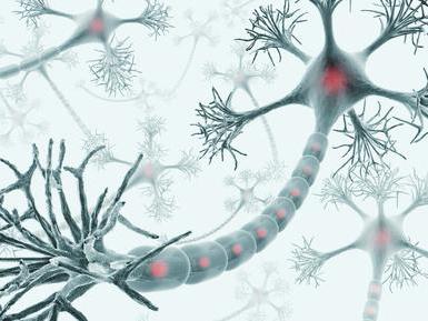
The structure of nerve cells. Structure of the nervous system
The human body is made up of trillions of cells, and the brain alone contains approximately 100 billion neurons, in a variety of shapes and sizes. The question arises: how is a nerve cell structured, and how does it differ from other cells in the body?
The structure of a human nerve cell
Like most other cells in the human body, nerve cells have nuclei. But compared to the others, they are unique because they have long, thread-like branches through which nerve impulses are transmitted.
The cells of the nervous system are similar to others because they are also surrounded by a cell membrane, have nuclei containing genes, cytoplasm, mitochondria and other organelles. They are involved in fundamental cellular processes such as protein synthesis and energy production.

Neurons and nerve impulses
Consists of a bundle of nerve cells. A nerve cell that transmits certain information is called a neuron. The data that neurons carry is called nerve impulses. Like electrical impulses, they carry information at incredible speeds. Fast signal transmission is ensured by neuron axons covered with a special myelin sheath.
This sheath covers the axon, similar to the plastic coating on electrical wires, and allows nerve impulses to travel faster. What is a neuron? It has a special shape that allows you to transmit a signal from one cell to another. A neuron consists of three main parts: a cell body, many dendrites, and one axon.

Types of neurons
Neurons are usually classified based on the role they play in the body. There are two main types of neurons - sensory and motor. Sensory neurons carry nerve impulses from the senses and internal organs to Motor neurons, on the contrary, carry nerve impulses from the central nervous system to organs, glands and muscles.

The cells of the nervous system are designed in such a way that both types of neurons work together. Sensory neurons carry information about the internal and external environment. This data is used to send signals through motor neurons to tell the body how it should respond to the information received.

Synapse
The place where the axon of one neuron meets the dendrites of another is called a synapse. Neurons communicate with each other through an electrochemical process. When this happens, chemicals called neurotransmitters react.

Cell body
The structure of a nerve cell presupposes the presence of a nucleus and other organelles in the cell body. Dendrites and axons connected to the cell body resemble rays emanating from the sun. Dendrites receive impulses from other nerve cells. Axons transmit nerve impulses to other cells.
A single neuron can have thousands of dendrites, so it can communicate with thousands of other cells. The axon is covered with a myelin sheath, a fatty layer that insulates it and allows signal transmission much faster.
Mitochondria
When answering the question of how a nerve cell is structured, it is important to note the element responsible for the supply of metabolic energy, which can then be easily utilized. Mitochondria play a primary role in this process. These organelles have their own outer and inner membrane.
The main source of energy for the nervous system is glucose. Mitochondria contain the enzymes needed to convert glucose into high-energy compounds, mainly adenosine triphosphate (ATP) molecules, which can then be transported to other areas of the body that need their energy.

Core
The complex process of protein synthesis begins in the cell nucleus. The nucleus of a neuron contains genetic information, which is stored as encoded strings of deoxyribonucleic acid (DNA). Each contains for all the cells in the body.
It is in the nucleus that the process of constructing protein molecules begins, by writing the corresponding part of the DNA code on complementary ribonucleic acid (RNA) molecules. Released from the nucleus into the intercellular fluid, they trigger the process of protein synthesis, in which the so-called nucleoli also take part. This is a separate structure within the nucleus that is responsible for building molecular complexes called ribosomes, which are involved in protein synthesis.

Do you know how a nerve cell works?
Neurons are the most tenacious and longest cells in the body! Some of them remain in the human body throughout life. Other cells die and are replaced by new ones, but many neurons cannot be replaced. With age there are fewer and fewer of them. This is where the expression comes from that nerve cells do not regenerate. However, research data from the late 20th century proves the opposite. In one area of the brain, the hippocampus, new neurons can grow even in adults.

Neurons can be quite large and several meters long (corticospinal and afferent). In 1898, the famous nervous system specialist Camillo Golgi announced his discovery of a ribbon-shaped apparatus specializing in neurons in the cerebellum. This device now bears the name of its creator and is known as the “Golgi apparatus.”

From the way a nerve cell is structured, it is defined as the main structural and functional element of the nervous system, the study of simple principles of which can serve as the key to solving many problems. This mainly concerns the autonomic nervous system, which includes hundreds of millions of interconnected cells.
Nervous tissue is a collection of interconnected nerve cells (neurons, neurocytes) and auxiliary elements (neuroglia), which regulates the activity of all organs and systems of living organisms. This is the main element of the nervous system, which is divided into central (includes the brain and spinal cord) and peripheral (consisting of nerve ganglia, trunks, endings).
Main functions of nervous tissue
- Perception of irritation;
- formation of a nerve impulse;
- rapid delivery of excitation to the central nervous system;
- information storage;
- production of mediators (biologically active substances);
- adaptation of the body to changes in the external environment.
Properties of nerve tissue
- Regeneration- occurs very slowly and is possible only in the presence of an intact perikaryon. Restoration of lost processes occurs through germination.
- Braking- prevents the occurrence of arousal or weakens it
- Irritability- response to the influence of the external environment due to the presence of receptors.
- Excitability— generation of an impulse when the threshold value of irritation is reached. There is a lower threshold of excitability at which the smallest influence on the cell causes excitation. The upper threshold is the amount of external influence that causes pain.
Structure and morphological characteristics of nerve tissues
The main structural unit is neuron. It has a body - the perikaryon (which contains the nucleus, organelles and cytoplasm) and several processes. It is the processes that are a distinctive feature of the cells of this tissue and serve to transfer excitation. Their length ranges from micrometers to 1.5 m. The cell bodies of neurons also vary in size: from 5 µm in the cerebellum to 120 µm in the cerebral cortex.
Until recently, it was believed that neurocytes were not capable of division. It is now known that the formation of new neurons is possible, although only in two places - the subventricular zone of the brain and the hippocampus. The lifespan of neurons is equal to the lifespan of an individual. Each person at birth has about trillion neurocytes and in the process of life, it loses 10 million cells every year.
Processes are divided into two types - dendrites and axons.
Axon structure. It starts from the neuron body as an axon hillock, does not branch throughout its entire length, and only at the end is it divided into branches. An axon is a long extension of a neurocyte that transmits excitation from the perikaryon.
Dendrite structure. At the base of the cell body, it has a cone-shaped extension, and then it is divided into many branches (this explains its name, “dendron” from ancient Greek - tree). The dendrite is a short process and is necessary for transmitting the impulse to the soma.
Based on the number of processes, neurocytes are divided into:
- unipolar (there is only one process, an axon);
- bipolar (both axon and dendrite are present);
- pseudounipolar (from some cells at the beginning one process extends, but then it divides into two and is essentially bipolar);
- multipolar (have many dendrites, and among them there will be only one axon).
Multipolar neurons predominate in the human body, bipolar ones are found only in the retina, and pseudounipolar ones are found in the spinal ganglia. Monopolar neurons are not found at all in the human body; they are characteristic only of poorly differentiated nervous tissue.
Neuroglia
Neuroglia are a collection of cells that surround neurons (macrogliocytes and microgliocytes). About 40% of the central nervous system consists of glial cells; they create the conditions for the generation of excitation and its further transmission, and perform supporting, trophic, and protective functions.

Macroglia:
Ependymocytes– formed from glioblasts of the neural tube, lining the spinal cord canal.
Astrocytes– stellate, small in size with numerous processes that form the blood-brain barrier and are part of the gray matter of the brain.
Oligodendrocytes- the main representatives of neuroglia, surround the perikaryon along with its processes, performing the following functions: trophic, isolation, regeneration.
Neurolemocytes– Schwann cells, their task is the formation of myelin, electrical insulation.
Microglia – consists of cells with 2-3 branches that are capable of phagocytosis. Provides protection from foreign bodies, damage, and removal of products of apoptosis of nerve cells.
Nerve fibers- these are processes (axons or dendrites) covered with a membrane. They are divided into myelinated and non-myelinated. Myelinous in diameter from 1 to 20 microns. It is important that myelin is absent at the junction of the membrane from the perikaryon to the process and in the area of axonal branches. Unmyelinated fibers are found in the autonomic nervous system, their diameter is 1-4 microns, the impulse moves at a speed of 1-2 m/s, which is much slower than myelinated ones, their transmission speed is 5-120 m/s.
Neurons are divided according to their functionality:
- Afferent– that is, sensitive, accept irritation and are able to generate an impulse;
- associative- perform the function of transmitting impulses between neurocytes;
- efferent- complete the transfer of impulses, performing motor, motor, and secretory functions.
Together they form reflex arc, which ensures the movement of the impulse in only one direction: from sensory fibers to motor fibers. One individual neuron is capable of multidirectional transmission of excitation, and only as part of a reflex arc does a unidirectional flow of the impulse occur. This occurs due to the presence of a synapse in the reflex arc - interneuron contact.
Synapse consists of two parts: presynaptic and postsynaptic, between them there is a gap. The presynaptic part is the end of the axon that brought an impulse from the cell; it contains mediators, which contribute to the further transmission of excitation to the postsynaptic membrane. The most common neurotransmitters are: dopamine, norepinephrine, gamma aminobutyric acid, glycine; there are specific receptors for them on the surface of the postsynaptic membrane.
Chemical composition of nervous tissue
Water is found in significant quantities in the cerebral cortex, less in the white matter and nerve fibers.
Protein substances represented by globulins, albumins, neuroglobulins. Neurokeratin is found in the white matter of the brain and axon processes. Many proteins in the nervous system belong to mediators: amylase, maltase, phosphatase, etc.
The chemical composition of nervous tissue also includes carbohydrates– these are glucose, pentose, glycogen.
Among fat Phospholipids, cholesterol, and cerebrosides were detected (it is known that newborns do not have cerebrosides; their amount gradually increases during development).
Microelements in all structures of the nervous tissue are distributed evenly: Mg, K, Cu, Fe, Na. Their importance is very great for the normal functioning of a living organism. Thus, magnesium is involved in the regulation of nervous tissue, phosphorus is important for productive mental activity, and potassium ensures the transmission of nerve impulses.
Nervous tissue controls all processes in the body.
Nervous tissue consists of neurons(nerve cells) and neuroglia(intercellular substance). Nerve cells have different shapes. A nerve cell is equipped with tree-like processes - dendrites, which transmit stimuli from receptors to the cell body, and a long process - an axon, which ends on the effector cell. Sometimes the axon is not covered by a myelin sheath.
Nerve cells are capable under the influence of irritation come into a state excitement, generate impulses and transmit their. These properties determine the specific function of the nervous system. Neuroglia are organically associated with nerve cells and perform trophic, secretory, protective and support functions.
Nerve cells - neurons, or neurocytes, are process cells. The dimensions of the neuron body vary widely (from 3-4 to 130 microns). Nerve cells are also very different in shape. The processes of nerve cells conduct nerve impulses from one part of the human body to another, the length of the processes is from several microns to 1.0-1.5 m.
Neuron structure. 1 - cell body; 2 - core; 3 - dendrites; 4 - neurite (axon); 5 - branched end of the neurite; 6 - neurilemma; 7 - myelin; 8 - axial cylinder; 9 - interceptions of Ranvier; 10 - muscle
There are two types of nerve cell processes. The processes of the first type conduct impulses from the body of the nerve cell to other cells or tissues of the working organs; they are called neurites, or axons. A nerve cell always has only one axon, which ends in a terminal apparatus on another neuron or in a muscle or gland. The processes of the second type are called dendrites; they branch in a tree. Their number varies among different neurons. These processes conduct nerve impulses to the body of the nerve cell. The dendrites of sensory neurons have special perceptive devices at the peripheral end - sensory nerve endings, or receptors.
Classification of neurons by function:
- perceiving (sensitive, sensory, receptor). Serve to perceive signals from the external and internal environment and transmit them to the central nervous system;
- contact (intermediate, interneurons, interneurons). Provide processing, storage and transmission of information to motor neurons. They are the majority in the central nervous system;
- motor (efferent). They generate control signals and transmit them to peripheral neurons and executive organs.
Types of neurons by number of processes:
- unipolar - having one process;
- pseudounipolar - one process extends from the body, which then divides into 2 branches;
- bipolar - two processes, one dendrite, the other an axon;
- multipolar - have one axon and many dendrites.

Neurons(nerve cells). A - multipolar neuron; B - pseudounipolar neuron; B - bipolar neuron; 1 - axon; 2 - dendrite
Axons covered with a sheath are called nerve fibers. There are:
- continuous- covered with a continuous membrane, are part of the autonomic nervous system;
- pulpy- covered with a complex, discontinuous membrane, impulses can move from one fiber to other tissues. This phenomenon is called irradiation.

Nerve endings. A - motor ending on a muscle fiber: 1 - nerve fiber; 2 - muscle fiber; B - sensitive endings in the epithelium: 1 - nerve endings; 2 - epithelial cells
Sensory nerve endings ( receptors) are formed by the terminal branches of the dendrites of sensory neurons.
- exteroceptors perceive irritations from the external environment;
- interoreceptors perceive irritations from internal organs;
- proprioceptors receiving irritations from the inner ear and articular capsules.
According to their biological significance, receptors are divided into: food, sexual, defensive.
Based on the nature of the response, receptors are divided into: motor- are located in the muscles; secretory- in the glands; vasomotor- in blood vessels.
Effector- executive link of nervous processes. There are two types of effectors - motor and secretory. Motor (motor) nerve endings are the terminal branches of neurites of motor cells in muscle tissue and are called neuromuscular endings. The secretory endings in the glands form neuroglandular endings. The named types of nerve endings represent a neurotissue synapse.
Communication between nerve cells is carried out using synapses. They are formed by the terminal branches of the neurite of one cell on the body, dendrites or axons of another. At a synapse, a nerve impulse travels in only one direction (from a neurite to the body or dendrites of another cell). They are arranged differently in different parts of the nervous system.
Nerve cell Not to be confused with neutron.
Pyramidal cell neurons in the mouse cerebral cortex
Neuron(nerve cell) is a structural and functional unit of the nervous system. This cell has a complex structure, is highly specialized and in structure contains a nucleus, a cell body and processes. There are more than one hundred billion neurons in the human body.
Review
The complexity and diversity of the nervous system depends on the interactions between neurons, which in turn represent a set of different signals transmitted as part of the interaction of neurons with other neurons or muscles and glands. Signals are emitted and propagated by ions that generate an electrical charge that travels along the neuron.
Structure

Cell body
A neuron consists of a body with a diameter of 3 to 100 μm, containing a nucleus (with a large number of nuclear pores) and other organelles (including a highly developed rough ER with active ribosomes, the Golgi apparatus), and processes. There are two types of processes: dendrites and axons. The neuron has a developed cytoskeleton that penetrates its processes. The cytoskeleton maintains the shape of the cell; its threads serve as “rails” for the transport of organelles and substances packaged in membrane vesicles (for example, neurotransmitters). A developed synthetic apparatus is revealed in the body of the neuron; the granular ER of the neuron is stained basophilically and is known as the “tigroid”. The tigroid penetrates the initial sections of the dendrites, but is located at a noticeable distance from the beginning of the axon, which serves as a histological sign of the axon.
There is a distinction between anterograde (away from the body) and retrograde (toward the body) axon transport.
Dendrites and axon

Neuron structure diagram
Synapse
Synapse- the site of contact between two neurons or between a neuron and the effector cell receiving the signal. It serves to transmit a nerve impulse between two cells, and during synaptic transmission the amplitude and frequency of the signal can be adjusted. Some synapses cause depolarization of the neuron, others cause hyperpolarization; the former are excitatory, the latter are inhibitory. Typically, stimulation from several excitatory synapses is necessary to excite a neuron.
Classification
Structural classification
Based on the number and arrangement of dendrites and axons, neurons are divided into axonless neurons, unipolar neurons, pseudounipolar neurons, bipolar neurons, and multipolar (many dendritic arbors, usually efferent) neurons.
Axonless neurons- small cells, grouped near the spinal cord in the intervertebral ganglia, which do not have anatomical signs of division of processes into dendrites and axons. All processes of the cell are very similar. The functional purpose of axonless neurons is poorly understood.
Unipolar neurons- neurons with a single process, present, for example, in the sensory nucleus of the trigeminal nerve in the midbrain.
Bipolar neurons- neurons having one axon and one dendrite, located in specialized sensory organs - the retina, olfactory epithelium and bulb, auditory and vestibular ganglia;
Multipolar neurons- Neurons with one axon and several dendrites. This type of nerve cells predominates in the central nervous system
Pseudounipolar neurons- are unique in their kind. One tip extends from the body, which immediately divides in a T-shape. This entire single tract is covered with a myelin sheath and is structurally an axon, although along one of the branches the excitation goes not from, but to the body of the neuron. Structurally, dendrites are branches at the end of this (peripheral) process. The trigger zone is the beginning of this branching (i.e., it is located outside the cell body).
Functional classification
According to their position in the reflex arc, afferent neurons (sensitive neurons), efferent neurons (some of them are called motor neurons, sometimes this not very accurate name applies to the entire group of efferents) and interneurons (interneurons) are distinguished.
Afferent neurons(sensitive, sensory or receptor). Neurons of this type include primary cells of the sensory organs and pseudounipolar cells, whose dendrites have free endings.
Efferent neurons(effector, motor or motor). Neurons of this type include the final neurons - ultimatum and penultimate - non-ultimatum.
Association neurons(intercalary or interneurons) - this group of neurons communicates between efferent and afferent, they are divided into comisural and projection (brain).
Morphological classification
Nerve cells are stellate and spindle-shaped, pyramidal, granular, pear-shaped, etc.
Neuron development and growth
A neuron develops from a small precursor cell, which stops dividing even before it releases its processes. (However, the issue of neuronal division currently remains controversial. (Russian)) As a rule, the axon begins to grow first, and dendrites form later. At the end of the developing process of the nerve cell, an irregularly shaped thickening appears, which, apparently, makes its way through the surrounding tissue. This thickening is called the growth cone of the nerve cell. It consists of a flattened part of the nerve cell process with many thin spines. The microspines are 0.1 to 0.2 µm thick and can reach 50 µm in length; the wide and flat region of the growth cone is about 5 µm in width and length, although its shape can vary. The spaces between the microspines of the growth cone are covered with a folded membrane. Microspines are in constant motion - some are retracted into the growth cone, others elongate, deviate in different directions, touch the substrate and can stick to it.
The growth cone is filled with small, sometimes connected to each other, membrane vesicles of irregular shape. Directly below the folded areas of the membrane and in the spines is a dense mass of entangled actin filaments. The growth cone also contains mitochondria, microtubules and neurofilaments found in the body of the neuron.
It is likely that microtubules and neurofilaments elongate mainly due to the addition of newly synthesized subunits at the base of the neuron process. They move at a rate of about a millimeter per day, which corresponds to the speed of slow axonal transport in a mature neuron. Since the average speed of advancement of the growth cone is approximately the same, it is possible that during the growth of the neuron process, neither the assembly nor destruction of microtubules and neurofilaments occurs at its far end. New membrane material is added, apparently, at the end. The growth cone is an area of rapid exocytosis and endocytosis, as evidenced by the many vesicles found here. Small membrane vesicles are transported along the neuron process from the cell body to the growth cone with a stream of fast axonal transport. The membrane material is apparently synthesized in the body of the neuron, transported to the growth cone in the form of vesicles and incorporated here into the plasma membrane by exocytosis, thus lengthening the process of the nerve cell.
The growth of axons and dendrites is usually preceded by a phase of neuronal migration, when immature neurons disperse and find a permanent home.
See also
| Histology: Nervous tissue | |
|---|---|
| Neurons (Gray matter) |
Soma Axon (Axon hillock, Axon terminal, Axoplasm, Axolemma, Neurofilaments) Dendrite (Nissl substance, Dendritic spine, Apical dendrite, Basal dendrite) types: Bipolar neurons · Pseudopolar neurons · Multipolar neurons · Pyramidal cells · Purkinje cells · Granule cells |
| Afferent nerve/ Sensory nerve/ Sensory neuron |
|
Nerve cells or neurons are electrically excitable cells that process and transmit information using electrical impulses. Such signals are transmitted between neurons through synapses. Neurons can communicate with each other in neural networks. Neurons are the main material of the brain and spinal cord of the human central nervous system, as well as ganglia of the human peripheral nervous system.
Neurons come in several types depending on their functions:
- Sensory neurons that respond to stimuli such as light, sound, touch, as well as other stimuli that affect the cells of the sensory organs.
- Motor neurons that send signals to muscles.
- Interneurons connect one neuron to another in the brain, spinal cord, or neural networks.
A typical neuron consists of a cell body ( soms), dendrites And axon. Dendrites are thin structures extending from the cell body; they have multiple branching and are several hundred micrometers in size. An axon, which in its myelinated form is also called a nerve fiber, is a specialized cellular extension that originates from the cell body at a place called the axon hillock and extends over a distance of up to one meter. Often, nerve fibers are bundled into bundles and into the peripheral nervous system, forming nerve filaments.
The cytoplasmic part of the cell containing the nucleus is called the cell body or soma. Typically, the body of each cell has dimensions from 4 to 100 microns in diameter and can be of various shapes: spindle-shaped, pear-shaped, pyramidal, and also much less often star-shaped. The nerve cell body contains a large spherical central nucleus with many Nissl granules containing a cytoplasmic matrix (neuroplasm). Nissl granules contain ribonucleoprotein and take part in protein synthesis. Neuroplasm also contains mitochondria and Golgi bodies, melanin and lipochrome pigment granules. The number of these cellular organelles depends on the functional characteristics of the cell. It should be noted that the cell body exists with a non-functional centrosome, which prevents neurons from dividing. This is why the number of neurons in an adult is equal to the number of neurons at birth. Along the entire length of the axon and dendrites there are fragile cytoplasmic filaments called neurofibrils, originating from the cell body. The cell body and its appendages are surrounded by a thin membrane called the neural membrane. The cell bodies described above are present in the gray matter of the brain and spinal cord.
The short cytoplasmic appendages of the cell body that receive impulses from other neurons are called dendrites. Dendrites conduct nerve impulses into the cell body. Dendrites have an initial thickness of 5 to 10 microns, but their thickness gradually decreases and they continue to branch abundantly. Dendrites receive an impulse from the axon of a neighboring neuron through the synapse and conduct the impulse to the cell body, which is why they are called receptive organs.
A long cytoplasmic appendage of the cell body that transmits impulses from the cell body to a neighboring neuron is called an axon. The axon is significantly larger than the dendrites. The axon originates at a conical height of the cell body called the axon hillock, which is devoid of Nissl granules. The length of the axon is variable and depends on the functional connection of the neuron. The axon cytoplasm or axoplasm contains neurofibrils, mitochondria, but does not contain Nissl granules. The membrane that covers the axon is called the axolemma. The axon can produce processes called accessory along its direction, and towards the end the axon has intensive branching ending in a brush, its last part has an increase to form a bulb. Axons are present in the white matter of the central and peripheral nervous systems. Nerve fibers (axons) are covered with a thin membrane that is rich in lipids called the myelin sheath. The myelin sheath is formed by Schwann cells that cover nerve fibers. The part of the axon that is not covered by the myelin sheath is a node of adjacent myelinated segments called the node of Ranvier. The function of the axon is to transmit an impulse from the cell body of one neuron to the dendron of another neuron through the synapse. Neurons are specifically designed to transmit intercellular signals. The diversity of neurons is associated with the functions they perform; the size of the neuron soma varies from 4 to 100 μm in diameter. The soma nucleus has dimensions from 3 to 18 microns. The dendrites of a neuron are cellular appendages that form entire dendritic branches.
The axon is the thinnest structure of a neuron, but its length can exceed the diameter of the soma by several hundred and thousand times. The axon carries nerve signals from the soma. The place where the axon emerges from the soma is called the axon hillock. The length of the axons can vary and in some parts of the body reaches a length of more than 1 meter (for example, from the base of the spine to the tip of the toe).
There are some structural differences between axons and dendrites. Thus, typical axons almost never contain ribosomes, with the exception of some in the initial segment. Dendrites contain granular endoplasmic reticulum or ribosomes, which decrease in size with distance from the cell body.
The human brain has a very large number of synapses. Thus, each of 100 billion neurons contains on average 7,000 synaptic connections with other neurons. It has been established that the brain of a three-year-old child has about 1 quadrillion synapses. The number of these synapses decreases with age and stabilizes in adults. In an adult, the number of synapses ranges from 100 to 500 trillion. According to research, the human brain contains about 100 billion neurons and 100 trillion synapses.
Types of neurons
Neurons come in several shapes and sizes and are classified according to their morphology and function. For example, anatomist Camillo Golgi divided neurons into two groups. He included neurons with long axons that transmit signals over long distances into the first group. He included neurons with short axons, which could be confused with dendrites, in the second group.
Neurons are classified according to their structure into the following groups:
- Unipolar. The axon and dendrites emerge from the same appendage.
- Bipolar. The axon and single dendrite are located on opposite sides of the soma.
- Multipolar. At least two dendrites are located separately from the axon.
- Golgi type I. A neuron has a long axon.
- Golgi type II. Neurons whose axons are located locally.
- Anaxon neurons. When the axon is indistinguishable from dendrites.
- Basket cages- interneurons that form densely woven endings throughout the soma of target cells. Present in the cerebral cortex and cerebellum.
- Betz cells. They are large motor neurons.
- Lugaro cells- cerebellar interneurons.
- Medium spiky neurons. Present in the striatum.
- Purkinje cells. They are large multipolar cerebellar neurons of the Golgi type I.
- pyramidal cells. Neurons with a triangular soma of Golgi type II.
- Renshaw cells. Neurons connected at both ends to alpha motor neurons.
- Unipolar racemose cells. Interneurons that have unique brush-shaped dendritic endings.
- Cells of the anterior corneal process. They are motor neurons located in the spinal cord.
- Spindle cages. Interneurons connecting distant areas of the brain.
- Afferent neurons. Neurons that transmit signals from tissues and organs to the central nervous system.
- Efferent neurons. Neurons that transmit signals from the central nervous system to effector cells.
- Interneurons, connecting neurons in specific areas of the central nervous system.
Action of neurons
All neurons are electrically excitable and maintain voltage across their membranes using metabolically conductive ion pumps coupled with ion channels that are embedded in the membrane to generate ion differentials such as sodium, chloride, calcium, and potassium. Changes in voltage in the cross-membrane lead to changes in the functions of voltage-dependent ionic cells. When the voltage changes at a sufficiently large level, the electrochemical impulse causes the generation of an active potential, which quickly moves along the axon cells, activating synaptic connections with other cells.
Most nerve cells are the basic type. A certain stimulus causes an electrical discharge in the cell, a discharge similar to the discharge of a capacitor. This produces an electrical impulse of approximately 50-70 millivolts, which is called the active potential. The electrical impulse travels along the fiber, along the axons. The speed of propagation of the pulse depends on the fiber; it is approximately on average tens of meters per second, which is noticeably lower than the speed of propagation of electricity, which is equal to the speed of light. Once the impulse reaches the axon bundle, it is transmitted to neighboring nerve cells under the influence of a chemical transmitter.
A neuron acts on other neurons by releasing a neurotransmitter that binds to chemical receptors. The effect of a postsynaptic neuron is determined not by the presynaptic neuron or neurotransmitter, but by the type of receptor activated. The neurotransmitter is like a key, and the receptor is a lock. In this case, one key can be used to open different types of “locks”. Receptors, in turn, are classified into excitatory (increasing the rate of transmission), inhibitory (slowing down the rate of transmission) and modulating (causing long-lasting effects).
Communication between neurons is carried out through synapses, in this place the end of the axon (axon terminal) is located. Neurons such as Purkinje cells in the cerebellum can have more than a thousand dendritic junctions, communicating with tens of thousands of other neurons. Other neurons (large neuron cells of the supraoptic nucleus) have only one or two dendrites, each of which receives thousands of synapses. Synapses can be either excitatory or inhibitory. Some neurons communicate with each other through electrical synapses, which are direct electrical connections between cells.
At a chemical synapse, when the action potential reaches the axon, voltage opens in the calcium channel, allowing calcium ions to enter the terminal. Calcium causes synaptic vesicles filled with neurotransmitter molecules to penetrate the membrane, releasing the contents into the synaptic cleft. The process of transmitters diffusing through the synaptic cleft occurs, which in turn activate receptors on the postsynaptic neuron. In addition, high cytosolic calcium at the axon terminal induces mitochondrial calcium uptake, which in turn activates mitochondrial energy metabolism to produce ATP, which supports ongoing neurotransmission.
