Comprehensive examination of the abdominal cavity. Ultrasound of the abdominal cavity, which organs are checked, diagnostics, results. Preparation for the procedure
Never had an ultrasound abdominal cavity? What organs are looked at during the procedure?
Modern medicine is developing rapidly and progressively, which makes it possible to successfully solve various problems human health. One of the most famous and popular procedures is the examination of the abdominal organs using ultrasound. The method is safe and in an efficient way For comprehensive examination, which allows you to promptly identify symptoms of any diseases. Today, this technology is very widespread and is used in various areas medicine.
IN in case of emergency Ultrasound results can be available quickly. Otherwise they are usually ready in 1-2 days. In most cases, results cannot be given directly to the patient or family at the time of the test. There are no risks associated with abdominal ultrasound. Unlike x-rays, radiation is not involved in this test.
Some younger children may be afraid of the equipment used for ultrasound. Explaining in simple words How an abdominal ultrasound is performed and why it is done can help ease any fears. You can tell your child that the equipment takes pictures of the belly and encourage him or her to ask the technician questions. Ask your child to try to relax during the procedure, as tense muscles may make it difficult to get accurate results.

The technology of this type of examination was first used in 1949. John Wild used this method to determine the thickness of intestinal tissue, and later the method developed very rapidly. So in 1962 the first scanner was developed composite type, operating in B-mode. The end of the twentieth century was marked by increased development and revolution in this area. The development of this technology has been continuous and today the research is the most accessible, simple, but very effective method.
If you have questions about abdominal ultrasound, talk to your doctor. You may also want to talk to a technician before your exam. Ultrasounds are non-invasive scans that allow doctors to take images and videos from internal parts your body. Ultrasounds use high frequency sound waves, which then capture your images of your organs. As you already know, these examinations are mainly used to examine fetuses in pregnant women without making any incisions. Ultimately, these tests allow doctors to understand what is going on in your body and various reasons, why a doctor might want to perform an ultrasound.
The use of ultrasound to diagnose diseases of the abdomen, for example, pancreas, allows us to identify all the symptoms and manifestations of the disease. Often similar procedures ultrasound is done for a routine examination of the abdominal cavity, which confirms excellent condition health. Also, organ research is carried out in various difficult situations, for example, in the emergency surgery department. This allows you to quickly and accurately determine the condition of the abdominal organs and a set of subsequent procedures. Thanks to this method it is possible to set accurate diagnosis and prescribe the correct treatment.
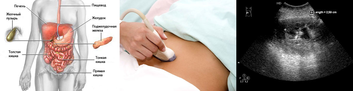
Read on to find out when an abdominal ultrasound is needed, what you can expect from the procedure, and how you can manage it. Abdominal ultrasounds are performed using an ultrasound machine, which uses sound waves to capture images of the abdomen. The test is safe, painless and can be used to evaluate various organs abdominal cavity. These include the spleen, gallbladder, liver, appendix, bladder and intestines. Ultrasound can also be used to evaluate all abdominal organs or selective organs.
Features of the ultrasound procedure
Diagnosis and monitoring of health status become much more effective if research is carried out internal organs and ultrasound is used. Safe method Ultrasound is also completely painless. Depending on the indications, a study is done with an emphasis on various organs.
When is an abdominal ultrasound examination required?
Captured images are black and white and documented on a computer monitor. There are various reasons, for which the doctor should request an abdominal ultrasound. In many cases, the doctor is concerned about the patient's symptoms, and this may include repeated vomiting, abdominal pain, or examining a swollen stomach, as well as kidney and liver function tests. These tests allow the doctor to examine the abdominal organs and evaluate them for disease or injury.
Ultrasounds are also used to guide physicians during procedures that require catheter insertions or biopsies, as it helps ensure good catheter or needle placement. Ultrasounds are also quite common during pregnancy and help monitor fetal development and placement.
The study includes the following points:
- comprehensive examination of the gallbladder;
- pancreas and assessment of its condition;
- special attention is paid to the liver;
- An ultrasound examination of the intestines and other elements is also performed.
When examining the gallbladder, its condition is determined, as well as the condition of the ducts. Their size, the presence of stones, patency and other medical indicators are recorded. An examination of organ tissues and their condition is also carried out.
How to prepare for an abdominal ultrasound
Some of the conditions that can be diagnosed using ultrasound. Pyloric stenosis Kidney or gallstones Appendicitis Abnormal abdominal fluid Abnormal masses in the abdominal cavity, such as abscesses, cysts, or tumors. Preparing for an abdominal ultrasound will ultimately depend on the reason for the test. Find out how you can prepare for various ultrasound tests below.
What happens in an abdominal ultrasound?
Aortic ultrasound: If you are scheduled for an aortic ultrasound, you will have to avoid eating for 8-12 hours before the test. Kidney ultrasound: Your doctor may ask you to drink a couple of glasses of water or juice to fill your bladder. When it comes to food, you will be asked not to eat for approximately 8-12 hours before the ultrasound, and it is recommended to avoid the formation of gas inside the intestines as this may interfere with the kidney assessment. Ultrasounds of the liver, spleen, pancreas and gallbladder: You will need to remain fat for the procedure, which means a low-fat dinner the evening before the test. You should also eat early, as you will have to stay out of food for 8-12 hours before your examination. As mentioned, ultrasounds are painless and simple.
An examination of the pancreas allows one to evaluate parameters such as size, shape, presence of any formations, and contour. Quite often it is difficult to see the organ, since it partially overlaps small intestine or stomach gases. A specialist’s conclusion may contain such a diagnosis as “ diffuse changes" All this suggests that the condition of the abdominal organ was affected by either age-related changes, or with chronic processes inflammatory in nature. Thus, at the moment is very important in the examination and determines many medical indicators.
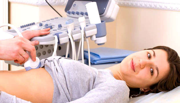
The procedure takes about 30 minutes, and in some cases you will need to wear a hospital gown for the examination. Ultrasound abdominal surgeries are performed by sonographers and are medical specialists who are specially trained to administer the test. You will need to lie on your back during the examination and a small amount of gel will be applied to your abdomen to eliminate any air pockets between the transducer. The transducer is an instrument used to hover over the abdomen and skin.
It is used to send images taken on a computer and the technician will monitor the patient's blood flow in the abdomen to check for any abnormalities or aneurysm. After your abdominal ultrasound, your doctor will evaluate the images and then discuss the results with you. If no aneurysms are found, no additional screening will be needed, but if you have an aneurysm, you may need additional tests. Your doctor will discuss various options treatments available for your condition.
Modern research where the liver enters allows you to get enough full information about the condition of the organ. Often, many ailments arise due to malfunction liver, and that is why it is given great attention. The specialist checks the size of the organ, condition, blood flow, structure and the presence of any changes. In this process, they are defined as focal changes, and diffuse. Thus, the method is quite effective for a comprehensive study and assessment of health status.
The procedure is mostly convenient. However, you may feel some pressure as the sensor hangs over your abdomen. The gel is not always warm and in some cases you will feel a cold sensation and you may also feel a little wet. If the procedure is performed on your young one, you will need to ask them to lie down while the test is taking place so that the sound waves reach all areas of the abdomen that require scanning. Sometimes the baby may need to be restrained during the procedure, and although the baby may cry, this will not interfere with the ultrasound.
Ultrasound is also used to examine other internal organs of the abdomen. An examination of the intestines is carried out, which determines the thickness of the walls, the presence of any formations and other indicators. The method is also effective for studying the kidneys, adrenal glands, and spleen, which provides accurate information about their work and condition. The modern procedure is a safe, high-accuracy determination of all indicators, which makes it possible to establish an accurate diagnosis. It is important that the ultrasound specialist’s conclusion must be analyzed in conjunction with outpatient test results, clinical and anamnestic data. Thus, it is possible to obtain a complete, correct and detailed picture of the health status of the patient and his organs.
Understanding results and other measures
The technician may also require you to shift positions and hold your breath slightly. Typically, ultrasound results are interpreted by a radiologist, who then forwards the results to your doctor. Radiologists are trained to read and interpret data from ultrasound and x-ray images. Your doctor will discuss the results ultrasound examination abdominal cavity with you and in cases where abnormal results were obtained; you may have to undergo further tests.
Preparing for an ultrasound
Ultrasound is a very popular method in medicine because it has high permeability and allows you to obtain the most accurate data. The effectiveness of the result depends not only on the ultrasound diagnostic doctor, but also on the thoroughness and correct preparation of the body. This process is quite important, because otherwise the results may be incomplete or incorrect. When doing an abdominal examination, several important rules should be taken into account:
Why an abdominal ultrasound is performed?
You don't always have to wait for ultrasound results and in emergency cases, these results are obtained quickly. However, in most cases, results are available within a day or two of the test. Ultrasound results are usually not immediately available. There are no risks associated with ultrasound testing, and unlike X-ray imaging, no radiation is required.
Helping your child cope with an abdominal ultrasound
Look next video to learn more about how abdominal ultrasound works and what to expect in the process by watching another person undergo the test. Younger children are sometimes afraid to have abdominal ultrasounds because of the technique used. You can alleviate these concerns by explaining to your child how an ultrasound is performed and why the test is necessary. Make it clear to your young one that the equipment takes pictures of his belly and also encourages questions. Explain that relaxing during the procedure makes it easier for the technique to explore his abdomen.
- First of all, it is important to follow a three-day diet that excludes the use of fresh vegetables and fruits, brown bread, legumes and carbonated drinks. These products cause gas formation and it will be very difficult to check the condition of some organs;
- to obtain a clear, high-quality image, you must not eat anything 7 hours before the organ examination procedure;
- The doctor should be informed before an abdominal ultrasound is performed if the patient is undergoing any course of therapy.
Simple recommendations will help you get a high-quality result and an accurate expert opinion. Most often, this procedure for examining internal organs is carried out in morning time and on an empty stomach. Thus, the condition of the gallbladder, pancreas and other organs will be optimal.
Ultrasound of the abdominal cavity for children and pregnant women
You will need to lie very quietly during the test. You may be asked to take a breath and hold it for a few seconds during the test. This allows the person performing the test to see organs and structures more clearly. The test usually takes 30 to 60 minutes.
You may be asked to wait while a radiologist checks the images. He or she may want to take a closer look at certain areas of your abdomen. You may not be able to take the test or the results may not be useful if. Other image tests may be done if gallstones suspected but not visible by ultrasound.
- Stool, air, or located in the stomach or intestines.
- You cannot remain seated during the test.
- You are extremely.
- You have an open or bandaged wound in the area being examined.
- To find out more, check out the section.
- IN in rare cases, gallstones cannot be detected by ultrasound.
The popular abdominal ultrasound procedure is safe and painless. A doctor can refer a patient to a similar process for the following indications:
- if pain occurs in the upper abdomen;
- if there is bitterness in the mouth, as well as unpleasant heaviness in the area of the left hypochondrium;
- increased gas formation;
- acute pain of a girdle nature.
Feelings like this could be a symptom various diseases, for example, exacerbation of chronic diseases. Under no circumstances is it recommended to delay seeing a doctor, but rather visit a specialist in a timely manner. This way you can avoid a lot of unpleasant situations and stay healthy.
Liver and blood vessels
An abdominal ultrasound to help your doctor see organs and structures inside the abdomen. Ultrasounds are safe and painless. They are also becoming more common. More and more ultrasounds are performed in the United States every year.
Ultrasound images are captured in real time. They can show the structure and movement of internal organs, as well as the blood flowing through blood vessels. This test is the most commonly used for viewing and studying the fetus in pregnant women, but it also has many other clinical uses.
Studying the body using this technique can be as follows: paid service, and free. In the first case, additional aspects are provided such as transferring all received data to digital media, printing a photo, additional examination, interpretation and others. In the second case, the specialist issues a full conclusion, which contains a description of the condition and characteristics of the organs. All this helps the doctor prescribe the correct course of treatment, optimal procedures and make an accurate diagnosis. If ultrasound is prescribed for children, then some preparation features should be taken into account. For example, babies under one year of age should skip one feeding and not drink liquids an hour before the procedure. Older children under the age of three should not eat for four hours before the examination and drink for one hour. Older children should not consume food for 6-7 hours and water for an hour. This way you can get the most accurate results and make the correct diagnosis.
If your doctor suspects that you have any of these conditions, an abdominal ultrasound may be in the near future. Increased kidney blockage or cancerous tumors. . Abdominal ultrasound may also be used to assist the doctor during certain procedures.
How to prepare for the test?
During an abdominal biopsy, your doctor may use ultrasound to determine where to place a needle to remove a small sample of tissue. Your doctor may use ultrasound to examine the blood flow inside your abdomen. Ultrasounds can help your doctor drain fluid from a cyst or abscess. . Ask your doctor if you can continue drinking water and taking your medications as usual before your ultrasound. Your doctor will usually tell you to try for 8-12 hours before your ultrasound.
Medical research, which involves the use of ultrasound, is safe and does not cause any discomfort. Moreover, this technique is very effective, since ultrasound has a high penetrating ability and transmits the most accurate data. Ultrasound includes many important elements. In some cases, before the procedure, the doctor may prescribe the use of special drugs, for example, reducing gas formation. All the specialist’s recommendations should be followed and this will ensure an accurate conclusion and correct therapy.
Abdominal ultrasound is important additional method examination of patients, both surgical and therapeutic. The indications for it are quite extensive. IN clinical guidelines Professor Palmer E.V. indicated following states requiring abdominal ultrasound:
- abdominal pain of unknown origin;
- fever of unknown origin;
- blunt abdominal trauma;
- confirmation of ascites - the presence of fluid (transudate) in the abdominal cavity;
- suspected presence of space-occupying processes (abscesses, hematomas, cysts, tumors).
Such different indications the procedure can be explained a large number vital organs in the abdominal cavity and ambiguity clinical picture, which occurs when they are defeated. In many diseases of these organs, the first symptoms are extremely nonspecific: pain, dyspeptic syndrome, intoxication, fever, and so on. Therefore, to assume correct diagnosis, the morphological structure of the formations that are located in the abdominal cavity should be assessed. The easiest way to do this is with ultrasound.
Ultrasound of the abdominal organs involves examining and assessing the condition of the following structures:
- liver and nearby vessels (portal and hepatic veins);
- gallbladder;
- biliary tract (cystic, common hepatic and common bile duct And);
- pancreas;
- walls of the stomach and intestines (extremely rare, since their visualization is almost always difficult)
- spleen;
- diaphragm;
- kidneys (despite the fact that they lie in the retroperitoneal space, clinicians assess their condition when performing an ultrasound scan of the abdominal cavity);
Each of these organs has its own morphological features, which are studied during ultrasound examination. With their help, you can determine the localization of the pathological process.
Liver and blood vessels
The normal structure of the liver implies a homogeneous structure of the parenchyma throughout its entire length. Diagnostic doctors look for the presence of any formations that manifest themselves as echo-negative foci. It is possible to estimate their size, density and suggest the nature of the focus.
The portal (portal) and hepatic veins are normally visible against the background of the liver. Walls portal vein and the branches extending from it have high echogenicity, while the hepatic veins do not conduct ultrasound at all. At portal hypertension, the walls and volume of these vessels can change significantly, which is manifested by a change in signal conductivity.
Gallbladder
In the absence of pathology, the gallbladder is an echo-negative formation - it does not conduct an ultrasound signal and appears on the monitor as a black structure. The presence of echogenic foci in it indicates the presence of stones. The size of the gallbladder is variable, so it is not assessed during ultrasound. Its shape is constant: on a transverse section it should be round, on a longitudinal section it should be pear-shaped. The condition of the wall and what formations are in its cavity should be assessed. Normally, the cavity of the bladder is filled only with bile, and the walls have a smooth and clearly defined surface.
Biliary tract
Hepatic bile ducts and cystic duct, as a rule, are not looked at during ultrasound of the abdominal organs, since their visualization is difficult due to the structural features of the mucous membrane and the low speed of bile flow.
The common bile duct on ultrasound looks like a straight tube. Its examination is often difficult due to the presence of gases in the intestines. This is due to improper preparation of the patient for the procedure. In this case, a discontinuous structure of the common bile duct can be detected.
Pancreas
This organ has greater echogenicity than surrounding tissues. In an unchanged state, it is visualized as a homogeneous, coarse-grained formation. Almost all diseases of the pancreas are reflected in its structure, shape and structure:
- pancreatitis – ultrasound can detect enlargement of the organ, a decrease in its ability to conduct an ultrasound signal, but clear contours remain;
- pseudocyst – an echo-negative formation with limited clear contours is determined in the structure of the pancreas;
- neoplasm (most often solid cancer) – indirect sign these are blurred boundaries of the head of the pancreas due to diffuse decrease in echo signal.
Diagnostic doctors must look for the presence of these signs during an ultrasound of the abdominal cavity.
Spleen
With a normal structure of the organ, you can see the parenchyma in the form of an oval formation with low echogenicity. As a rule, the vessels are not visible. Increased conduction signal and visualization of blood vessels is a direct sign of splenomegaly - an increase in the size of the spleen.
The edges of the spleen differ significantly on ultrasound. The superior and lateral edges are more convex, while the inferior and medial have depressions of varying sizes from nearby organs. The medial surface of the spleen is always concave. Irregular or blurred contours of the organ may indicate a rupture of the spleen or the formation of a space-occupying process (most often a cyst). The formation of a double circuit is absolute sign formation of a subcapsular hematoma.
The hilum of the spleen (the place where the vessels enter and exit) has a higher echogenicity than the parenchyma.
Kidneys
Despite the fact that the kidneys are an organ of the retroperitoneal space, they are often examined together with the abdominal organs. The examination plan includes assessment of the location of the kidneys, its parenchyma, capsule, pelvis and renal veins. Normal indicators the following:
- the kidneys are located between the bispinarum line (the segment connecting the anterosuperior spines iliac bones) and diaphragm;
- clear contour of the renal capsule;
- the parenchyma is heterogeneous: the cortex is more echogenic than the medulla;
- the collecting system (pelvis) has an irregular asymmetrical shape and a denser structure.
An ultrasound can determine an additional kidney, the presence of cystic processes, severe glomerulonephritis or long-term chronic pyelonephritis.
Evaluation of the abdominal organs using ultrasound has great value V clinical practice. Using it, you can determine the affected organ, in the case of non-localized or nonspecific symptoms, assess the condition of organs and some vessels. The pathology of each organ on ultrasound has its own echographic signs, which make it possible to determine the clinical diagnosis.
