Osteochondropathy - causes, symptoms, diagnosis and treatment. Osteochondropathies of short bones
Many ailments occur in humans life path. Some of them begin to bother us in childhood. And these include Perthes disease in children. If you do not start treating your child in time or miss the first symptoms of this disease, dire consequences- the child will lose the opportunity to live a normal life.
Perthes disease
Perthes disease in children is a disease characterized by poor circulation in the pelvic area. Because of this, there is a malfunction of the head pelvic bone, which leads to the death of bone cells, accompanied by irritation and inflammatory processes.
This disease was first described more than a century ago and meant a special case of arthritis of the hip joint. Another name for this disease is osteochondropathy of the head hip joint, that is, the destruction of bone cells under the influence of various factors. Perthes in children most often manifests itself between the ages of 4 and 10 years, and this pattern is typical in in most cases for boys than for girls.
Symptoms
The first signs of perthes disease in children appear in the form of minor pain in the articulation of the femur with the pelvis. Often they develop on only one side. Sometimes pain can originate in the knee. In this case, the child’s gait first changes - he begins to fall on one leg. Usually, at this moment the child is not bothered by severe pain, his gait is almost unimpaired, which is why parents postpone going to the doctor.
Then the symptoms begin to bother the children more and more. Due to impaired blood circulation, the tissue of the femoral head begins to die, under the influence of normal stress - running, walking, more severe pain appears, lameness increases. These symptoms indicate that upper part The femur began to deform. In this case, symptoms such as weakness, pain, limited leg mobility and even shortening of the lower limb appear.
If you do not catch it in time, the symptoms of the disease can lead to much more harmful consequences.
The child’s bone becomes irreversibly deformed, which will result in chronic pain in the leg and a significant change in gait.
Culprits
IN present moment Scientists identify two causes of the disease in a child:
- Birth defects that occur due to underdevelopment in the womb.
- Acquired factors are the basis on which the disease develops.
TO congenital factors include myelodysplasia, or underdevelopment, lumbar region backs. As a result, the child experiences deteriorated innervation of tissues in the lumbar region, the connection of the pelvic bone with the thigh. Myelodysplasia is also characterized by fewer blood vessels in the area. The causes of underdevelopment are hereditary predisposition.

Acquired factors can be infections, bruises, injuries, which lead to compression of the blood vessels of the hip joint and, as a result, disruption or complete cessation of bone nutrition. As a result of infection, an inflammatory process develops that pinches the supplying vessels. The causes of inflammation can be a common cold. The same thing happens with injuries, and they can be so minor that they will not cause concern to either the child or the parents.
Diagnostic measures
X-ray examination allows identifying Perthes in children. This method of diagnosing the disease is decisive and allows us to accurately say at what stage the disease is, as well as to identify the prerequisites for its development.
Other diagnostic methods that can be used are examination of the femoral head with magnetic resonance or ultrasound imaging. Moreover, such diagnostic methods are not decisive in making a diagnosis, and are additional diagnostic methods.
Treatment
The method by which Perthes disease will be treated depends on what stage of development of the disease you discovered the first symptoms on X-ray or ultrasound diagnostics. In cases where this happened in a child aged 4 to 6 years, treatment may consist of observing the femoral head, with the hope that the body will return to normal on its own.

If osteochondropathy of the femoral head has been identified for more than late stage, then treatment will vary based on accompanying symptoms:
- If there is a strong inflammatory process in the head area, then non-steroidal anti-inflammatory drugs can be taken.
- If osteochondropathy significantly limits movement, or significant tissue deformation is observed, then a method of holding the bone is used - special splints are applied for this.
- The method of surgical intervention is used for more serious complications. In this case, the head is surgically returned to the acetabulum and its position is secured with bolts, and a corset is placed on the child’s body from the chest to the hip.
In all cases, osteochondropathy of the femoral head can be treated with a method of treatment such as exercise therapy (therapeutic physical culture) and massage. This helps improve blood circulation in the tissues, and also increases muscle tone and develops the joint.
Therapeutic physical and massage procedures
Exercise therapy is used at all stages of the disease. It is designed to increase activity in the joint, improve blood flow and stretch stagnant muscles. In cases where osteochondropathy was eliminated by surgery, exercise therapy is necessarily included in the rehabilitation course. Physical education in this case should be carried out under the strict supervision of a doctor and constant monitoring changes on x-rays. At first, exercise therapy should only influence the affected part at half strength, and the load should increase gradually.

Massage is indicated in any case, regardless of when pertheses were discovered in children. It produces an analgesic effect, prevents muscle atrophy, improves blood circulation and increases muscle tone.
Massage provided active influence on the disease in several directions:
- massage stretches muscles, which is a natural way to increase sensitivity skin And muscle tissue, and in addition, it tones the muscles;
- massage breaks down stuck together muscle fibers that occur during prolonged inactivity;
- massage improves blood circulation, literally disperses blood through the vessels;
- massage improves tissue nutrition.
The method by which osteochondropathy will be eliminated depends primarily on when the disease was discovered. In cases where diagnostic methods have made it possible to identify the early stage of development of the disease, the treatment will pass much easier and painless. Therefore, do not neglect periodic consultations with your child’s doctor.
Perthes disease of the hip joint: causes, stages, symptoms and treatment
This disease is aseptic necrosis of the femoral head. It can occur in the hip joint, but can also spread to the articular area, bone tissue, blood vessels, and nerves. This disease is osteochondropathy of the hip joint. The right joint is most often affected. With a bilateral disease, the left joint is less affected.
- Reasons
- Stages of Legg–Calvé–Perthes disease
- Diagnostics
- Symptoms of Perthes disease of the hip joint
- Treatment of Perthes disease
The disease develops when there is a temporary cessation of blood supply, which results in necrosis of the tissue of the head of the bone. In this case, a fracture and recovery occurs in the future. bone tissue. This disease most often affects boys aged 4-14 years. According to statistics, there is one case of this disease per thousand boys. Almost 10% of patients have damage to both hip joints. It is believed that patients under 6 years of age have a greater chance of full recovery than other children. In them, the head of the bone is restored over a longer period. Moreover, its structure is almost the same as that of an intact head.
Reasons
The exact causes of this disease have not yet been established. Despite the existence of several theories of its development, they all boil down to the fact that it is the disruption of blood flow that leads to necrosis of bone tissue cells. Among the most possible reasons the occurrence of this disease should be highlighted:
- injuries caused by impact or displacement of the head of the bone;
- hormonal surge;
- infections followed by the body's immune response;
- disturbances in the exchange of macro- and microelements involved in bone formation.
After normal blood flow is restored, new cells are formed instead of dead ones. They start the process of restoring the structure of the bone head. One of important conditions progression of this pathology is considered to be the presence of a predisposing factor, namely myelodysplasia (congenital underdevelopment spinal cord in the area of the lumbar spine). It is he who controls the innervation of the joints in the pelvic area and is responsible for blood flow in it. With this pathology, the size and number of vessels supplying this part of the body is less than that of healthy child. All these circumstances lead to disturbances in the normal state of the joint head.
Experts highlight following features of this disease:
- The likelihood of the disease is determined by the constitution of the child.
- The disease most often affects weakened, malnourished and long-term ill children.
- Male children are 5 times more susceptible to this disease than girls. However, in boys the disease is somewhat milder than in girls.
- Children 4-10 years old get sick more often than other ages.
- The likelihood of developing this pathology is also influenced by hereditary (genetic) factors. There are cases of several pathologies in one family.
- Children who have had rickets are more susceptible to this disease.
- Short children who experience physical stress get sick much more often than their tall peers.
- Most often, children in whose family someone smokes get sick.
Stages of Legg–Calvé–Perthes disease
There are 5 stages in children:
- The blood supply to the femoral head begins to stop, necrosis begins, that is, necrosis of the femur.
- This is followed by a secondary fracture of the femoral head.
- Necrotic bone tissue is resorbed, and the femoral neck shortens, that is, fragmentation occurs.
- Connective tissue will grow into the space vacated by the dead femur.
- Ossification occurs connective tissue, the fracture heals, the correct structure of the joint is disrupted.

Possible complications and consequences of Perthes disease
Osteochondropathy of the hip joint can be accompanied by the following complications:
- Permanent residual deformation of the head of the bone (most often observed in children over 6 years of age).
- Osteoarthritis of the head resulting from its deformation, injury to the acetabulum and thinning cartilage tissue. With this complication, patients need surgery to replace the hip joint. After such an operation, it will take from 2 to 5 years to fully restore health.
Diagnostics
The diagnosis is made based on:
- medical examination;
- symptoms of the disease;
- diagnostics using special tools.
To confirm the diagnosis, the child must undergo x-rays of the hip joint and pelvis. If necessary, magnetic resonance imaging and osteoscintigraphy can be performed. Thanks to hardware research, it is possible to establish a diagnosis of early stages diseases.
Symptoms of Perthes disease of the hip joint
When the first signs of this disease appear, you should immediately consult a doctor. The main symptoms of this disease include:
- pain and tension in the hip joint, groin area, regions knee joint;
- leg muscle atrophy;
- lameness;
- restriction of movements in the joint;
- in severe cases, shortening of the leg on the affected side is observed.
This pathology is characterized by spontaneous disappearance of the above symptoms and their reappearance after 3-4 months. In some cases, pain decreases at rest and increases significantly with physical activity.
Due to the ability articular cartilage to rapid regeneration in children under 10 years of age during the course of the disease, recovery processes. Perthes disease is characterized by phasicity. Experts distinguish the following phases of the disease:
- necrosis;
- impression fracture;
- resorptive-restorative;
- outcome.
Treatment of Perthes disease
Therapy for this disease is primarily aimed at preventing changes in the spherical shape of the femoral head. At the same time, maintaining its anatomical position in the joint is also important. If therapy is started in a timely manner, complete restoration of the femoral head is most often observed. Otherwise, the disease leads to its deformation. Therapy consists of the following activities:
- Maximum unloading of the legs, including bed rest, use of crutches, immobilization of limbs.
- Bringing blood flow to the hip joint to normal condition through physiotherapy and massage.
- Surgical treatment in particularly severe cases.
 During the treatment of Perthes disease, non-steroidal anti-inflammatory drugs (Ibufen, Neurofen) are used. With their help, they eliminate pain and reduce inflammation in the joint. Thanks to these medications, the motor function hip joint.
Physiotherapy also plays an important role in the treatment of this disease. With its help, joint mobility improves and the anatomical spherical shape bone heads. To reduce pain When walking, it is recommended to use crutches, which help reduce the load on the inflamed head of the bone.
Sometimes it is necessary to immobilize the hip joint using a cast or a special splint. Thanks to temporary immobilization, the optimal position of the head in the joint cavity is maintained, which leads to faster healing.
In cases where lameness has led to shortening of the limb muscles and limited joint mobility or displacement of the head of the bone, surgical intervention is required. During the operation, muscle fibers are lengthened. After this, the hip joint is fixed by applying a plaster cast. It is left for 1-2 months (depending on the severity of the patient’s condition). During this period, the muscles are restored to normal length.
Unfortunately, preventive measures There is no cure for Perthes disease. To return children to normal life required early diagnosis and proper complex therapy. Treatment and recovery period for this disease can last 18-24 months. Without proper therapy, Perthes disease leads to deforming arthrosis of the hip joint.
- Clinical picture
- Surgical and conservative therapy
- Complications
 Shoulder dislocation is one of the most common injuries. It accounts for more than half of all such damage, as well as 3% of total number injuries This frequency can be explained very simply - by the peculiarity of the anatomical structure shoulder joint and the large volume of movements that he performs. According to ICD 10, this diagnosis is encrypted as S43.
Depending on what caused the injury, it is divided into:
- Traumatic.
- Free.
- Congenital.
- Familiar.
- Pathological.
Habitual dislocation begins to develop when, after the initial dislocation, the recovery rules were not followed and the injury was not completely treated. The pathological form occurs when the tissues of the shoulder joint are affected, for example, by a tumor, osteomyelitis occurs in the bones, and there are also diseases such as osteochondropathy, tuberculosis, osteodystrophy and some others.
In some cases, this injury may be combined with others, for example, with a fracture of the head humerus, separation of the greater or lesser tubercle, damage to the tendons that are located nearby.
Clinical picture
 All dislocations of the shoulder joint are accompanied by severe pain in the affected area and deformation of the shoulder area. Any hand movements become simply impossible. When attempting passive movements, they are characterized by significant resistance. These symptoms can be considered basic.
In an anterior dislocation, the head of the bone moves forward and downward. The hand is moved to the side. During palpation, the head of the humerus cannot be detected in its place, but is felt in the armpit or below the scapula.
An anterior dislocation is often accompanied by a separation of the greater tubercle of the bone and a fracture of the processes of the scapula. With the lower one, the head of the humerus also goes into the armpit, where many vessels and nerves pass through. If the head begins to put pressure on the neurovascular bundle, this manifests itself in the form of numbness of the skin, paralysis of the muscles in the area where the compression occurred.
To determine whether a shoulder is dislocated and to understand whether certain collateral damage, it is necessary to conduct an x-ray examination in two projections. In case of old injury, it is more effective to use MRI in diagnosis.
Surgical and conservative therapy
First aid - immobilization, introduction non-narcotic analgesics and transportation to medical institution. During transportation, the arm should be fixed using a Kramer splint; a Deso bandage can also be used for a dislocated shoulder. It is important to remember that as little time as possible should pass from the moment of injury to hospitalization, since delay can cause unsuccessful reduction, after which it is necessary to resort to surgery.
Only a doctor should correct a dislocated shoulder. Before the procedure, an anesthetic, for example, lidocaine, is injected into the joint cavity, after which one of the popular and widespread methods is used. After the procedure, the arm is suspended on a scarf or fixed with a plaster cast with further X-ray monitoring.
Sometimes it is not possible to correct the situation under local anesthesia; most often this is due to long period time that has passed since the injury. In this case, intravenous anesthesia is given. But if even then the joint does not fall into place, then a full-fledged surgery with fixation of the head of the bone in the articular cavity.
After reduction, immobilization should be at least 3 weeks, but since the patient does not feel pain and considers himself healthy, the bandage is often removed on its own much earlier, which can lead to re-injury. This situation ultimately leads to habitual dislocation, which cannot be cured conservatively.
Treatment after reduction is based on wearing a Deso bandage, which limits movement in the affected joint. It can be made either from plaster, or from a bandage or scarf, but it is best to use a special orthopedic bandage that does not restrict movement and at the same time securely holds the arm.
Complications
 If the shoulder dislocation is not corrected, it may result in the development of some serious complications. In addition to the usual dislocation, damage to blood vessels, nerves, and tendons can occur. In this case, the hand ceases to perform its function, and the person subsequently becomes disabled.
If the reduction is carried out incorrectly, further problems may arise. dystrophic changes in the joint, which can cause a chronic disease such as arthrosis. Another irreversible change that can occur is called fibrosis.
Often, if the injury occurs as a result of a car accident, there may be severe pain shock, therefore, the administration of analgesics is immediately required here.
In the article you will learn what osteochondropathy is, why this disease appears in children, what the first symptoms are and how to treat this disease.
One of the joint diseases that can be found most often in adolescents is osteochondropathy. The disease is associated with metabolic disorders with joint tissues as a result of impaired blood supply to a certain area. Dead areas of bone form, for example, during physical activity. With osteochondropathy, the bones become brittle and the child is susceptible to spontaneous fractures, so it is very important to consult a doctor at the first signs of the disease. The main symptom of the pathology is pain in the affected area; the child also becomes lethargic and may complain of weakness.
Osteochondropathy main causes
The etiology of the disease is not fully understood, but doctors say that the following factors underlie osteochondropathy:
- Hereditary predisposition. If one of the family members suffered from bone pathology, the child has an increased risk of getting sick.
- Hormonal disorders. Often osteochondropathy is associated with pathology endocrine system.
- Injuries. Children who are often injured, and also play sports and undergo heavy physical activity are more susceptible to getting sick.
- Metabolic disorder. Pathology can be caused by a lack of calcium and vitamin D in the body, which is why it is so important to monitor the child’s nutrition.
- Circulatory disorders. In this case, lack of nutrition provokes bone necrosis.
The disease most often affects the legs, since they bear the main load. The onset of the disease practically does not manifest itself, but when the first symptoms appear, it is important to consult a doctor. By taking a picture, a specialist can determine the extent of the lesion and the complexity of the disease. Typically, treatment can be complex (bed rest, therapy, medications) or surgical.
Symptoms of the disease
The first signs of bone tissue damage can be determined by the following factors:
- Swelling appears at the site of the lesion, but signs of the inflammatory process may be absent.
- Pain occurs. It can be either constant or occur under a certain load.
- In the presence of such a disease, fractures can often occur.
- There are visual signs of changes in posture. There may be lameness.
Despite the danger of the disease, it can be brought under control and even completely eliminated. Proper treatment in a timely manner will avoid bone deformation and additional difficulties will not arise in the future.
Classification of osteochondropathy
Distinguish the following types osteochondropathy:
- Pathology of short tubular bones. This type of disease includes Köhler I, Kienbeck, Calve, and Praiser diseases.
- Pathology of tubular bones. The second type includes Legg-Calvé-Perthes and Köhler II disease.
- In the apophyses. This type includes Haglund-Schinz, Osgood-Schlatter, and Scheuermann-Mau disease.
- Osteochondropathy of the joint surface. The latter type is called Koening's disease.
Vertebral osteochondropathy
Diseases that affect the spine are called Sherman-Mau disease and Calvet disease. Sherman-Mau disease is characterized by destruction of the apophyses of the vertebrae; this disease occurs most often in boys during adolescence.
In this case, there are often no symptoms, patients do not complain of pain, but there is a pronounced deformation of the spine, the back becomes round. This condition often forces parents to consult a specialist.
With Calvet disease, damage to the vertebral body occurs, and this condition usually occurs before the age of 7 years. The disease is accompanied by pain in the spine, and the child becomes lethargic, weak, and capricious. For osteochondropathy of the spine, treatment should be carried out in a hospital setting, since the patient needs complete rest.
Osteochondropathy of the hip joint
This pathology is called Perthes disease, the disease occurs most often in boys under the age of 9 years, and the head of the femur is affected. Often the disease occurs after a pelvic injury, the child suddenly begins to limp and complains of pain in the pelvis, which radiates to the leg.

The disease gradually progresses, the function of the hip joint is impaired and motor activity is impaired, the muscles atrophy. If hip subluxation occurs, the affected leg may become shorter.
This pathology requires compulsory treatment, otherwise the head of the hip joint is formed incorrectly, which leads to the occurrence of deforming arthrosis. In most cases, the disease is treated conservatively, but in advanced stages of the pathology, surgical intervention may be necessary.
Osteochondropathy of the tibia
Schlatter's osteochondropathy most often occurs in boys during adolescence, it is characterized by pain in the area tibia.

Unpleasant sensations intensify when the patient gets up from a chair, or climbs stairs, as well as in a kneeling position. Sometimes there may be dysfunction of the knee joint.
In most cases, this pathology does not leave complications; the disease is treated on an outpatient basis, surgical methods do not apply.
Osteochondropathy of the foot
In Köhler's disease II, destruction of 2 and 3 occurs metatarsal bones. The disease occurs more often in girls during adolescence, develops gradually, the first symptoms of the disease are mild pain in the foot area. Over time, the pain intensifies, the child limps, and the skin around the affected area swells slightly.

The disease can be cured at home by prescribing limb immobilization and then physical therapy. If you consult a doctor in a timely manner, the prognosis is favorable.
In Köhler's disease I, the scaphoid bone is affected and occurs mainly in boys under 7 years of age. The symptoms of the pathology are similar to the previous one, the child limps and complains of pain, there is swelling of the foot with outside.
Schinz's disease is characterized by lesions of the tubercle calcaneus, children aged 6 to 14 years are susceptible to pathology. The child complains of pain in the heel, swelling of the affected area is observed, the patient limps and tries to take care of the affected heel.
Koenig's disease
This pathology is called osteochondropathy dissecans and is more common in boys during adolescence. When the disease occurs, the knee and hip joints are affected, a section of cartilage undergoes necrosis and peels off, sometimes fragments penetrate into the joint.

Patients who are faced with this diagnosis are interested in how arthrosis differs from osteochondropathy. With arthrosis, deformation of the joint occurs, and with osteochondropathy, destruction of cartilage tissue occurs.
The disease is accompanied by characteristic symptoms: joint pain, impaired motor activity. The advanced stage of the disease is characterized by atrophy of muscle tissue, and a complication is arthrosis deformans.
Most often, diseases are provoked by increased physical activity, while the blood circulation of any part is disrupted, and its destruction occurs due to lack of nutrition. The disease is treated surgically, since the dead section of cartilage must be removed.
Stages and symptoms of the disease
The following stages of osteochondropathy are distinguished:
- The disease begins with gradual necrosis of bone tissue, while symptoms may not be observed at all, or the patient may be bothered aching pain in the affected area. The pain intensifies with movement and when pressing on the affected area, and may also be disrupted motor activity affected limb.
- On next stage disease, a fracture of the affected bone occurs, with corresponding symptoms.
- At the next stage, the affected tissue is resorbed and replaced by granulation tissue.
- On last stage bone tissue is restored. Without treatment full recovery does not occur, complications arise in the form cosmetic defects and deforming arthrosis.
The average duration of the disease is 3 to 4 years.
Echo signs of osteochondropathy
Only a doctor can correctly diagnose the disease, therefore, at the first signs of the disease, it is necessary to show the child to an orthopedist as soon as possible. During the examination, the doctor interviews the patient and examines him; based on the data obtained, an experienced specialist can immediately make a preliminary diagnosis.
To confirm the diagnosis, the patient is referred for radiography and ultrasound, or computed tomography. In the initial stages of the disease, it is difficult to identify pathology using radiography, and with the help of ultrasound, the doctor detects echo signs of the disease even in the initial stages.
Treatment of osteochondropathy
There are 2 stages of treatment for osteochondropathy. First, the patient is prescribed complete rest and non-steroidal anti-inflammatory drugs to stop the destruction of bone tissue and relieve pain. The next step is for the patient to therapeutic massage, physical therapy, physiotherapy.
In case of severe pain, a plaster cast may be applied, and if the foot is affected, wearing special orthopedic insoles. The operation is performed for severe deformation of the spine or limb, and if the hip joint is affected, skeletal traction may be indicated.
(No ratings yet)
Bones perform the most important supporting function in the human body; they are its frame. If bone tissue in any part of the body is destroyed, the function of the entire limb is impaired and the person becomes disabled.

One of the serious childhood bone diseases is osteochondropathy of the femoral head, in which bone tissue is destroyed due to lack of nutrition. This disease causes a lot of inconvenience to the patient and can provoke serious complications, therefore, treatment of osteochondropathy should be started immediately.
Osteochondropathy of the femoral head in children
Perthes disease, or osteochondropathy of the femoral head, occurs mainly in boys aged 5 to 14 years; girls are affected much less frequently, in about 15 cases out of 100. The pathology affects not only bone tissue, but also nearby joints, which complicates the patient’s condition.
The exact cause of osteochondropathy in children is still not known, but doctors are confident that the disease is associated with a hereditary predisposition to it. Experts have also identified a number of negative factors that can provoke necrosis of the femoral head:
- hip injuries in which the femur is displaced;
- some infectious diseases;
- inflammation of the hip joint, such as synovitis;
- metabolic disorders;
- hormonal disorders;
- weak immunity;
- vitamin deficiency, poor nutrition;
- rickets;
- congenital defects associated with poor circulation in the lower part of the spine.
Perthes disease develops slowly and gradually progresses, but in the initial stages the pathology is asymptomatic, so the disease cannot be detected immediately. Doctors distinguish 5 stages of osteochondropathy of the femoral head:
- The initial stage of osteochondropathy of the femoral head is characterized by poor circulation; the bone begins to deteriorate due to lack of nutrition. Pain on initial stage weak, the child may limp slightly when walking.
- At the second stage, a depressed fracture occurs, which is accompanied by severe pain, severe lameness, and swelling in the affected area.
- At the next stage, bone necrosis begins to resolve and be overgrown with connective tissue, the femoral neck shortens, but the pain gradually goes away. If treatment is not carried out, the limb shortens and the child continues to limp.
- The disease usually ends with healing of the fracture. But due to damage to the femoral neck, the joint suffers, which, if left untreated, ultimately leads to the formation of arthrosis of the hip joint.
Osteochondropathy of the 2nd metatarsal bone
Osteochondropathy can affect not only the femur; necrosis can also affect the metatarsal heads. The disease most often occurs due to injury, wearing tight and uncomfortable shoes, increased load on the feet. The consequence of such a negative impact is impaired blood circulation in the joint and destruction of the head of the metatarsal bone.

Osteochondropathy of the head of the 2nd or 3rd metatarsal bone is accompanied by the following symptoms:
- Pain appears, which initially bothers you during physical activity, but over time becomes constant and unbearable.
- Swelling occurs with back side feet.
- Because of the pain, the patient limps and takes care of his sore foot.
- The motor activity of the metatarsal joint is disrupted, it is deformed, and the finger is shortened.
Osteochondropathy of the head of the 2nd metatarsal bone occurs in several stages, as does necrosis of the femur. If untreated, the pain goes away 2 years after the onset of the disease, but the pathology is complicated by arthrosis of the affected joint.
Osteochondropathies of the metatarsal bones are treated conservatively in most cases. The patient is prescribed to wear orthopedic shoes and insoles, it is also recommended not to put any weight on the sore foot, so as not to injure the cartilage tissue of the joints. In complex therapy, the doctor prescribes painkillers and physiotherapy.
Treatment of osteochondropathy of the femur
Treatment of osteochondropathy of the femur is complex and long-term. First of all, immobilization of the affected hip is prescribed to prevent further destruction of bone and cartilage tissue. The patient will be placed on bed rest and will wear a brace or plaster cast. If a fracture is present, skeletal traction may be prescribed.

The next step is to take painkillers and drugs to normalize blood circulation in the affected area. To maintain muscle tone, special physical therapy and massage are prescribed. To prevent weight gain during bed rest, the patient must adhere to a special diet.
In complex therapy, the child is prescribed to attend physiotherapeutic treatment. This can be electrophoresis with drugs, magnetic therapy, UHF and other procedures that are individually selected by a specialist in each case. To restore cartilage tissue and prevent its destruction, injections with chondroprotectors may be indicated.
Throughout the entire period of treatment, the child must be regularly shown to a pediatrician and orthopedist to monitor the condition of the joint. To prevent relapse of the disease, the patient is prohibited from putting too much strain on the diseased joint, but daily exercise is indicated. therapeutic exercises and proper nutrition.
Osteochondropathy is a disease of children and adolescents in which a degenerative-dystrophic process develops in the bones.
With osteochondropathy, the calcaneus, femur, apophyses of the vertebral bodies, and the tibial tuberosity are most often affected.
Reasons for appearance
Today, the causes of the disease are not fully understood, but several decisive factors are identified:
- congenital or family predisposition;
- hormonal factors - the disease develops in patients with pathology of the function of the endocrine glands;
- metabolic disorders of essential substances. Osteochondropathy is often caused by impaired absorption of calcium and vitamins;
- traumatic factors. Osteochondropathy occurs after excessive physical exertion, incl. increased muscle contractions, frequent injuries. Initially, these types of loads lead to progressive compression, and then to narrowing small vessels spongy bones, especially in areas of greatest pressure.
Symptoms of osteochondropathy
Osteochondropathy of the calcaneus (Haglund-Schinz disease) develops most often in girls 12-16 years old, characterized by gradually increasing or acute pain in the tubercle of the calcaneus that occurs after exercise. There is swelling at the site of attachment of the Achilles tendon, above the calcaneal tubercle. Patients begin to walk, leaning on their toes, and playing sports and jumping become physically impossible.
Spinal osteochondropathy (Scheuermann-Mau disease) develops most often in boys 11-18 years old. The first stage is characterized by increased thoracic kyphosis (curvature of the spine in its upper part), the second - by back pain (especially with prolonged walking or sitting), rapid fatigue and weakness of the spinal muscles, and increased thoracic kyphosis. At the third stage of spinal osteochondropathy, complete fusion of the apophyses with the vertebrae is observed. Over time, osteochondrosis develops with increasing pain.
Osteochondropathy of the femur (Legg-Calvé-Perthes disease) develops in most cases in boys 4-12 years old. At the beginning of the disease there are no complaints, after which pain appears in the hip joint, radiating to the knee. Pain occurs after exercise and goes away after rest, so children do not always complain about it. The movements of the hip joint are gradually limited, muscle atrophy develops, and the thigh on the affected side loses weight.
Osteochondropathy of the tibial tuberosity (Schlatter's disease) develops in boys 12-16 years old, especially in those who engage in ballet, competitive dancing, and sports. The patient complains of pain under the patella and swelling. When the quadriceps femoris muscle is tense, when squatting, or climbing stairs, the pain intensifies.
Diagnosis of the disease
To determine osteochondropathy of the calcaneus, they are based on clinical data and results X-ray examination(fragmentation, compaction of the apophysis, “roughness” on the tubercle of the calcaneus are noted). Also carried out differential diagnosis osteochondropathy with heel spur(in older patients), achillobursitis.
Diagnosis of spinal osteochondropathy occurs on the basis of examination data (increased thoracic kyphosis) and x-ray examination (the pictures show that the shape of the vertebrae has been changed - they become wedge-shaped).
 Osteochondropathy of the femur is also determined by X-ray images. Five stages of change in the femoral head have been identified.
Treatment of osteochondropathy
Therapy for osteochondropathy of the calcaneus consists of prescribing non-steroidal anti-inflammatory drugs (if severe pain), physiotherapeutic procedures, and reducing physical activity. To relieve the load on the heel bone, special insoles-instep supports are used.
Spinal osteochondropathy is treated with massage, swimming, underwater stretching, and physical therapy. In some cases, when severe violation posture surgery is prescribed.
Treatment of osteochondropathy of the femur can be surgical or conservative. Various osteoplastic surgeries are prescribed depending on the stage of the disease. Conservative treatment of osteochondropathy consists of bed rest (the patient should not sit), foot massage, and physiotherapeutic procedures. They practice skeletal traction on both hips.
To treat osteochondropathy of the tibial tuberosity, physiotherapeutic procedures and heat are prescribed. If the pain is severe, apply a plaster cast. Sometimes they resort to surgery - a fragment of the tuberosity is removed. Loads on the quadriceps femoris muscle are excluded.
Prevention of disease
To prevent osteochondropathy of the calcaneus, it is recommended to wear loose shoes.
Prevention of spinal osteochondropathy involves exercise physical therapy to create a muscle corset. Strenuous physical activity should be limited. Wearing a corset for this disease is ineffective.
A good prevention of osteochondropathy of the femur is massage and swimming.
To prevent osteochondropathy of the tibial tubercle, athletes are recommended to sew 2-4 cm thick foam pads into their uniform during training.
Video from YouTube on the topic of the article:
Perthes disease (or osteochondropathy of the femoral head) is one of the most common diseases of the hip joint in children aged 3 to 14 years and is the most common in childhood a type of aseptic necrosis of the femoral head.
The term " aseptic necrosis"in this case means necrosis of the bone tissue of the epiphysis (that is, the upper hemisphere) of the femoral head non-infectious nature(Fig. 1).
Rice. 1. Anatomical structure hip joint (R.D. Sinelnikov. “Atlas of normal human anatomy.” 1967):
1. Epiphysis of the femoral head.
2. Femoral neck.
3. Acetabular cavity.
4. Greater trochanter.
5. Lesser trochanter.
6. Articular capsule.
Boys aged 5-12 years are more susceptible to the disease, but in girls it is more severe. The pathological process has several stages and can last for many months, in approximately 5% of cases both hip joints are affected.
The views of different researchers on the etiology and pathogenesis (that is, the origin and development) of Perthes disease still differ. The most common pathogenetic theory of the disease in a “compact” and simplified form is presented below.
A prerequisite for the development of Perthes disease is the presence in the child of certain predisposing (congenital) and producing (acquired) factors. The predisposing factor for Perthes disease (or the background for its appearance) is the so-called myelodysplasia of the lumbar spinal cord, which is responsible for the innervation of the hip joints.
The term “myelodysplasia” means a developmental disorder (in this case, underdevelopment) of the spinal cord. Underdevelopment of the spinal cord is congenital - hereditary in nature; it occurs in a significant proportion of children and varies in severity. Mild myelodysplasia may go undetected throughout life, while more significant disorders of spinal cord development manifest themselves in a variety of ways. orthopedic diseases, which includes Perthes disease.
The anatomical structure of the hip joint area in children with myelodysplasia differs, in particular, in that the number and caliber of vessels and nerves that innervate and supply the joints is less than in a healthy child (Fig. 2).

Rice. 2. Blood supply to the head and neck of the femur (G.H. Thompson, R.B. Salter. “Legg-Calve-Perthes Disease.” 1986):
1. External and internal arteries that bend around the femur.
2. Ascending branches of the circumflex arteries entering the neck and head of the femur.
3. Cartilaginous growth zone of the femoral head.
Extremely simplified this situation one can imagine as follows- instead of 10-12 relatively large arteries and veins entering and exiting the femoral head, the patient has 2-4 small (congenitally underdeveloped) arteries and veins, so the blood flow in the joint tissues is chronically reduced, and they experience nutritional deficiency. The presence of myelodysplasia also negatively affects the tone of the vascular wall.
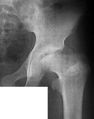
Meanwhile, Perthes disease develops only when the blood supply to the femoral head completely stops, which occurs under the influence of the so-called producing factors. The producing factors of Perthes disease are most often inflammation of the hip joint or minor trauma, leading to compression (squeezing) of the above-described small and underdeveloped vessels from the outside. Inflammation of the hip joint (in this case, transient synovitis) occurs under the influence of an infection entering the joint, for example, from the nasopharynx during colds. That is why the latter often precede the onset of Perthes disease, as noted by the parents themselves. A minor injury, such as a result of jumping from a chair, or simply an awkward movement may go unnoticed by both parents and children. As a result, there is a complete blockage of blood flow (or a heart attack) of the femoral head, leading to its partial or complete necrosis, that is, the formation of a focus of necrosis (Fig. 3).

Rice. 3. X-ray of the hip joint of a child with Perthes disease:
1. A focus of necrosis in the head of the right femur.
Clinical manifestations of the disease at this stage are very scarce or absent altogether. Most often, children periodically complain of minor pain in the thigh, knee or hip joint. Parents may notice some gait disturbances in the form of “falling” on one leg or “dragging” of the leg. Severe pain are observed much less frequently; severe lameness, as a rule, does not occur - therefore, visiting a doctor in the first stage of the disease (stage of osteonecrosis) is quite rare.
Subsequently, the dead bone tissue of the upper hemisphere of the femoral head loses its normal mechanical strength, as a result of which, under the influence of normal everyday load - walking, jumping, etc. or even in the absence of it, deformation of the femoral head gradually develops, which is the main and most complex problem in the treatment of a child (Fig. 4).

Rice. 4. Saddle deformity of the femoral head in a child with Perthes disease:
A. Diagram of the hip joint.
B. Arthropneumogram of the hip joint.
At the time of completion of Perthes disease, the deformation of the femoral head can have varying degrees of severity - from insignificant and barely noticeable on an x-ray to a rough “mushroom-shaped” or “saddle-shaped” one. The degree of deformation of the femoral head is determined by the size and localization (location) of the necrosis focus in the epiphysis and, in turn, directly determines the outcome of the disease - favorable or unfavorable. An unfavorable outcome of the disease is the appearance clinical signs so-called deforming coxarthrosis (steadily progressing degenerative changes in the joint) in the form of pronounced pain syndrome and gait disturbances at an age when it is necessary to arrange a personal life and receive an education. A favorable outcome means a situation where the joint functions for many years into old age without making itself felt.
However, according to modern research, the first stage of the disease is reversible, which means that under favorable circumstances (small volume of necrosis and rapid restoration of blood flow in the epiphysis), the disease can be completed before the development of deformation of the femoral head, without moving into the second stage. Appearance radiological signs The onset of deformation of the femoral head indicates the transition of the disease to the stage of impression fracture and the beginning of an irreversible and long-term multi-stage pathological process.
During the course of the disease, the structure of the femoral head undergoes significant changes - after flattening in the second stage, it undergoes fragmentation (third stage), that is, the existing focus of necrosis “breaks up” into several separate parts as a result of the ingrowth of connective tissue containing blood vessels and nerves into it ( Fig. 5).

Rice. 5. Picture of Perthes disease in the fragmentation stage (G.H. Thompson, R.B. Salter. “Legg-Calve-Perthes Disease.” 1986):
1. Fragments of the epiphysis of the femoral head.
Gradually, the processes of “destruction” begin to be dominated by the processes of reparation (healing), which consist in the new formation of bone tissue in the focus of necrosis - the stage of fragmentation is replaced by the stage of restoration. Newly formed bone tissue, as a result of ongoing restructuring, over time acquires a beam structure and architectonics approaching normal (outcome stage), however, the mechanical strength of the bone remains reduced.
In addition to the new formation of bone tissue in the area of necrosis, the recovery stage is characterized by the resumption of growth of the femoral head. With a large volume of necrosis and the absence of adequate treatment, the growth of the femoral head is the cause of progression of the deformity. The femoral head, being spherical (convex) before the onset of the disease, becomes flat or concave, its anterior-outer quadrant can significantly stand out from the glenoid cavity, so subluxation in the hip joint often occurs (Fig. 6).

Rice. 6. Scheme of the formation of deformity and subluxation of the femoral head as a result of continued growth in the absence of treatment (K.-P. Schulitz, H.-O. Dustmann. "Morbus Perthes". 1998).
There is a discrepancy between the shape of the articular surfaces of the acetabulum, which maintains sphericity, and the femoral head, which plays a decisive role in the fate of the affected joint.
The femoral head of a child contains a cartilaginous growth zone - this is the part of the femur due to which the latter grows in length (Fig. 2). With extensive foci of necrosis in the epiphysis, the growth zone is involved in the pathological process, completely or partially collapsing. As a result, over the years, deformation of the entire upper end of the femur is formed, characterized by shortening of the femoral neck and high standing of the greater trochanter, and shortening of the lower limb, which are the causes of gait disturbance (Fig. 7).
Rice. 7. Typical deformity of the proximal end of the femur in Perthes disease.
Fortunately, in the majority of children with Perthes disease (at least two-thirds of cases), the focus of necrosis in the femoral head is small and does not include outer section epiphysis and its growth zone, therefore the development of severe deformation of the femoral head and significant discrepancy in the shape of the articular surfaces is excluded. In this case, the deformation of the upper end of the femur as a whole and the shortening of the limb are also slightly expressed.
The appearance of pain in the hip, knee or hip joint, as well as gait disturbances, is a reason to contact an orthopedic doctor, who, after ascertaining the history (history of the onset and development) of the disease and a clinical examination of the child, prescribes radiographs of the hip joints in the direct projection and Lauenstein projection (Fig. 8).

Rice. 8. Minimum required x-ray examination if Perthes disease is suspected:
A. X-ray of the hip joint in the anteroposterior projection.
B. X-ray of the hip joint in the Lauenstein projection.
This is the necessary minimum examination, which allows high accuracy confirm the presence of Perthes disease even in the first stage or exclude this disease. In such cases, magnetic resonance imaging may also be recommended and ultrasound examination hip joints, but the latter are carried out only as additional methods studies, since they are not decisive in making the diagnosis of Perthes disease.
There are some pathological conditions that, in their clinical manifestations, resemble the onset of Perthes disease, but are much more favorable in terms of prognosis and less burdensome in terms of treatment. Such diseases include, in particular, neurodysplastic coxopathy and infectious-allergic arthritis of the hip joint. Treatment for these diseases does not require surgical intervention, long-term exclusion of leg support and walking, or the use of orthopedic devices.
Neurodysplastic coxopathy, as in the case of Perthes disease, is based on dysplasia of the lumbosacral spine and spinal cord, which causes a decrease in arterial inflow and outflow venous blood in the area of the hip joint and disruption of its innervation. X-rays show signs of degeneration (malnutrition) of the bone tissue of the pelvic and, predominantly, femoral components. The fundamental difference from Perthes disease is the absence of complete blockage of blood flow (infarction) and, consequently, the formation of a focus of necrosis in the femoral head.
Infectious-allergic arthritis (or transient synovitis) is an inflammation of the inner (synovial) membrane of the hip joint, associated with the appearance and short-term residence in the joint of an infectious agent brought from other organs (foci of infection) with the bloodstream, and characterized by impaired blood flow of varying degrees , but also without complete blocking and formation of a necrosis focus.
When diagnosing Perthes disease, the doctor must completely exclude reliance on the “sick” leg - depending on the age of the child and its development, strict bed rest or semi-bed rest with the possibility of limited walking on crutches is prescribed. The same measures are taken if Perthes disease is suspected during the examination before diagnosis. final diagnosis. It is necessary to maintain a certain position in bed - on the back and on the stomach with moderate separation of the legs; a position on the side (both on the “sick” and on the “healthy”) is not advisable. The child is allowed to sit up in bed with support for his back and sit to a limited extent. Subject to availability inflammatory phenomena in the joint, often accompanying diseases Perthes, and the associated limitation of the range of motion of the hip, the patient is necessarily prescribed anti-inflammatory therapy as the first stage of treatment.
The main goal of treating children with Perthes disease is to bring the anatomical structure of the affected hip joint closer to normal (initial), only in this case can we hope that during the next years of life the patient will not experience pronounced gait disturbances and pain. In this case, the most important is the restoration of the shape of the femoral head (round head), which should correspond to the shape of the acetabulum and the prevention of the formation (or elimination) of subluxation in the affected joint.
The treatment of children with Perthes disease is traditionally based on conservative measures - therapeutic exercises, massage, physiotherapeutic procedures, as well as drug therapy which are carried out after ensuring the centration of the femoral head (that is, its complete “immersion” in the acetabulum) through the use of one of the orthopedic devices. Such devices include functional splints (Mirzoeva splint or Vilensky splint), plaster bandages (Lange spacer bandage or coxite bandage) and various types of traction for the thigh or lower leg (adhesive plaster, skeletal or cuff) and other devices (Fig. 9).

Rice. 9. Orthopedic devices used to treat children with Perthes disease:
A. Plaster bandage-spacer according to Lange.
B. Shina Doctor of Medical Sciences Professor I.I. Mirzoeva.
All these devices should give the “sick” leg a constant (during the entire period of treatment) position of abduction and internal rotation, or at least only abduction, which ensures centering in the affected joint.
Constantly maintaining the position of centration (or complete “immersion”) of the femoral head in the acetabulum is prerequisite when treating Perthes disease with a large focus of necrosis, because in this case only with the help of centration can the progression of the deformity and the development of subluxation of the femoral head be stopped. In addition, complete “immersion” of the femoral head into the glenoid cavity ensures optimal conditions to correct a deformity that already existed at the start of treatment. The use of conservative treatment methods that do not involve centering the femoral head is permissible only when the focus of necrosis is small and includes only the anterior or anterior central departments epiphysis, and the child belongs to the younger age group. At the same time, the doctor, when prescribing control radiographs, constantly makes sure that the size of the necrosis focus has not increased.
Therapeutic gymnastics is carried out with the aim of stimulating the reparative process (healing) in the femoral head and increasing the range of motion in the affected joint; it is possible only with the use of removable centering devices, while in conditions of skeletal traction or plaster casts, exercises are performed in to a large extent limited. Toning massage and electrical stimulation of the muscles surrounding the hip joint allows you to maintain them functional activity and prevent the progression of malnutrition (decrease in muscle volume), which is one of the integral clinical manifestations of Perthes disease and characteristic of long-term bed rest.
An important component of a comprehensive conservative treatment is the use medications and physiotherapeutic devices with angioprotective action against which osteoprotectors and chondroprotectors are prescribed. Such devices and drugs (angioprotectors) improve the flow of arterial and outflow of venous blood in the area of the hip joint, while drugs with osteo- and chondroprotective action stimulate the formation of new bone tissue in the area of necrosis and have a positive effect on the structure of the cartilage tissue that forms articular surfaces. These drugs are usually prescribed in the form of electrophoresis to the area of the lumbosacral spine and the affected joint, as well as in oral forms (capsules, tablets, powders) and intramuscular injections. In particular, trace elements have a pronounced osteoprotective effect - calcium, phosphorus and sulfur, which are prescribed, as a rule, in the form of electrophoresis using a three- or two-pole technique, in combination with each other or with ascorbic acid. Microelements can also be prescribed in the form of warm (mineral) mud or baths, most often available in a sanatorium. Among the physiotherapeutic devices that help normalize blood flow deep in tissues, the Vitafon, which has a vibroacoustic effect on the vascular wall, has become widespread.
The listed therapeutic measures are prescribed in courses lasting from two to four weeks with breaks of at least one month - usually four or five courses of massage and physiotherapy are carried out per year. An exception is therapeutic exercises, which are performed daily (usually twice a day) throughout the entire period of treatment and, if necessary, combined with hip adjustments. In addition, it is advisable to include swimming in the pool in the treatment plan - no more than twice a week, and during the recovery stage - an exercise bike. Thermal procedures (paraffin, ozokerite and hot mud), the temperature of which is more than 40°C, are considered contraindicated, as they contribute to the obstruction of the outflow of venous blood and the associated increase in intraosseous pressure, which slows down the course of the reparative process. Children receiving treatment for Perthes disease are advised to spa treatment. There are orthopedic resorts in many regions of Russia - in the Novgorod region (“Old Russia”), in Kaliningrad region(“Pionersk”), in the Ryazan region (“Kiritsy”), as well as on the Black Sea coast (Gelendzhik, Anapa, Evpatoria).
The duration of the conservative treatment under consideration, carried out in conditions of complete and then partial exclusion of support on the “sick” leg, averages two and a half years - from one to four years. It depends mainly on the age of the child at the time of onset of the disease, the stage of the pathological process at the time of treatment and the volume of the necrosis focus in the femoral head. That's why similar treatment It is most often indicated for children of the younger age group (up to 6 years) with a small focus of necrosis. The timing of the beginning of dosed weight bearing on the affected leg, and then walking without improvised means, is determined by an orthopedic doctor based on X-ray data of the hip joints, carried out no more than once every three or four months, or magnetic resonance imaging.
In addition to the long duration and associated isolation of the child from peers and lag in physical development, negative side conservative treatment occurs in many children overweight, which subsequently becomes the cause of constant overload of the affected joint. In this regard, treatment is carried out against the background special diet and necessarily includes acceptable physical activity. But the main disadvantage of conservative treatment is, perhaps, the need to constantly maintain the correct position in bed and the use of the above-mentioned centering devices, which cause significant inconvenience to children and their parents. Meanwhile, the use of orthopedic splints, various types traction or plaster casts in many cases are mandatory and absolutely necessary conditions for treatment.
The advantage of conservative treatment is the absence of the need for surgical intervention and related activities, which include general anesthesia(anesthesia), dressing the surgical wound and removing sutures, intramuscular injections analgesics and antibiotics, and in some cases blood transfusions. Waiting for surgery can be a serious stress for the child, and the early postoperative period is associated with pain.
However, in some of the most severe cases of Perthes disease, instead of using centering orthopedic devices, it is preferable to perform reconstructive surgery on the hip joint. The latter ensures complete “immersion” of the femoral head into the acetabulum not by giving the lower limb a certain position, but by instantly changing the spatial position of the pelvic or femoral component of the joint after crossing the corresponding (pelvic or femoral) bone. Therefore in postoperative period the child is spared from wearing orthopedic splints for many months or being in traction. Important positive effect Such operations also stimulate the reparative process in the focus of necrosis and, consequently, reduce the time of treatment for the patient.
Surgical intervention is included in the treatment plan only in cases of Perthes disease in children aged at least 6 years with a large focus of necrosis, which is the cause of the development of severe deformity, and, often, subluxation of the femoral head. The cases under consideration are also characterized by long periods of the disease - without surgery, a child can be treated for three years and even up to five years. Unfortunately, the incidence of severe cases of Perthes disease in recent years has increased significantly.
Typical reconstructive surgical interventions used to treat children with Perthes disease in world practice are corrective medializing osteotomy of the femur and rotational transposition of the acetabulum according to Salter, which are characterized by relatively low trauma and lasting no more than one hour (Fig. 10).

Rice. 10. Anatomical structure of the hip joint in a child with Perthes disease (candidate of medical sciences D.B. Barsukov, candidate of medical sciences I.Yu. Pozdnikin “Surgical treatment of children with Legg-Calvé-Perthes disease):
A. Before surgical treatment.
B. After performing corrective (varius) medializing osteotomy of the thigh.
B. After performing rotational transposition of the acetabulum according to Salter.
1. Metal structures.
2. Lines of osteotomy (intersection) of the pelvic and femoral bones.
Larger interventions are used much less frequently. When performing these operations, bone fragments are fixed in the correction position with special metal structures, which are usually removed after a few months. On operating table the child is imposed plaster cast of one type or another - depending on the nature of the intervention performed, the period of stay in it is four or five weeks.
In addition to reducing the time required to exclude support from the “affected” leg, which averages 12 months (from 9 to 15 months), a serious advantage of surgical treatment is a more complete restoration of the height of the affected femoral head and, consequently, its shape as a whole.
Great influence The prognosis of the disease is, of course, influenced by the quality of the surgical intervention performed. The likelihood of successful completion of the operation increases significantly if the patient is operated on by surgeons who specialize in the pathology of the pediatric hip joint and who frequently perform such operations. Most likely, such specialists can be found in specialized hip pathology departments of research institutes or in children's orthopedic departments of regional, republican and regional hospitals.
The prognosis of the disease is no less influenced by the conscientiousness of the child and his parents following the recommendations for further (postoperative) treatment of the data by the orthopedic doctor. The main ones are the elimination of excessive physical activity (jumping, running, lifting weights) and excess body weight throughout the rest of life. In this regard, the child is prohibited from playing almost all types of sports and physical education at school - in best case scenario You are allowed to visit a special group without passing the standards. Physical activity For such children, it should be manifested in the form of some acceptable types of exercise - regular swimming in the pool, therapeutic exercises to maintain a normal range of motion in the joint, exercise on an exercise bike and a sports bike. Otherwise, developing physical inactivity almost always leads to excess weight, which becomes a serious additional problem for the patient. To maintain normal weight Often a special diet is required, and sometimes drug treatment in consultation with an endocrinologist.
It is difficult to overestimate the correctness of employment of people who have suffered Perthes disease - the chosen profession should not be associated with severe physical activity and even constant (during the entire working day) stay on your feet. Regularity of maintenance courses is important rehabilitation treatment(physio- and drug therapy), including in sanatorium conditions.
Neglect of the listed recommendations, even with the best immediate treatment results, leads to the appearance of clinical signs of deforming coxarthrosis in the form of pain and gait disturbance. Usually with Perthes disease clinical symptoms coxarthrosis appears relatively late in comparison with other, more severe, diseases of the hip joint or does not appear at all. However, in cases where the patient forgets that the joint must be protected, they can develop into at a young age, then, depending on the severity of the pain syndrome, a decision is made about hip replacement (replacing an organ that is unfit for function with an artificial one). Endoprosthesis replacement surgery at a young age is extremely undesirable - it should be performed as late as possible. Fortunately, not all people treated for Perthes disease require endoprosthetics. The same can be said about surgical interventions aimed at equalizing the length lower limbs - big difference in leg length is not common.
Thus, the determining factors in the prognosis of Perthes disease are the age of the child at the time of the onset of the pathological process, the size and location of the necrosis focus in the femoral head, early diagnosis of the disease and the adequacy of the therapeutic measures. Therefore, the appearance of even minor leg pain or gait disturbances in a child is a reason to consult an orthopedic doctor. The right choice places of treatment and integrity of implementation medical recommendations provide favorable outcome Perthes disease even in the most severe cases of the disease.
