Medial bone of the foot. Signs of a broken foot
The human foot is the most important part of the musculoskeletal system. It functions like an elastic arch. It is man who is considered the only organism in the world that has a vaulted foot structure. This anatomy of the foot is due to upright posture. As soon as man, with the course of evolution, began to walk on two legs, the foot needed to perform new functional tasks, due to which the structure of this part lower limbs vaulted.
The foot and its joints very often suffer due to many negative mechanical factors. Among them:
- excessive stress on the joints;
- injuries, fractures, sprains;
- violations metabolic processes in the body;
- lack of nutrients in the body;
- heavy work and standing work;
- freezing feet and more.

The most common symptoms of foot disease are:
- pain syndrome;
- swelling of tissues;
- feeling of stiffness.
In order to cope with the disease as effectively as possible and identify its cause, it is worth understanding the anatomical structure of the foot.
Anatomy of the foot
The human foot includes 3 main components: bones, ligaments, and muscles. Each of these elements performs a number of essential functions. This allows you to maintain musculoskeletal system in working order. If the integrity of one of the structures is violated, dysfunction of the entire joint is observed.
The human foot has a rather complex bone structure. The articulation includes three sections, namely:
- The tarsus is the leading part of the foot, which has 7 main bones in its structure - the calcaneus, talus, wedge-shaped, cuboid, navicular.
- Metatarsus is middle section, consisting of 5 leading bones, shaped like a tube and leading to the beginning of the phalanges of the fingers. At the ends of these bones there is an articular surface. This ensures the mobility of the bones. It is this part of the foot that contributes to the correct arch of the feet.
- Fingers - this section has 14 bones. Thanks to the correct functioning of the phalanges of the fingers, a person is able to properly maintain balance and evenly distribute body weight. The human thumb consists of 2 bones, the remaining fingers have 3 bones in their structure in the standard version.

The dice play extremely important role in the structure of the skeleton of the foot and its joints. Special attention should be paid to their location and main functions:
- The largest bone in the foot is the heel. It takes on the maximum load and is responsible for distributing balance. It is located in the back of the foot. This bone does not apply to the ankle, but due to its work it occurs correct distribution weight and pressure.
- The talus bone is smaller. It is covered with cartilage tissue and at the same time enters the ankle part of the joint. Responsible for the functioning of the ligamentous apparatus. The bone itself has as many as 5 articular surfaces. All of them are covered with hyaline cartilage, which significantly reduces the friction process.
- The cuboid bone is located on back side feet. By external signs reminds geometric figure- a cube, which allows you to quickly distinguish it from other bones.
- The navicular bone is responsible for the arch of the foot. The element is located on the body of the foot itself, moving parallel to the talus bone.
- The sphenoid bones are as close to each other as possible, providing maximum mobility. There are 3 such bones in total. Directly behind them is the scaphoid bone, and in front of them are the metatarsals.

It is worth noting that the structure and functioning of the metatarsal bones in a person at any age is the same. The initial appearance is tubular in shape with a characteristic angular bend. It is this that forms the arch of the foot.
The anatomy of human feet is not limited to just joints, bones, and ligaments. The full structure of the ankle is ensured thanks to the proper functioning of blood vessels, nerve fibers and muscles.
The mobility of the feet is provided by joints. The following varieties are distinguished:
- Ankle - formed by the tibia and talus bone. The ankle is presented in the form of a block. Along its edges there are ligaments, and the joint is attached to cartilage. Thanks to the mobility of this joint, a person is able to freely perform any rotational movements.
- Subtalar - represented by a low-moving joint located in the posterior compartment. It performs the work of the arch of the calcaneus and talus.
- Talo-calcaneal-navicular - all 3 bones are a universal joint that has a specific axis of rotation. Rotational movements inward and outward are performed around this axis.
- The tarsometatarsals are small joints, which have a specific flat shape. They have extremely limited and poor mobility. Due to the presence of multiple ligaments present in the tarsal bone, the remaining bones are actually immovably connected to each other. This helps form a solid base for the foot.
- Metatarsophalangeal joints are low-moving joints that have a streamlined spherical shape. Responsible for bending and unbending fingers.
- Interphalangeal - secured on the sides by ligaments, which helps ensure optimal fixation and immobility of the joint.

If we consider all the parts and components of the joint of the foot, then ankle joint It is considered the largest, since it combines 3 bones at once. It is also this joint that takes the greatest load. As for other joints, they are smaller. Additionally, they provide the foot with flexibility and mobility.
Structure
The skeleton of the foot and joints is considered incomplete without muscle function. The main operating and actively working muscles are located in the ankle, foot, and lower leg. Together, the work of all muscles allows a person to fully move.
- Calf muscles - in the front of the calf is the tibialis muscle, which is responsible for flexion and extension of the feet. Thanks to proper operation these muscles, a person has the ability to make extension movements with his fingers. This section also includes the following types of muscles: peroneus brevis and peroneus longus. They take on the job that is responsible for performing lateral flexion of the foot. The back of the tibia is responsible for plantar flexion. The triceps, gastrocnemius, and soleus muscles are involved here. It is this part that is subjected to serious daily stress.
- Foot muscles - are a dorsal muscle group that is responsible for the extension of the small toes (all four small toes, except the big one). Additionally, there are several small muscles located on the sole of the foot. They are responsible for abduction, adduction and full flexion of the toes.
The human foot performs 3 main functions:
- Support. This function explains the ability to easily resist and hinder the reaction when vertical loads are applied. When walking, this function is pushing. This task of the foot is the most difficult, since it simultaneously uses both functions - balancing and springing. As this function worsens, a person begins to suffer from pain in the ankle when running or jumping.
- Spring. Aimed at smoothing out shocks during exercise physical actions(running, jumping, walking). With low arches, a person may suffer from diseases of the lower extremities and spine. Internal organs can also be injured.
- Balancing. Aimed at adjusting the posture of the human body during movement. A healthy foot can spread out and embrace the underlying surface, thereby giving a person the opportunity to feel the area where the foot is placed.
All functions of the foot interact with each other during active physical activity. If one of the functions is violated, the remaining two are automatically violated.

Foot diseases
There are several main ailments of the feet and joints:
- arthrosis is a chronic disease of the joints leading to deformation and low mobility;
- arthritis - inflammation in a joint;
- gout is a disease of tissues and joints that develops against the background of metabolic failure;
- flat feet is a disease that involves a person having a flat foot that does not have a characteristic notch.
Diagnostics is necessary when the patient begins to feel any unpleasant symptoms in the form of pain, stiffness or swelling of tissues. The diagnosis is made only on the basis of clinical signs and the picture obtained during an x-ray examination. This is the minimum diagnostic basis necessary to identify the problem.
To find out a more complete picture of the disease, the doctor may prescribe a series of tests. This will help identify the inflammatory process, which can be a sign of a wide variety of ailments. The following instrumental studies may also be prescribed:
- CT scan of joints. This allows you to determine the condition of tissues, identify anatomical structure foot and its features, pathologies, as well as injuries. The doctor can get a complete picture of what the foot looks like thanks to the layer-by-layer images provided by the tomograph.
- MRI of joints. With the help this study the doctor can determine the presence of an inflammatory process in tissues, as well as identify the first signs of such serious diseases as osteoarthritis, gout and much more.
Other diagnostic methods, if the patient has undergone CT or MRI, are not prescribed as unnecessary.

To prevent the development of diseases of the feet and joints, patients must observe preventive measures, which are recommended by doctors.
- If you feel pain or fatigue in your foot, you need to rest.
- Warm-up of the feet is necessary before each excessive effort and upcoming load.
- It is useful to walk barefoot on the grass, the main thing is to choose the safest places.
- Comfortable shoes too prerequisite foot health The risk of disease increases significantly when wearing heels and unstable stilettos.
- Feet need to be kept warm. Frequently freezing feet can lead to arthritis and other ailments.
- Almost all doctors recommend walking more, regardless of their specialization. The ideal solution would be to not only go for walks, but also sometimes go swimming, cycling or skiing.
- Nutrition is the basis of the health of the whole body. It is important to eat properly and nutritiously as a preventive measure for foot diseases.

Observe some preventive rules much easier than treating foot diseases. Maintaining healthy feet from a young age will allow you to enjoy life and maintain mobility into your later years.
The foot is the distal part of the lower part, which performs a supporting function when moving. The top part of the foot that a person sees when looking down is called the dorsum. Bottom part, in contact with a horizontal support - the foot (sole).
The specific anatomy of the foot is due to the phylogenetic development of evolutionary adaptive mechanisms associated with upright walking.
The foot as part of the human skeleton
Man is the only one biological species having a complex arched structure of the foot.
Also an adaptation to upright walking are such features of the foot as: 
- shorter and more massive finger bones, forced to withstand constant load;
- long elongated predigital Part;
- significantly less flexibility and mobility of joints compared to a brush;
- high bone density, thick skin and fat layer to protect bones and joints from injury;
- abundance and high density of nerve endings, allowing you to respond to information about environment and appropriately adjust the nature of the movement.
Physiological features and functions of the foot
Physiology and excessive stress on the feet is the cause of arthrosis: this is the price that a person is forced to pay for the benefits brought by upright walking. It is natural that most often people who suffer from arthrosis are those who are overweight and have a profession that requires them to stand on their feet for a long time and not walk much.
The constituent elements of the anatomy of the foot are bone structure(supporting frame), connecting elements - joints and ligaments, and muscles that ensure mobility of the foot.

Mammalian and human feet in comparison
The occurrence of a structural and functional disorder in any group of elements has a negative impact on the others.
The main functions of the foot are:
- support during movement;
- leveling body shocks when running, physical work and exercises (provided by the arch), which protects bones and visceral organs from injury when moving;
- assistance in adjusting postures and positions of body parts when walking upright.

Human foot bones
The foot integrates the following sections:
- tarsus(the back part connected to the tibia), the tarsus consists of 5 bones;
- metatarsus(middle part, forming the elastic arch), includes 5 bones;
- phalanges of fingers, include 14 dice.
Thus, the foot is formed 26 dice and each bone has its own name.
Most people also have 2 small sesamoid bones. IN in rare cases the foot includes 1-2 additional, anatomically not provided bones, which often cause foot health problems to their owners.
Tarsals
The talus is the highest bone of the foot and its upper side forms the ankle joint:
- The bone has no tendons or muscles attached.
- It has 5 articular surfaces on which a layer of hyaline cartilage is located.
- The heel also has many articular surfaces (6 pieces), multiple ligaments are tied to it, the weakening of which is often associated with the formation of flat feet.
- The Achilles tendon is attached to the convex rear part.

Talus of the foot
Scaphoid forms inner part by palpating the joint, the doctor determines the degree of flatfoot:
- Participates in the formation of the anatomical vault.
- Connected by a joint to the talus.
- Three wedge-shaped bones are attached to it in front.
- The cuneiform bones have articular surfaces at their proximal ends for connection with the first three metatarsals.
Cuboid included in the upper tarsal part inside.

Navicular bone of the foot
Metatarsal or metatarsal bones
Even though these five tubular bones differ in diameter and length (the thickest and shortest is the first bone, the most elongated is the second), their structure is identical.
They include:
- head;
- body;
- base.
The bodies of these bones have the shape of a pyramid with three ribs, and the heads have rounded anterior ends. The articular surfaces on the heads of the metatarsal bones are connected with the lower phalanges of the fingers, and on the bases of the bones - with the anterior tarsal bones.

Metatarsal bones of the foot
Phalanges of fingers
By analogy with the hand, the big toes have only proximal (lower) and distal (upper) phalanges, and the remaining fingers have three phalanges (intermediate, proximal and distal), connected by movable joints. These are generally small and thin tubular bones.
Sometimes the two phalanges of the little toes grow together (which is not a pathology).
The phalanges of the feet are noticeably shorter and thicker than those of the hands. This is due to the fact that the foot is not required to have the flexibility and development of fine motor skills like the fingers, but it does require strength and the ability to withstand long-term loads.

Phalanges of fingers
Like the metatarsal bones, the bones of the phalanges of the toes are protected by a fairly meager amount of soft tissue, so they are easily palpated, especially in lean, wiry people.
Two such bones are located in the thickness of the tendons thumbs in the area where the metatarsal bones meet the proximal phalanges of the big toes. They affect the severity of the metatarsal arch.
When X-raying the foot, they appear on the image as grains of a foreign substance in the thickness of the ligaments. Sometimes these bones have a bifurcated shape (this can be either a given from birth or a consequence of injury).

Sesamoid bones
Accessory or supernumerary bones
Most common outer tibia(12% of the population, almost twice as often in women), which is connected to the scaphoid cartilage or ligaments. Its dimensions are variable; in people with large bones, it protrudes strongly downward, which entails constant rubbing of this area with shoes. Sometimes it is also found in professional athletes.
For those who have been diagnosed with external tibia, it is recommended to wear arch supports or special insoles (for large bones, also orthopedic shoes). Treatment of the consequences caused by the bone is determined by the particular case of the clinical picture.
In 7% of the population - triangular bone. On x-ray it can be confused with a fracture. An uneven border line and clearly focused pain indicate a fracture, a smooth, even border line indicates the presence of a triangular bone.

Diagram of foot bones with captions
Features of joints, ligaments and cartilage
Complexes of joints are responsible for the mobility of the foot - intertarsal, tarsometatarsal, metatarsophalangeal and interphalangeal.
Intertarsal joints
They realize the connection between the bones of the tarsus.
The ankle joint is highest point feet:

Subtalar joint has the shape of a cylinder, formed rear parts and the talus and calcaneus, short ligaments are present.
The spherical one works synchronously with it talocaleonavicular joint. The axis formed by this pair of joints serves as the center of supination and pronation of the foot.
Tarsometatarsal joints
The joints of this group connect the parts of the tarsus with each other and with the bones of the metatarsus. Most of them have flat articular surfaces and very little mobility.
In addition to the joints, numerous ligaments are responsible for the stability of this part of the foot, most of which are attached to the heel and outer parts of the foot. The largest of these connects the calcaneus to the proximal portions of all the tarsals (except those associated with the big toes).

Tarsometatarsal joints of the foot
Intermetatarsal joints
They have flat shape surfaces and connect the lateral sides of the metatarsal bones.
They are connected by ligaments:
- plantar;
- interosseous;
- rear
Metatarsophalangeal joints
Formed by the posterior parts of the proximal phalanges and the rounded heads of the metatarsal bones. Despite rounded shapes, these joints have rather low mobility (but still superior to the tarsometatarsal joints).
In older people, deforming is quite common, which usually manifests itself on the inner lateral side of the proximal phalanx thumb(thus the metatarsophalangeal joint is affected).

Metatarsophalangeal joints of the foot
For inflammation of the joints (), in addition to visible signs swelling in the affected joint, are also manifested by an increase in body temperature (both general and in the area of the affected joint) and very sharp pains, drawing all the patient’s attention to themselves, especially when the load on the foot increases. The pain can even make it difficult to fall asleep.
Interphalangeal joints
They connect the phalanges of the fingers, have fairly high mobility, but are inferior similar to joints fingers. They are responsible for the ability to flex and extend the fingers.
Muscles and nerves of the foot
The muscular system of the foot includes the muscles plantar surface and dorsal surface. The muscles that connect the foot to the lower leg qualify as calf muscles.
The plantar muscles are divided into several groups:
- The outer group includes two, providing flexion and abduction of the little finger (they are attached to its lower phalanx).
- In the inner - three muscles, responsible for the movement of the thumb (flexion, protrusion and adduction). They connect the lower phalanx of the finger with the bones of the tarsus and metatarsus.
- IN middle group several muscles are included, whose function is to bend, protrude and adduct the fingers. The plantaris muscles responsible for flexing the toes are called the flexor brevis muscles. The plantar muscles are much stronger and more resilient than the dorsal muscles, since they also bear a greater load in maintaining the arch.

Foot muscles
The dorsal surface includes two muscles called extensor brevis:
- One of them is associated with thumb, the second - with the rest.
- When the leg is directed forward during movement, the short extensors work.
- At one end they are attached to the lower phalanges of the fingers, at the other - to the heel bone.
Physiology of the circulatory system
The medial plantar artery divides into two grooves: one of which supplies blood to the flexor digitorum muscle, and the other to the abductor pollicis muscle. The wider and more branched lateral plantar artery supplies many muscles of the foot.
The dorsal artery is divided into two branches - one goes between the great index fingers, the other - deep to the sole, merging with the plantar arch.

Veins and arteries of the foot
The metatarsal arteries are divided into 4 plantar (continued by the plantar digital arteries, extending to the lateral sides of the fingers) and 4 dorsal.
The veins of the foot are divided into:
- deep;
- rear;
- perforating.
Structure of the lower leg
The anatomy of the lower leg includes two tubular tibia bones - big and small.
The body of the tibia is formed in the form of a triangular prism, and its lower epiphysis is covered with cartilage and forms an articular connection with the talus bone of the foot. The upper epiphysis is branched into two cup-shaped condyles that form connections with the femoral condyles.
The body of the fibula also has an elongated triangular shape, but much thinner. Its upper diaphysis is attached to the tibia.
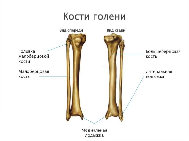
The structure of the bones of the lower leg
Foot diseases
Arthrosis or deforming osteoarthritis
- This degenerative disease joints, in which nutritional deficiency articular cartilage provokes bone deformation and inflammation in the cartilage shell. Main medication Treatments are non-steroidal anti-inflammatory drugs.
It is advisable to combine medications with treated physical education and physiotherapeutic procedures. In any case, treatment is prescribed after an x-ray of the foot.

Arthrosis and arthritis
Arthritis or joint inflammation
- characterized by an inflammatory process in the cartilage tissue of the joints in combination with swelling. The disease can have different causes, but most often they are either associated with metabolic diseases (diabetes) or are of an infectious nature.
Drug treatment for arthritis is aimed at eliminating inflammation and includes:
- antibiotics;
- chondroprotectors;
- and non-steroidal anti-inflammatory drugs.
For successful treatment the patient should monitor his diet, excluding foods high in uric acid, as well as fatty and salty.
Foot deformity
There are different types foot deformities:

Flat feet
- Clubfoot usually has a cause in insufficient tone of the foot muscles or incorrect positioning of the legs when learning to walk, but it can also be congenital.
- Hollow foot– a consequence of paralysis, characterized by hypertrophy of the longitudinal arch and visual shortening of the foot. Treatment – special gymnastics and.
- – widening of the metatarsus and flattening of the arch. Occurs with increased load in combination with insufficient elasticity of the arch muscles. Accompanied by an increase in the transverse distance between the metatarsal bones.
- Horse foot– a consequence of paralysis of the triceps surae muscle, characterized by the position of the foot at an obtuse angle to the lower leg. In this condition, the regulating function of the foot is impaired.
- Heel foot– in contrast to the horse’s, the foot forms an acute angle with the lower leg. The condition can be either congenital or a consequence of paralysis. In the first case, its cause is a violation of the position of the fetus in the womb. Such feet are corrected with plaster casts.
Growths and other formations on the bones of the foot:
- on the bones (exostosis)– pathology of unknown origin, the appearance of a growth on the lower part of the heel. At first it consists only of cartilage; over time, solid calcium salts are deposited around the cartilage.
- Osteophytes of bones - spine-like projections on bones. The most common are osteophytes of the calcaneus, developing in parallel with the inflammatory process in the Achilles tendon. Probably involved in the occurrence of pathology hereditary factor(frequent occurrence in direct relatives).

Bone growths on the bones of the foot
Foot injuries
Foot bone fracture
Regarding the symptoms of a fracture, it must be said that due to large number bones in the foot and large differentiation of functional load, symptoms manifest themselves variably depending on the anatomy of the injury.
But there are also universal manifestations:
- foot position shift(visible inner surface when viewed from above + displacement in the horizontal plane);
- pain(the nature is variable depending on the nature of the damage);
- rush of blood to the foot and swelling of the foot.

Ankle injury
Most often, the victims of fractures are the metatarsal bones (due to their characteristics - tubular structure, thinness, as well as the need to maintain an elastic arch, which can be a problem with poorly trained flaccid muscles of the foot).
The patient may sometimes be unaware of damage to the small bones of the tarsus (obvious pain and abnormal shape of the foot are not always present).
Fractures of the talus take the longest to heal (3-6 months) due to the underdeveloped blood flow in this area and the fact that this bone accounts for largest percentage body weight. The fastest time (a month to a month and a half) the finger phalanges grow together.
According to ICD-10, foot fractures are classified into:
- thumb fracture(and open);
- fracture of another finger(closed and open);
- unspecified fracture(closed and open);
- multiple foot injuries(closed and open).
If a suspected fracture is detected, it is necessary to call an ambulance and, if possible, apply a cold object (for example, food from the freezer) wrapped in two layers of towel to the site of injury.
Displaced fracture
Its signs are:
- shooting pain at the site of deformation;
- swelling of the entire limb, and not just the affected area;
- change in shape.
Closed foot fracture
Most often it affects the metatarsal bones (mechanical compression from above) and heels (both legs together) when landing unsuccessfully. Less commonly, it affects the talus in combination with the tibia. It is often splintered and may be accompanied by displacement.

Calcaneal fracture
Jones's fracture
Affects the outer metatarsals. Due to poor blood flow, about 20% of Jones fractures do not heal (and in general this type of injury is characterized by slow healing).
Risk groups include people who dance professionally and women who wear high heels a lot. If there is no displacement, the injured limb is bandaged for up to 3-4 weeks; in case of sensitive displacement, surgical intervention is used.

Fracture of the little toe
Stress fracture
Occurs with regular excessive physical exertion on unprepared feet. It differs from other fractures in its ease of detection by palpation and increased pain when putting weight on the leg.
Fractures of the foot bones in children
Most often, foot bones in children are broken as a result of jumping and landing on straightened legs. Due to the greater elasticity of children's bones, the incidence of fractures is lower than in adults. Usually the bones of the phalanges or heels are damaged. Treatment is traditional and includes a combination of plaster and physiotherapy.
Since a person moves in an upright position, the lion's share of the load falls on the lower extremities. Therefore, it is important to monitor your body weight, making it easier for the bones of the foot to work.
The structure of the ankle joint in humans is represented by the articulation of the bones of the foot with the shin bones, ensuring the performance of complex functions.
Human ankle joint
The bones are clearly shown in the diagram and classified into groups.
These include:
- Articulation of the bones of the lower leg with the bones of the foot.
- Internal articulation of the tarsal bones.
- Articulations between the bones of the metatarsus and tarsus.
- Articulations of the proximal phalanges with the metatarsal bones.
- Articulation of the phalanges of the fingers with each other.
The anatomical abilities of the foot suggest high level motor activity. For this reason, a person can perform heavy physical activity.
Both the foot and the entire leg are designed to help a person move freely in the environment.
 The structure of the foot is divided into 3 working parts:
- Bones.
- Ligaments.
- Muscles.
The skeletal base of the foot includes 3 sections: toes, metatarsus and tarsus.
The design of the toes includes phalanges. Just like the hand, the big toe consists of 2 phalanges, and the remaining 4 fingers - of 3.
There are often cases when the 2 components of the 5th fingers grow together, forming a finger structure of 2 phalanges.
The structure has proximal, distal and middle phalanges. They differ from the phalanges of the hand in that their length is shorter. A clear expression of this is seen in the distal phalanges.
The tarsal bones of the posterior section have talus and calcaneal components, and posterior section divided into cuboid, scaphoid and sphenoid bones.
The talus lies at a distance from the distal end of the tibia, becoming the bony meniscus between the bones of the foot and knee.
It consists of a head, neck and body, and is designed to connect with the shin bones, ankles and calcaneus.
The calcaneus is part of the posterior lower lobe of the tarsus. It is the largest part of the foot and has a laterally flattened, elongated appearance. At the same time, the calcaneus is the connecting link between the cuboid and talus bones.
The navicular bone is located on the inside of the foot. It has a convex forward appearance with articular components connecting to nearby bones.
The cuboid is located on the outer side of the foot, articulating with the calcaneus, navicular, cuneiform and metatarsal bones. At the bottom of the cuboid bone there is a groove in which the tendon of the elongated peroneus muscle is laid.
The composition of the sphenoid bones includes:
- Medial.
- Intermediate.
- Lateral.
They lie in front of the scaphoid, inboard of the cuboid, behind the first 3 metatarsal fragments and represent the anterior inner part of the tarsus.
The skeleton of the metatarsus appears in tubular segments, consisting of a head, body and base, where the body is similar to a triangular prism. At the same time, the most long bone- the second, and thickened and short - the first.
Bases of the metatarsal bones equipped with articular surfaces, serving as a connection with the bony components of the tarsus. In addition, it articulates with the adjacent bones of the metatarsus. At the same time, the heads equipped with articular surfaces are connected to the proximal phalanges.
The metatarsal bones are easily palpated due to the fairly thin covering of soft tissue. They are placed in multi-angle planes, creating a vault in a transverse line.
Circulatory and nervous systems of the foot
An important component of the foot is considered nerve endings and blood arteries.
Distinguish 2 main arteries of the foot:
- Rear.
- Posterior tibial.
 Also circulatory system includes small arteries that distribute to all tissue areas.
Due to the distance of the arteries of the feet from the heart, circulatory disorders are often recorded due to oxygen deficiency. The results of this manifest themselves in the form of atherosclerosis.
The longest vein that carries blood to the heart area is located at the point of the big toe, extending inside the leg. It is commonly called the great saphenous vein. In this case, the small saphenous vein runs along the outside of the leg.
Placed deep into the legs tibial anterior and posterior veins, and small ones drive blood into large veins. Moreover, small arteries supply tissues with blood, and tiny capillaries connect veins and arteries.
A person suffering from circulatory disorders notes the presence of edema in the afternoon. In addition, it may appear varicose veins veins
As in other parts of the body, nerve roots in the foot read all sensations and transmit them to the brain, controlling movement.
The nervous system of the foot includes:
- Superficial fibular.
- Deep fibula.
- Posterior tibial.
- Calf.
Tight shoes can compress any nerve, causing swelling, which will lead to discomfort, numbness and pain.
Diagnostic measures
At the moment when alarming symptoms arise in the foot area, a person comes to an orthopedist and traumatologist, who, knowing the complete structure of the ankle joint, can determine a lot by external signs. But at the same time, specialists prescribe the examination necessary for a 100% correct diagnosis.
Examination methods include:
- X-ray examination.
- Ultrasound examination.
- Computed and magnetic resonance imaging.
- Athroscopy.
 Detection of pathologies using x-rays is the most budget option. Pictures are taken from several sides, recording possible dislocations, tumors, fractures and other processes.
Ultrasound helps detect blood concentrations, find foreign bodies, possible edema process in the joint capsule, and also check the condition of the ligaments.
Computed tomography provides full examination bone tissue, with neoplasms, fractures and arthrosis. Magnetic resonance imaging is an expensive research technique that provides maximum reliable information about the Achilles tendon, ligaments and articular cartilage.
Athroscopy– a minimally invasive intervention that involves inserting a special camera into the joint capsule, through which the doctor will be able to see all the pathologies of the ankle joint.
After collecting all the information using instrumental and hardware tools, examining doctors and receiving the results laboratory tests put accurate diagnosis with the determination of treatment methods.
Pathologies of the ankle and feet
Frequent painful sensations, external changes, swelling and impairment motor functions can serve as signs of foot ailments.
Typically, a person may experience the following diseases:
- Arthrosis in the ankle joint.
- Arthrosis of the toes.
- Valgus change of the thumb.
Arthrosis of the ankle joint is characterized by crunching, pain, swelling, and fatigue during running and walking. This is due to the course of the inflammatory process, which spoils cartilage tissue, leading to typical deformation of joint tissues.
The causes of the disease can be constant increased loads and injuries, provoking the development of dysplasia, osteodystrophy and negative changes in statics.
Treatment is carried out based on the degree of arthrosis with means that reduce pain, restore blood circulation and block the spread of the disease. In difficult cases carried out surgery , relieving the patient of damaged joint segments, restoring mobility and eliminating pain.
Arthrosis of the toes is noted as a result of disruption of metabolic processes and typical blood circulation in the metatarsophalangeal joints. This is facilitated by a lack of moderation in exercise, uncomfortable narrow shoes, injuries, excess weight and frequent hypothermia.
Symptoms of the disease include swelling, deformation of the structure of the fingers, pain during movement and crunching.
 On initial stage arthrosis of the fingers, measures are taken to avoid deformation and relieve pain. If an advanced stage is detected, in most cases the doctor prescribes arthrodesis, endoprosthesis replacement or surgical arthroplasty, which should completely solve the problem of the disease.
Hallux valgus, better known as a “bump” at the base of the big toe. This disease is characterized displacement of the head of one phalangeal bone, inclination of the big toe towards the other four, weakening of the muscles and resulting deformation of the foot.
Treatment that inhibits the development of the disease is determined by prescribing baths, physiotherapy, and physical therapy. When the form of changes becomes obvious, an operation is performed, the method of which is determined by the attending orthopedist, taking into account the stage of the disease and general health patient.
The bones of the foot represent 26 related friend with other small elements, fractures or bruises of which will harm the entire body. The parts are connected to each other by ligaments and have significant functions. When you first look at a limb after an injury, you can roughly determine which bone is damaged if you know the anatomy.
Foot structure
The foot is divided into three parts: tarsus, metatarsus and toes.
Tarsus
This upper part, connects to the tibia and fibula, participates in the formation of the ankle joint and consists of seven bones:
- ram;
- calcaneal, forming the heel;
- the cuboid, forming a joint with the fourth and fifth metatarsal bones, is located on the outer edge of the foot;
- scaphoid;
- three wedge-shaped, which are connected to the base of the metatarsal bones - medial, intermediate, lateral.

Metatarsus
Located between the tarsus and fingers, it consists of five tubular metatarsal bones, the heads of which are connected to the phalanges.
Toes
The five toes of the foot consist of phalanges - the first toe is of two, and the rest of three.

Foot bone injuries
- The bones of the foot are connected by tight joints, so flip flop legs to the right or left, strong bending forward or backward can lead to dislocations, fractures, or both.
- A foot fracture will occur when a massive object falls on the leg or jumps from a great height, hits, or runs over the leg of a car.
- Stress fractures of the foot bones are found in athletes or people who constantly exercise. physical labor. Because of increased load the bones of the foot may crack, a non-displaced injury that is difficult to diagnose appearance, but the damage is clearly visible on x-rays.
- Injury occurs when light loads are placed on the legs in the presence of diseases of the musculoskeletal system, for example, a lack of calcium in the blood, bone tuberculosis or osteoporosis.

- All bone fractures are characterized by crepitus of bone fragments - the appearance of a crunch when turning or moving the injured part.
- A foot fracture is accompanied by severe pain when the victim does not allow the limb to be touched.
- The appearance of swelling at the site of injury. Edema develops due to damage to the blood and lymphatic vessels, the fluid from which flows under the skin. Increases during the day and decreases at night.
- Damage to blood vessels causes the development of a hematoma (bruise), the resorption of which takes a long time.
- A characteristic symptom is the behavior of a patient who cannot step on a limb.
- Deformation of the damaged area.
- The patient says that he heard a click or crunch at the time of injury.
- When one of the tarsal bones is injured, a characteristic symptom occurs - the spread of swelling to the ankle joint and above.
- When the base of the metatarsal bones is fractured, a characteristic symptom will be that the pain subsides after rest and resumes after physical activity.
- Subungual hematoma due to injury to the phalanges of the fingers.
Signs of a foot fracture are varied, but only one of the symptoms may appear, so correct diagnosis only a doctor. For example, a non-displaced injury will not lead to a violent reaction on the part of the victim.
Always go to the hospital if you suspect a fracture or after a severe injury.

Fracture of sphenoid bones
The medial sphenoid bone is most often susceptible to injury due to least protection ligamentous apparatus and soft tissues. A foot fracture will be accompanied by dislocation of the metatarsal bones.
Caused by falling heavy objects, characteristic symptoms no, the diagnosis is confirmed by x-ray. To restore joint function after removing the cast, it is recommended to wear an instep support for about a year.

Metatarsal fractures
They take first place in terms of frequency of occurrence; the causes are falling heavy objects or compression. Can be single or multiple. The metatarsal bones consist of a head, neck and base, so there are three types of bone damage according to their parts.
- Symptoms of a single injury: swelling on the back of the foot, slight pain on palpation.
- Symptoms of multiple trauma: swelling of the entire foot, severe pain, .
One type of injury to the metatarsal bones is stress fractures, which occur with constant and excessive load, for example, when practicing ballroom dancing, running, football.

A common fracture of the fifth bone is a Jones fracture, which is difficult to diagnose, and incorrect treatment will cause the fracture to persist. This type Fractures occur with repetitive stress.
Always contact a traumatologist for injuries, and do not refuse an X-ray examination so that the doctor can make a correct diagnosis.
A fracture of the base of an unprotected bone occurs when the leg is turned inward; it may be accompanied by a sprain of the ligaments, so it is often not noticed. The separation of a bone fragment occurs under the influence of traction force from the attached tendons. The base of the bone has a poor blood supply, which ensures prolonged healing and nonunion.

Complications
A foot fracture if not properly treated will lead to foot deformation, the development of arthrosis, and the appearance of the following symptoms:
- chronic pain when walking;
- inability to stand in one place for a long time;
- quickly after walking;
- difficult to wear tight shoes.
In the absence of medical manipulations, improper fusion of bone fragments may occur, which will lead to limitation or complete absence movements due to pain and deformity.

Treatment
- The most important thing in treatment is rest.
- To reduce swelling, apply cold compress and raise the limb, which will help get rid of the unpleasant symptom - hematoma.
- Fractures without displacement of bone fragments are treated conservatively - by applying a plaster splint. It protects the foot from movement, from infections and promotes anatomically correct fusion of bones. It is prohibited to remove the splint yourself.
- When bone fragments are displaced, surgical intervention is indicated, during which the fragments are compared with each other, avoiding trauma to surrounding tissues. After the procedure, the torn tissues, blood vessels and skin are stitched together. Then apply plaster cast to ensure immobilization of the limb.
- If surgical intervention is impossible due to the patient’s health, then the patient is prescribed a traction, which ensures the comparison of fragments without the intervention of a surgeon. Longer method.
- To improve blood flow in the area of injury and to prevent the development of muscle atrophy, moderate physical activity, physiotherapy and massage are prescribed. Blood supplies nutrients and oxygen, which promotes rapid tissue healing.
- If the bones do not heal properly, the bones are broken again and the fragments are aligned correctly, so do not self-medicate.
- For better bone healing, follow a diet: more protein and calcium, vitamin D, water, minerals.

Perform exercises as recommended by your doctor (10-15 times each exercise):
- flexion and extension of fingers;
- sitting on a chair, stand on your tiptoes and sit on your heels;
- roll a bottle or stick;
- pull your leg towards you;
- pull out your toes;
- turn the leg to the right;
- turning the leg at the ankle joint to the left.
A foot fracture is characterized by severe pain and limited mobility. There may be fractures of different bones, but they have similar symptoms Therefore, consultation with a traumatologist and orthopedist is always necessary. To prevent fractures, you need to follow one rule - take care of yourself and your loved ones!
Human feet are a part of the body thanks to which a person moves, maintains balance, and with the help of the foot the body can provide resistance while performing many movements. The process of evolution has made the structure of the foot complex, due to which modern man can walk straight.
The foot consists of 26 bones, which are connected by ligaments and joints. There are also many muscles and tendons there. In anatomy, there are three sections of the foot, which will be discussed below.
Foot bones
As is known, human foot It resembles the hands, there are sections similar in structure, but they are called differently.
The feet have:
- Tarsal bones. This part of the foot consists of seven bones - the calcaneus and talus are large, the rest are wedge-shaped, club-shaped and navicular. The talus is located in the area between the bones of the lower leg and is part of the ankle.
- Metatarsus - middle part of the foot. Consists of five tube-shaped bones, they go to the beginning of the fingers. At the end of these bones there is a joint surface that helps the fingers move. This group of bones also provides correct level vault.
- The end of the foot is the phalanges of the fingers (rib formation); they are mobile due to the presence of joints between them. There are 14 bones in this part. The thumb consists of two bones, and the rest have 3 in each finger. Due to this part, a person can maintain body balance and perform simple movements. However, there have been many cases where, as a result of the loss of arms, a person maintains his vital functions with the help of his toes.
The bones are connected to each other by joints. The correct structure of the ankle and foot bones is ensured by nerves, blood vessels, ligaments, muscles and joints.

Location of bones
As is known, important element responsible for the structure are bones. They need to be considered in more detail.
The largest bone is the calcaneus, located in the back of the foot and bears a lot of load, this bone partly contributes to the flexibility of both arches. The bone is not part of the ankle, but it distributes pressure. It is shaped like a three-dimensional rectangle with a long axis.
In the front part there are joints that are needed for the strongest connection between the heel and heel, which ensures normal form feet. There is a small protrusion at the back of the bone where the Achilles tendon is attached. Bottom side a man steps on the ground.
There is also a tubercle in the front for connection to the joint. The entire surface is covered with protrusions and depressions for the attachment of nerves, blood vessels, muscles and ligaments.
Slightly smaller is the talus, which enters the ankle. Almost all of it is covered with cartilage, and what is most interesting is that nothing except ligaments is attached to it. Bone has five surfaces covered thin layer hyaline cartilage.

It consists of a body, head and neck:
- body - is part of the ankle, connected to the foot through ligaments and joints;
- The head is the front of the bone that has an articular surface. The head provides a strong connection to the boat.
- The neck is the thin part located between the head and the body.
Cuboid. Located on the outside of the foot behind the fourth and fifth metatarsal bones. Outwardly, it looks like a cube, which gave it its name.
Scaphoid. Its peculiarity is that it is located on the foot itself and, through joints, is brought together with the talus bone, forming.
Sphenoid bones. There are three such bones on the human foot; they are small in size and located close to each other (in rib order). Behind them is the navicular bone, and in front of them are the metatarsal bones.
The structure and functions of the metatarsal bones are the same in both adults and children. Anatomical appearance - tube-shaped with an angled bend. This bend forms the arches of the feet. There are tubercles on the surface for attaching ligaments, muscles and joints.

The bones of the phalanges of the fingers are identical to those on the hands, differing only in size. The big toe has two phalanges, the other four toes have three.
Due to the load on the feet, the phalanges of the big toe are thick, while the rest are thin and short. They are connected to each other by joints, thanks to which a person can bend and straighten his fingers.
The structure of the joints
The feet have many joints that move several bones together at the same time. Regarding size, the ankle joint is considered the largest; it connects three big bones. Thanks to this connection, a person can raise and lower the foot, as well as rotate it. All other joints are smaller, but perform the same function, which together makes the foot flexible and mobile.
The ankle joint consists of a large talus and two smaller tibia bones. The latter have ankles that fix the talus. There are strong ligaments along the edges, and the joint itself is attached to the cartilage that covers the surface of the bone.

An important component is the subtalar (transverse) joint, which consists of a low-moving joint and performs the function of the arch of the talus and calcaneus. It connects three bones - the scaphoid, calcaneus and talus; ligaments are also involved in the connection process, contributing to a tighter fixation.
The cuboid and calcaneus bones are connected by the joint of the same name. Together with the subtalar, they form a practical type of education. This connection is sometimes called the "Greek cavity" and is known medically as "".
As far as surgical practice is concerned, smallest value have joints that are located on the scaphoid and sphenoid bones. But the metatarsals are connected by low-moving joints; they are surrounded by elastic ligaments and are part of the transverse and longitudinal arches of the foot. The intermetatarsal joints are located costally in the space between the metatarsal bones.
One of the most important joints are those called metatarsophalangeal joints; they are involved in almost every step or movement of the body when walking.

Foot ligaments
The most important of all is the longitudinal (or long) plantar ligament. The ligament extends from the heel bone and reaches the beginning of the metatarsal bones. It has many branches that perform the function of strengthening and fixing the longitudinal and transverse arches, and also maintains them in normal condition throughout life. But, as you know, violation of the arches of the feet can indicate flat feet, the treatment of which sometimes takes more than one year, especially if it concerns an adult.
The remaining, smaller ligaments also fix and strengthen the bones and joints of the foot, which helps a person maintain body balance and withstand dynamic and static loads during long walking or running.
Any movements of the feet are possible only with the help of the muscles that are located in the area of the foot, ankle and lower leg. The important thing is that the calf muscles help make many movements of the feet, both when walking and in an upright position.

Calf muscles
In the anterior part there is a group of long extensor muscles, the tibialis muscle. A person uses them when performing dorsal extension or flexion of the feet. Thanks to these muscles, a person can straighten and bend his fingers.
The external or lateral group includes the short and long peroneus muscles. With their help, it is possible to perform pronation, as well as lateral flexion of the foot.
The back part is distinguished by massive muscle groups consisting of many layers. They have a huge daily workload. This includes the triceps muscle, which consists of the gastrocnemius and soleus muscles. This area contains the flexor digitorum longus muscle, as well as part of the tibialis muscle. These muscle groups allow plantar flexion to be performed using the Achilles tendon. They are also involved in the process of extension and flexion of the fingers.

The dorsal muscle group contains the extensor digitorum brevis. It originates from the heel and is responsible for the motor activity of the four toes, but does not control the big toe.
On the sole of the foot there are several small muscles responsible for adduction, abduction and flexion of the toes.
Vessels and nerves
The posterior and anterior tibial arteries are responsible for the flow of blood into the human feet. On the foot itself, these arteries continue with the external internal and dorsal arteries located on the plantar part. They also form a small number of arterial connections and circles. And in case of injury of varying severity, when damage occurs to one of the circles, the rest will be able to ensure normal blood flow to the feet.
As for the outflow of blood, it is carried out by the veins of the same name, which are located on the back side. These veins form the weave. Thanks to them, blood flows into the small and large saphenous veins located in the lower leg.

Nerve impulses from the central nervous system are transmitted along the sural, deep peroneal, superficial and posterior tibial nerves. Thanks to nerve innervation, a person feels movement in space, vibration, painful sensations, touch, distinguishes between cold and heat. All nerve impulses are processed in the spinal cord.
These same nerves provide signal transmission from the brain to muscle groups. Such impulses are called reflexes, which can be involuntary or voluntary. As for the latter, this is observed when muscle tissue contracts, which does not always depend on the will of the person. The reason for this phenomenon may be the work of sweat and sebaceous glands, increase or decrease in the tone of the vascular walls.
The top layer is the skin. The skin on the feet differs depending on the area of the foot. On the very sole it has high density, but in the heel area it is thicker. The skin has the same structure as on the palms, but as a result high loads with age it begins to layer. In the dorsal area, the skin is quite smooth and elastic, there are nerve endings here.
So, based on everything that has been said above, it becomes clear that nature has made sure that the feet can withstand enormous pressure. The formation of the foot is rarely influenced by a person's nationality or the conditions in which he lives.
If at least one element of the foot is injured, a hyperkeratotic form of mycosis of the feet, deforming osteoarthritis, flat feet, heel spur and other serious diseases.
