The structure and function of the heart and the circulatory system. The structure of the human heart and its functions
Circulation circles in humans: evolution, structure and work of large and small, additional features
In the human body, the circulatory system is designed to fully meet its internal needs. An important role in the movement of blood is played by the presence of a closed system in which arterial and venous blood flows are separated. And this is done through the presence of blood circulation circles.
Historical background
In the past, when scientists did not yet have information instruments at hand capable of studying physiological processes on a living organism, the greatest scientists were forced to search anatomical features at the corpses. Naturally, a deceased person’s heart does not contract, so some of the nuances had to be figured out on their own, and sometimes simply fantasized. So, back in the second century AD Claudius Galen, self-learner Hippocrates, assumed that the arteries contained air instead of blood in their lumen. Over the next centuries, many attempts were made to combine and link together the existing anatomical data from the standpoint of physiology. All scientists knew and understood how the circulatory system works, but how does it work?
Scientists have made a tremendous contribution to the systematization of data on heart function. Miguel Servet and William Harvey in the 16th century. Harvey, scientist who first described the systemic and pulmonary circulation , in 1616 determined the presence of two circles, but he could not explain in his works how the arterial and venous beds were connected to each other. And only later, in the 17th century, Marcello Malpighi, one of the first to use a microscope in his practice, discovered and described the presence of tiny capillaries, invisible to the naked eye, which serve as a connecting link in the blood circulation.
Phylogeny, or the evolution of blood circulation
Due to the fact that as animals of the vertebrate class evolved, they became more and more progressive in anatomical and physiological terms, they needed complex device And cordially- vascular system. So, for faster movement of liquid internal environment In the body of a vertebrate animal, the need for a closed blood circulation system arose. Compared to other classes of the animal kingdom (for example, arthropods or worms), the rudiments of a closed vascular system appear in chordates. And if the lancelet, for example, does not have a heart, but there is an abdominal and dorsal aorta, then fish, amphibians (amphibians), reptiles (reptiles) have a two- and three-chambered heart, respectively, and birds and mammals have a four-chambered heart, the peculiarity of which is is the focus in it of two circles of blood circulation that do not mix with each other.

Thus, the presence in birds, mammals and humans, in particular, of two separated circulatory circles is nothing more than the evolution of the circulatory system, necessary for better adaptation to conditions environment.
Anatomical features of the blood circulation
Circulation circles are a set of blood vessels, which is closed system for admission to internal organs oxygen and nutrients through gas and nutrient exchange, as well as for removing carbon dioxide and other metabolic products from cells. The human body is characterized by two circles - systemic, or big circle, as well as pulmonary, also called the small circle.

Video: blood circulation circles, mini-lecture and animation
Systemic circulation
The main function of the large circle is to ensure gas exchange in all internal organs except the lungs. It begins in the cavity of the left ventricle; represented by the aorta and its branches, the arterial bed of the liver, kidneys, brain, skeletal muscles and other organs. Further, this circle continues with the capillary network and venous bed of the listed organs; and through the entry of the vena cava into the cavity of the right atrium it ends in the latter.

So, as already said, the beginning of the great circle is the cavity of the left ventricle. This is where the arterial blood goes blood flow, containing most of oxygen rather than carbon dioxide. This flow enters the left ventricle directly from the circulatory system of the lungs, that is, from the small circle. Arterial flow from the left ventricle through aortic valve pushes into the largest main vessel- into the aorta. The aorta can be figuratively compared to a kind of tree, which has many branches, because arteries extend from it to the internal organs (liver, kidneys, gastrointestinal tract, to the brain - through the system of carotid arteries, to skeletal muscles, to subcutaneous fat, etc.). Organ arteries, which also have numerous branches and bear names corresponding to their anatomy, carry oxygen to each organ.
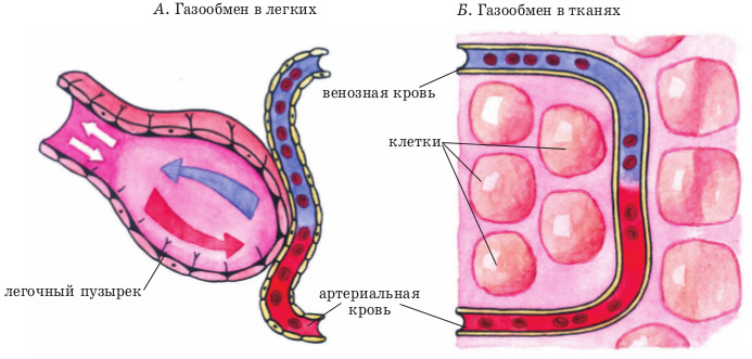
In the tissues of internal organs arterial vessels are divided into vessels of smaller and smaller diameter, and as a result a capillary network is formed. Capillaries are the smallest vessels, practically without a middle muscular layer, and are represented by an inner membrane - intima, lined with endothelial cells. The gaps between these cells at the microscopic level are so large compared to other vessels that they allow proteins, gases and even shaped elements into the intercellular fluid of surrounding tissues. Thus, intense gas exchange and exchange of other substances occurs between the capillary with arterial blood and the liquid intercellular medium in a particular organ. Oxygen penetrates from the capillary, and carbon dioxide, as a product of cell metabolism, enters the capillary. The cellular stage of respiration occurs.
After it has passed into the tissue more oxygen, and all carbon dioxide has been removed from the tissues, the blood becomes venous. All gas exchange occurs with each new influx of blood, and during the period of time while it moves along the capillary towards the venule - a vessel that collects venous blood. That is, with each cardiac cycle, in one or another part of the body, oxygen enters the tissues and carbon dioxide is removed from them.
These venules unite into larger veins, and a venous bed is formed. Veins, similar to arteries, are named according to the organ in which they are located (renal, cerebral, etc.). From large venous trunks, tributaries of the superior and inferior vena cava are formed, and the latter then flow into right atrium.
Features of blood flow in the organs of the systemic circle

Some of the internal organs have their own characteristics. So, for example, in the liver there is not only a hepatic vein, which “carries” the venous flow away from it, but also a portal vein, which, on the contrary, brings blood to the liver tissue, where the blood is purified, and only then the blood collects in the tributaries hepatic vein to get to the big circle. The portal vein brings blood from the stomach and intestines, so everything that a person eats or drinks must undergo a kind of “purification” in the liver.
In addition to the liver, certain nuances exist in other organs, for example, in the tissues of the pituitary gland and kidneys. Thus, in the pituitary gland the presence of the so-called “wonderful” capillary network, because the arteries that bring blood to the pituitary gland from the hypothalamus divide into capillaries, which then collect into venules. The venules, after the blood with the molecules of releasing hormones are collected, are again divided into capillaries, and then veins are formed that carry the blood from the pituitary gland. In the kidneys, the arterial network is divided twice into capillaries, which is associated with the processes of excretion and reabsorption in the kidney cells - in the nephrons.
Pulmonary circulation
Its function is to carry out gas exchange processes in lung tissue in order to saturate the “waste” venous blood with oxygen molecules. It begins in the cavity of the right ventricle, where venous blood flow with an extremely small amount of oxygen and with high content carbon dioxide. This blood moves through the pulmonary valve into one of the large vessels, called the pulmonary trunk. Next, the venous flow moves along the arterial bed in the lung tissue, which also breaks up into a network of capillaries. By analogy with capillaries in other tissues, gas exchange occurs in them, only oxygen molecules enter the lumen of the capillary, and carbon dioxide penetrates into the alveolocytes (cells of the alveoli). With each act of breathing, air enters the alveoli from the environment, from which oxygen through cell membranes penetrates into blood plasma. With the exhaled air, when exhaling, the carbon dioxide that enters the alveoli is expelled.

After being saturated with O2 molecules, the blood acquires the properties of arterial blood, flows through the venules and ultimately reaches the pulmonary veins. The latter, consisting of four or five pieces, open into the cavity of the left atrium. As a result, through right half venous blood flows through the heart, and through left half- arterial; and normally these flows should not mix.
Lung tissue has a double network of capillaries. With the help of the first, gas exchange processes are carried out in order to enrich the venous flow with oxygen molecules (relationship directly with the small circle), and in the second, the lung tissue itself is supplied with oxygen and nutrients (relationship with the large circle).
Additional circulation circles
These concepts are used to distinguish blood supply individual organs. For example, to the heart, which needs oxygen more than others, arterial inflow is carried out from the branches of the aorta at its very beginning, which are called the right and left coronary (coronary) arteries. Intense gas exchange occurs in the capillaries of the myocardium, and venous drainage carried out in coronary veins. The latter collect in the coronary sinus, which opens directly into the right atrial chamber. In this way it is carried out cardiac or coronary circulation.

coronary (coronary) circle of blood circulation in the heart
Circle of Willis is a closed arterial network from the cerebral arteries. The medulla provides additional blood supply to the brain in case of disruption cerebral blood flow along other arteries. This protects so much important organ from lack of oxygen, or hypoxia. The cerebral circulation is represented by the initial segment of the anterior cerebral artery, initial segment of the posterior cerebral artery, anterior and posterior connecting arteries, internal carotid arteries.

circle of willis in the brain ( classic version buildings)
Placental circulation functions only during pregnancy by a woman and performs the function of “breathing” in a child. The placenta is formed starting from 3-6 weeks of pregnancy, and begins to function fully from the 12th week. Due to the fact that the fetal lungs do not work, oxygen enters its blood through the flow of arterial blood into umbilical vein child.

fetal circulation before birth
Thus, the entire human circulatory system can be divided into separate interconnected sections that perform their functions. The proper functioning of such areas, or blood circulation circles, is the key to the healthy functioning of the heart, blood vessels and the entire body as a whole.
The heart has a complex structure and performs no less complex and important work. Contracting rhythmically, it ensures blood flow through the vessels.
The heart is located behind the sternum, in the middle section chest cavity and is almost completely surrounded by lungs. It may move slightly to the side as it hangs loosely on the blood vessels. The heart is located asymmetrically. Its long axis is inclined and forms an angle of 40° with the axis of the body. It is directed from top to right, forward, down to the left, and the heart is rotated so that its right section is tilted more forward, and the left one – back. Two-thirds of the heart is to the left of the midline and one-third (the vena cava and right atrium) is to the right. Its base is turned towards the spine, and the apex is facing the left ribs, to be more precise, the fifth intercostal space.
Anatomy of the heart

Sternocostal surface the hearts are more convex. It is located behind the sternum and cartilages of the III-VI ribs and is directed forward, upward, and to the left. The transverse coronary groove runs along it, which separates the ventricles from the atria and thereby divides the heart into top part, formed by the atria, and the lower one, consisting of the ventricles. Another groove of the sternocostal surface - the anterior longitudinal - runs along the border between the right and left ventricles, with the right forming the largest part of the anterior surface, the left - the smaller one.
Diaphragmatic surface flatter and adjacent to the tendon center of the diaphragm. A longitudinal posterior groove runs along this surface, separating the surface of the left ventricle from the surface of the right. In this case, the left one makes up the majority of the surface, and the right one makes up the smaller part.
Anterior and posterior longitudinal grooves they merge at their lower ends and form a cardiac notch to the right of the cardiac apex.
There are also side surfaces located on the right and left and facing the lungs, which is why they are called pulmonary.
Right and left edges hearts are not the same. The right edge is more pointed, the left is more blunt and rounded due to the thicker wall of the left ventricle.
The boundaries between the four chambers of the heart are not always clearly defined. The landmarks are the grooves in which the blood vessels of the heart are located, covered with fatty tissue and the outer layer of the heart - the epicardium. The direction of these grooves depends on how the heart is located (obliquely, vertically, transversely), which is determined by the body type and the height of the diaphragm. In mesomorphs (normosthenics), whose proportions are close to the average, it is located obliquely, in dolichomorphs (asthenics) with a thin physique - vertically, in brachymorphs (hypersthenics) with wide short forms - transversely.

The heart seems to be suspended by the base on large vessels, while the base remains motionless, and the apex is in a free state and can move.
Structure of heart tissue
The heart wall is made up of three layers:
- Endocardium - inner layer epithelial tissue, lining the cavities of the heart chambers from the inside, accurately repeating their relief.
- The myocardium is a thick layer formed by muscle tissue (striated). The cardiac myocytes of which it consists are connected by many bridges that link them into muscle complexes. This muscle layer ensures rhythmic contraction of the heart chambers. The myocardium is thinnest at the atria, the greatest is at the left ventricle (about 3 times thicker than the right), since it needs more force to push blood into the systemic circulation, in which the resistance to flow is several times greater than in the small circle. The atrial myocardium consists of two layers, the ventricular myocardium - of three. The atrial myocardium and ventricular myocardium are separated by fibrous rings. The conduction system that provides rhythmic contraction of the myocardium is one for the ventricles and atria.
- Epicardium is the outer layer, which is the visceral lobe of the heart sac (pericardium), which is serosa. It covers not only the heart, but also the initial parts of the pulmonary trunk and aorta, as well as the final parts of the pulmonary and vena cava.

Anatomy of the atria and ventricles
The heart cavity is divided by a septum into two parts - right and left, which do not communicate with each other. Each of these parts consists of two chambers - the ventricle and the atrium. The septum between the atria is called the interatrial septum, and the septum between the ventricles is called the interventricular septum. Thus, the heart consists of four chambers - two atria and two ventricles.
Right atrium
In shape it looks like an irregular cube, in front there is additional cavity, called the right ear. The atrium has a volume of 100 to 180 cubic meters. cm. It has five walls, 2 to 3 mm thick: anterior, posterior, superior, lateral, medial.
The inferior vena cava (below) also flows into the right atrium (from above at the back). On the lower right is the coronary sinus, where the blood of all the cardiac veins drains. Between the openings of the superior and inferior vena cava there is an intervenous tubercle. In the place where the inferior vena cava flows into the right atrium, there is a fold of the inner layer of the heart - the valve of this vein. The sinus of the vena cava is the posterior dilated section of the right atrium, into which both of these veins flow.
The chamber of the right atrium has a smooth internal surface, and only in the right appendage with the adjacent anterior wall the surface is uneven.
Many pinpoint openings of the small veins of the heart open into the right atrium.
Right ventricle
It consists of a cavity and an arterial cone, which is a funnel directed upward. The right ventricle has the shape trihedral pyramid, the base of which faces up and the top faces down. The right ventricle has three walls: anterior, posterior, medial.
The front is convex, the back is flatter. The medial is the interventricular septum, consisting of two parts. The larger one, the muscular one, is located at the bottom, the smaller one, the membranous one, is at the top. The pyramid faces the atrium with its base and has two openings: posterior and anterior. The first is between the cavity of the right atrium and the ventricle. The second comes out in pulmonary trunk.
Left atrium
It has the appearance of an irregular cube, is located behind and adjacent to the esophagus and the descending aorta. Its volume is 100-130 cubic meters. cm, wall thickness – from 2 to 3 mm. Like the right atrium, it has five walls: anterior, posterior, superior, literal, medial. The left atrium continues anteriorly into an additional cavity called the left appendage, which is directed towards the pulmonary trunk. Four pulmonary veins flow into the atrium (back and above), in the openings of which there are no valves. Medial wall is interatrial septum. Inner surface the atrium is smooth, the pectineus muscles are found only in the left appendage, which is longer and narrower than the right one, and is noticeably separated from the ventricle by an interception. It communicates with the left ventricle via the atrioventricular orifice.
Left ventricle
It is shaped like a cone, the base of which faces upward. The walls of this chamber of the heart (anterior, posterior, medial) have the greatest thickness - from 10 to 15 mm. There is no clear boundary between the front and back. At the base of the cone are the openings of the aorta and the left atrioventricular opening.
The round opening of the aorta is located in front. Its valve consists of three valves.
Heart size
The size and weight of the heart differs among different people. The average values are as follows:
- length is from 12 to 13 cm;
- greatest width – from 9 to 10.5 cm;
- anteroposterior size – from 6 to 7 cm;
- weight in men - about 300 g;
- weight in women is about 220 g.

Functions of the cardiovascular system and heart
The heart and blood vessels make up the cardiovascular system, the main function of which is transport. It consists of supplying tissues and organs with nutrition and oxygen and returning metabolic products.
The heart acts as a pump - it ensures continuous circulation of blood in the circulatory system and delivery of nutrients and oxygen to organs and tissues. When stressed or physical activity his work is immediately restructured: the number of layoffs increases.
The work of the heart muscle can be described as follows: his right side(venous heart) receives waste blood from the veins, saturated carbon dioxide and gives it to the lungs for oxygen saturation. From the lungs, O2-enriched blood is sent to left side heart (arterial) and from there is forcefully pushed into the bloodstream.
The heart produces two circles of blood circulation - large and small.
The large one supplies blood to all organs and tissues, including the lungs. It begins in the left ventricle and ends in the right atrium.
The pulmonary circulation produces gas exchange in the alveoli of the lungs. It begins in the right ventricle and ends in the left atrium.
Blood flow is regulated by valves: they prevent it from flowing in the opposite direction.
The heart has such properties as excitability, conductivity, contractility and automaticity (excitation without external stimuli under the influence of internal impulses).
Thanks to the conduction system, sequential contraction of the ventricles and atria occurs, and the synchronous inclusion of myocardial cells in the contraction process.
Rhythmic contractions of the heart ensure a portioned flow of blood into the circulatory system, but its movement in the vessels occurs without interruption, which is due to the elasticity of the walls and the resulting small vessels resistance to blood flow.
The circulatory system has a complex structure and consists of a network of vessels for different purposes: transport, shunting, exchange, distribution, capacitance. There are veins, arteries, venules, arterioles, capillaries. Together with the lymphatic ones, they maintain the constancy of the internal environment in the body (pressure, body temperature, etc.).
Arteries move blood from the heart to the tissues. As they move away from the center, they become thinner, forming arterioles and capillaries. The arterial bed of the circulatory system transports necessary substances to organs and maintains constant pressure in the vessels.

The venous bed is more extensive than the arterial bed. Veins move blood from tissues to the heart. Veins are formed from venous capillaries, which merge to first become venules, then veins. They form large trunks near the heart. Distinguish superficial veins, located under the skin, and deep, located in the tissues near the arteries. Main function venous section circulatory system - blood outflow, rich in products metabolism and carbon dioxide.
For evaluation functionality cardiovascular system and load tolerance, special tests are carried out, which make it possible to assess the body’s performance and its compensatory capabilities. Functional tests cardiovascular system are included in the physical examination to determine the degree of fitness and general physical training. The assessment is given based on such indicators of the functioning of the heart and blood vessels as blood pressure, pulse pressure, blood flow speed, minute and stroke volumes of blood. Such tests include Letunov's tests, step tests, Martinet's test, Kotov's - Demin's test.
The heart begins to beat from the fourth week after conception and does not stop until the end of life. It does a gigantic job: per year it pumps about three million liters of blood and makes about 35 million heartbeats. At rest, the heart uses only 15% of its resource, and under load – up to 35%. For average duration During its lifetime it pumps about 6 million liters of blood. Another interesting fact: The heart supplies blood to 75 trillion cells human body except the cornea of the eyes.
Circulation- blood circulation in the body. Blood can perform its functions only by circulating in the body.
Circulatory system: heart(central circulatory organ) and blood vessels(arteries, veins, capillaries).
Structure of the heart
Heart- a hollow four-chambered muscular organ. The size of the heart is approximately the size of the fist. The average weight of the heart is 300 g. The outer lining of the heart is pericardium. It consists of two leaves: one forms pericardial sac, the other - the outer shell of the heart - epicardium. Between the pericardial sac and the epicardium there is a cavity filled with fluid to reduce friction during heart contraction. Middle layer of the heart - myocardium. It consists of a striated muscle tissue special structure (cardiac muscle tissue). In it, neighboring muscle fibers are interconnected by cytoplasmic bridges. Intercellular connections do not interfere with the conduction of excitation, due to which the heart muscle is able to contract quickly. In nerve cells and skeletal muscles each cell is excited separately. Inner lining of the heart - endocardium. It lines the cavity of the heart and forms the valves - valves
The human heart consists of four chambers: 2 atria(left and right) and 2 ventricle(left and right). The muscular wall of the ventricles (especially the left) is thicker than the wall of the atria. Venous blood flows in the right half of the heart, arterial blood flows in the left.
Between the atria and ventricles there are flap valves(between the left - two-leaf, between the right - three-leaf). Between the left ventricle and the aorta and between the right ventricle and the pulmonary artery there are semilunar valves(consist of three sheets resembling pockets). Heart valves allow blood to flow in only one direction: from the atria to the ventricles, and from the ventricles to the arteries.
Work of the heart
The heart contracts rhythmically: contractions alternate with relaxations. Contraction of parts of the heart is called systole, and relaxation - diastole. Cardiac cycle- a period covering one contraction and one relaxation. It lasts 0.8 s and consists of three phases: Phase I- contraction (systole) of the atria - lasts 0.1 s; II phase- contraction (systole) of the ventricles - lasts 0.3 s; III phase- general pause - both the atria and ventricles are relaxed - lasts 0.4 s. At rest, the heart rate of an adult is 60-80 times per minute. The myocardium is formed by a special striated muscle tissue that contracts involuntarily. Characteristic of the heart muscle automatic- the ability to contract under the influence of impulses arising in the heart itself. This is due to special cells located in the heart muscle, in which excitations rhythmically appear -
Rice. 1. Diagram of the structure of the heart (vertical section):
1 - muscular wall of the right ventricle, 2 - papillary muscles, from which tendon threads arise (3), attached to valve (4), located between the atrium and ventricle, 5 - right atrium, 6 - opening of the inferior vena cava; 7 - superior vena cava, 8 - septum between the atria, 9 - openings of the four pulmonary veins; 10 - right atrium, 11 - muscular wall of the left ventricle, 12 - septum between the ventricles
The automatic contraction of the heart continues even when isolated from the body. In this case, the excitation received at one point passes to the entire muscle and all its fibers contract simultaneously.
There are three phases in the work of the heart. First - atrial contraction, second - contraction of the ventricles - systole, third - simultaneous relaxation of the atria and ventricles - diastole, or a pause in the last phase, both atria are filled with blood from the veins and it freely passes into the ventricles. Blood entering the ventricles presses on the atrial valves with bottom side, and they close. When both ventricles contract, blood pressure increases in their cavities and it enters the aorta and pulmonary artery (into the systemic and pulmonary circulation). After contraction of the ventricles, their relaxation occurs. The pause is followed by contraction of the atria, then the ventricles, etc.
The period from one atrial contraction to another is called cardiac cycle. Each cycle lasts 0.8 s. Of this time, contraction of the atria accounts for 0.1 s, contraction of the ventricles accounts for 0.3 s, and the total pause of the heart lasts 0.4 s. If the heart rate increases, the time of each cycle decreases. This occurs mainly due to a shortening of the overall cardiac pause. With each contraction, both ventricles eject the same amount of blood into the aorta and pulmonary artery (on average about 70 ml), which is called stroke volume of blood.
The work of the heart is regulated by the nervous system depending on the influence of the internal and external environment: the concentration of potassium and calcium ions, thyroid hormone, resting state or physical work, emotional stress. Two types of centrifugal nerve fibers belonging to the autonomic nervous system approach the heart as a working organ. One pair of nerves (sympathetic fibers) when irritated, it increases and increases heart rate. When another pair of nerves is irritated (branches of the vagus nerve) impulses entering the heart weaken its activity.
The work of the heart is connected with the activities of other organs. If excitation is transmitted to the central nervous system from working organs, then from the central nervous system it is transmitted to the nerves that enhance the function of the heart. Thus, through a reflexive process, a correspondence is established between the activities of various organs and the work of the heart. The heart beats 60-80 times per minute.
The walls of arteries and veins consist of three layers: interior(thin layer of epithelial cells), average(a thick layer of elastic fibers and smooth muscle cells) and outer(loose connective tissue and nerve fibers). Capillaries consist of a single layer of epithelial cells.
Arteries- vessels through which blood flows from the heart to organs and tissues. The walls consist of three layers. The following types of arteries are distinguished: elastic type arteries (large vessels closest to the heart), muscular type arteries (medium and small arteries that resist blood flow and thereby regulate blood flow to the organ) and arterioles (the last branches of the artery that turn into capillaries).
Capillaries - thin vessels, in which fluids, nutrients and gases are exchanged between blood and tissues. Their wall consists of a single layer of epithelial cells.
Vienna- vessels through which blood flows from organs to the heart. Their walls (like those of arteries) consist of three layers, but they are thinner and poorer in elastic fibers. Therefore, the veins are less elastic. Most veins have valves that prevent blood from flowing back.
141 142 ..Circulation circles (human anatomy)
The pattern of blood movement in the circulatory circles was discovered by W. Harvey (1628). Since that time, the doctrine of the anatomy and physiology of blood vessels has been enriched with numerous data that revealed the mechanism of general and regional blood supply. In the process of development in circulatory system, especially in the heart, certain structural complications occurred, namely in higher animals the heart was divided into four chambers. The heart of fish has two chambers - the atrium and the ventricles, separated bicuspid valve. Flows into the atrium venous sinus, and the ventricle communicates with the conus arteriosus. In this two-chambered heart, venous blood flows, which is discharged into the aorta and then to the branchial vessels for oxygenation. In animals with the appearance pulmonary respiration(double-breathing fish, amphibians) a septum with holes is formed in the atrium. In this case, all venous blood enters the right atrium, and arterial blood enters the left atrium. Blood from the atria enters the common ventricle, where it mixes.
In the heart of reptiles, due to the presence of incomplete interventricular septum(except for the crocodile, which has a complete septum), a more perfect separation of arterial and venous blood flows is observed. Crocodiles have a four-chambered heart, but the mixing of arterial and venous blood occurs at the periphery due to the connection of arteries and veins.
Birds, like mammals, have a four-chambered heart and there is a complete separation of blood flows not only in the heart, but also in the vessels. A feature of the structure of the heart and large vessels in birds is the presence right arch aorta, the left arch atrophies.
In higher animals and humans, which have a four-chambered heart, the greater, lesser, and cardiac circles of blood circulation are distinguished (Fig. 138). Central to these circles is the heart. Regardless of the composition of the blood, all vessels coming to the heart are considered to be veins, and those leaving it are considered to be arteries.
Rice. 138. Blood circulation diagram (according to Kishsh-Sentagotai).1 - a. carotis communis; 2 - arcus aortae; 3 - a. pulmonalis; 4 - v. pulmonalis; 5 - ventriculus sinister; 6 - ventriculus dexter; 7 - truncus coeliacus; 8 - a. mesenterica superior; 9 - a. mesenterica inferior; 10 - v. cava inferior; 11 - aorta; 12 - a. iliaca communis; 13 - vasa pelvina; 14 - a. femoralis; 15 - v. femoralis; 16 - v. iliaca communis; 17 - v. portae; 18 - vv. hepaticae; 19 - a. subclavia; 20 - v. subclavia; 21 - v. cava superior; 22 - v. jugularis interna
Pulmonary circulation (pulmonary). Venous blood from the right atrium through the right atrioventricular opening passes into the right ventricle, which, contracting, pushes blood into the pulmonary trunk. The latter is divided into the right and left pulmonary arteries, passing through the hilum of the lungs. In the lung tissue, the arteries divide into capillaries surrounding each alveolus. After red blood cells release carbon dioxide and enrich them with oxygen, venous blood turns into arterial blood. Arterial blood through four pulmonary veins (there are two veins in each lung) it is collected in the left atrium, and then through the left atrioventricular opening it passes into the left ventricle. The systemic circulation begins from the left ventricle.
Systemic circulation . Arterial blood from the left ventricle is ejected into the aorta during its contraction. The aorta breaks up into arteries that supply blood to the head, neck, limbs, torso and all internal organs, in which they end in capillaries. From the blood capillaries into the tissues come nutrients, water, salts and oxygen, metabolic products and carbon dioxide are resorbed. The capillaries gather into venules, where it begins venous system vessels, representing the roots of the superior and inferior vena cava. Venous blood through these veins enters the right atrium, where the systemic circulation ends.
The pattern of blood movement in circulatory circles was discovered by Harvey (1628). Subsequently, the doctrine of the physiology and anatomy of blood vessels was enriched with numerous data that revealed the mechanism of general and regional blood supply to organs.
In goblin animals and humans, which have a four-chambered heart, the greater, lesser, and cardiac circles of blood circulation are distinguished (Fig. 367). The heart occupies a central place in blood circulation.
367. Blood circulation diagram (according to Kishsh, Sentagotai).
1 - general carotid artery;
2 - aortic arch;
3 - pulmonary artery;
4 - pulmonary vein;
5 - left ventricle;
6 - right ventricle;
7 - celiac trunk;
8 - top mesenteric artery;
9 - inferior mesenteric artery;
10 - inferior vena cava;
11 - aorta;
12 - total iliac artery;
13 - general iliac vein;
14 - femoral vein. 15 - portal vein;
16 - hepatic veins;
17 - subclavian vein;
18 - superior vena cava;
19 - internal jugular vein.
Pulmonary circulation (pulmonary)
Venous blood from the right atrium passes through the right atrioventricular orifice into the right ventricle, which contracts and pushes blood into the pulmonary trunk. It is divided into right and left pulmonary arteries penetrating into the lungs. In the lung tissue, the pulmonary arteries are divided into capillaries surrounding each alveolus. After red blood cells release carbon dioxide and enrich them with oxygen, venous blood turns into arterial blood. Arterial blood flows through four pulmonary veins (there are two veins in each lung) into the left atrium, then passes through the left atrioventricular orifice into the left ventricle. The systemic circulation begins from the left ventricle.
Systemic circulation
Arterial blood from the left ventricle is ejected into the aorta during its contraction. The aorta splits into arteries that supply blood to the limbs and torso. all internal organs and ending with capillaries. Nutrients, water, salts and oxygen are released from the blood capillaries into the tissues, metabolic products and carbon dioxide are resorbed. Capillaries gather into venules, where the venous system begins vascular system, representing the roots of the superior and inferior vena cava. Venous blood through these veins enters the right atrium, where the systemic circulation ends.
Cardiac circulation
This circle of blood circulation begins from the aorta with two coronary cardiac arteries, through which blood enters all layers and parts of the heart, and then collects through small veins into the venous coronary sinus. This vessel opens with a wide mouth into the right atrium. Some of the small veins of the heart wall directly open into the cavity of the right atrium and ventricle of the heart.
