On what day of the menstrual cycle should an ultrasound be done? Time for an ovarian ultrasound: when is the best time to do it?
Ultrasound is used to diagnose diseases in gynecology. It is a very accessible way to study the condition of internal organs. However, it is harmless to patients due to ultrasonic vibrations and can be performed during pregnancy. But on what day of the cycle you need to do a pelvic ultrasound depends on various circumstances.
To conduct standard ultrasound diagnostics, this procedure is prescribed in the first half of the cycle.
To perform an ultrasound of the pelvic organs, you should contact a specialist as soon as menstrual bleeding stops on the 5-7th day of the cycle. There is no point in doing it earlier, because at this time the uterus is filled with blood and the examination will not be able to show accurate results. If the menstrual cycle is short, then the optimal study is on the 5th day, and if it is long, from 5 to 10.
 Doctors also do not recommend carrying out a study before the onset of menstruation. The reason for this is the active formation of progesterone. This hormone leads to thickening of the endometrium. Therefore, when contacting a specialist during this period, it is possible to do an ultrasound, but it will not be impossible to identify pathologies and defects in detail (for example,). That is why it is necessary to perform an ultrasound of the pelvic organs in the first half of the cycle.
Detection of a cyst
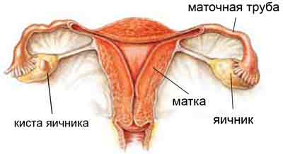
When there is a certain discomfort and pain upon palpation, this indicates the presence of a neoplasm. Before diagnosing an ovarian cyst, it is necessary to understand what it is. It is a formation that is filled with fluid and is located in the middle of the ovary. The pathology does not pose any particular extreme danger, especially if its size is small. But its presence may be the result of the development of serious disorders in the body.
An ovarian cyst on ultrasound looks like a hollow formation. Also, a transabdominal and transvaginal (vaginal) ultrasound sensor is used to study it. This device is a small device. In some cases, the study can be performed with a transrectal ultrasound sensor.
Before this procedure, it is necessary to fill the bladder by first drinking a large amount of liquid approximately one hour before the examination. On an ultrasound, the pathology will be visible, even if its size is very small.

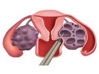
According to the descriptions, it is a liquid formation in the shape of a ball, which can have a different structure, as well as the level of coloring. Often, the presence of a follicular cyst is diagnosed, as well as cysts that go away on their own and do not pose a serious danger. Their size is usually small. There are also other types that are pathological. They usually require surgical treatment:
After detection of pathology
 Biopsy is a procedure for removing cells from formations for further examination.
 After pathology is detected and the development of a malignant ovarian tumor is suspected, a histological examination can be performed. This procedure is necessary to obtain accurate information about the origin of the pathology. The study allows not only to recognize the cancer cells themselves, but some methods make it possible to determine the reasons for their appearance. To do this, a biopsy of the ovarian cyst is first performed. It allows you to take a specific piece of education to study it. This procedure is necessary to obtain accurate information about the origin of the pathology.
Histology of an ovarian cyst is carried out when the presence of cancer is suspected and consists of studying the structure of the tissue of the formation. The histology of an ovarian cyst consists of studying the structure of the tissue of the formation. On average, the procedure takes about 10 days. With its help, you can understand the nature of the pathology and whether it is malignant. Once the type of formation has been established, it is recommended to remove it. For this purpose, therapeutic and surgical methods are used.
We offer you to watch a short informational video on the topic of the article:
On what day of the cycle is folliculometry done?
Folliculometry is a test to detect ovulation in the current cycle. When there are pregnancy plans, doctors recommend diagnosis at the beginning of the second half of the cycle. But there is also a certain dependence of time on the individual structural characteristics of each woman’s body. In cases where the average duration of the second phase lasts 2 weeks, then using basic calculations you can calculate the approximate day of ovulation.
Therefore, diagnosis must be carried out several days before its onset. In this case, it is carried out after menstruation, but during the period when bleeding has not yet begun. The final stage of this diagnosis is confirmation that ovulation has completed.
Ultrasound after childbirth or abortion
In cases where we are talking about ultrasound of the pelvic organs after an abortion, then such a study must be carried out at least 5-7 days after curettage. If bleeding is observed, then this is normal, because it does not apply to menstruation. The cycle itself will gradually, over several months, be restored. Ultrasound of the ovaries, in this case, is necessary in order to identify the remains of the removed fetus in the uterine cavity. And if they are detected, additional medical procedures are prescribed to eliminate them.
After the birth of a child, it is mandatory for every woman to undergo an ultrasound scan. Often it is carried out in maternity hospitals on the 5th day after the birth. In addition, the menstrual cycle in this case will not be restored soon (especially if breastfeeding is carried out).
Therefore, after childbirth, this type of examination is carried out before the onset of menstruation.
Many studies that are carried out to assess the functioning of the body as a whole, and individual organs and systems in particular, require certain preparation. Patients sometimes hear that it is better to do them on some days than on others. After all, test results can depend on many factors - diet, the influence of aggressive substances (including nicotine or alcohol), lifestyle, etc. Gynecological ultrasound is considered one of the most common studies. It is carried out with a referral from the attending physician to identify various pathological conditions or to refute their presence. Let’s clarify which day of the cycle is best to do a gynecological ultrasound.
Gynecological ultrasound allows you to assess the general condition of the genital organs and the correctness of their structure, examine the periuterine space and the ligaments that support the uterus. There are three ways to carry it out, each of which requires certain preparation. Gynecological ultrasound can be performed on different days of the cycle, depending on the purpose of such a study. Thus, the first thing to remember is that the day of the ultrasound determines the purpose of it.
What types of gynecological ultrasound are known??
1. Diagnostic testing of non-pregnant patients
Transvaginal ultrasound is performed in order to more accurately diagnose diseases of the genital area. It is performed by inserting a special sensor into the patient’s vaginal cavity.
Transabdominal ultrasound is performed for virgins or patients in whom it is necessary to detect or refute gross pathologies of organs located in the pelvis. It is performed through the abdominal wall.
Transrectal ultrasound is performed on virgins as a replacement for transvaginal ultrasound. It is performed through the rectum.
2. Folliculometry
With this ultrasound diagnosis of the female genital organs, only the ovaries are examined to assess the maturation of the follicles in them. Typically this examination is performed transvaginally.
3. Ultrasound during pregnancy
In the first trimester of pregnancy, such an ultrasound is usually performed transvaginally, and in the second and third trimester - transabdominal. This research method is aimed not only at examining the fetus, but also at examining the uterus, cervix, fallopian tubes and appendages.
On what day is a gynecological ultrasound informative??
Only the attending physician can determine the optimal day for a gynecological ultrasound. Typically, routine diagnostic tests outside of pregnancy are carried out in the first half of the cycle. Indeed, at this stage, the mucous membranes of the uterus are still quite thin, so it will be easier for the doctor who performs the ultrasound to consider the possibility of the presence of some formations - polyps, condylomas, small tumors, etc. Usually 3-5 days after the end of the next menstruation cycle, a gynecological ultrasound will be able to tell the doctor more than at other times.
Among other things, in the second phase of the menstrual cycle, a small cyst, also known as a follicle, forms inside the ovary. Then it bursts. The corpus luteum, which appears at the site of a ruptured follicle from which an egg emerges, also looks like a cyst. Both described structures disappear by the time menstruation begins, and only pathological cysts remain. Therefore, when performing a gynecological ultrasound at the beginning of the cycle, only pathological formations can be seen.
If a patient consults a gynecologist with acute symptoms, represented by pain in the lower abdomen, heavy discharge (especially purulent), discharge of blood outside of menstruation, excessively heavy menstruation, pain during sexual intercourse, she may undergo an ultrasound examination in any day of the cycle.
If menstruation is delayed, it is allowed to conduct an ultrasound diagnostic examination transvaginally on the fifth to tenth day after the delay.
Sometimes in the middle of the cycle or in the second half of the cycle, an ultrasound is performed for the purpose of folliculometry. In other words, to monitor the maturation of the follicle in order to confirm the fact of ovulation. This diagnostic method may be necessary for women who are being seen by a specialist due to infertility.
How to properly prepare for a gynecological examination?
If you are to undergo a transabdominal ultrasound, then three days before the examination, refrain from consuming foods that can cause fermentation in the gastrointestinal tract. This applies to fatty foods, carbonated drinks, fresh sweet berries and fruits, cabbage and black bread.
If the study was scheduled for the morning, then have dinner in advance - at about seven in the evening, and in the morning only drink, but do not eat. If you have to undergo an ultrasound in the evening, you can take dietary food until noon. About an hour before the planned test, you need to drink about a liter of ordinary still water. This will help fill the bladder and make the ultrasound as informative as possible.
If the ultrasound is performed transvaginally, you should adhere to a diet for one or two days. Four hours before the examination you should not eat anything, and before the procedure itself it would be a good idea to visit the restroom briefly.
Before performing a transrectal ultrasound, it is also recommended to adhere to a diet, and the night before you should perform a cleansing enema or use a microenema, glycerin suppository, etc.
The method and on what day to do the study are determined by the attending physician on an individual basis.
The question of when to do a gynecological ultrasound after menstruation interests many women. It cannot be answered unambiguously. Each situation needs detailed consideration.
Brief introduction to the problem
Ultrasound examination is an accessible, simple and informative technique that allows you to diagnose various abnormalities within the body. Doctors prescribe ultrasound in gynecology if the patient has problems with the genitourinary system. This procedure has no contraindications, and therefore anyone can do it.
However, ultrasound examination, like any similar medical procedure, requires some preparation. This is done so that the doctor can detect any abnormalities and obtain a reliable clinical picture, on the basis of which the appropriate treatment regimen will be selected. The patient should always have a menstrual cycle calendar with her so that the gynecologist can determine the date of the study.
As for preventive research, it can be carried out regardless of the day of the cycle. Menstruation deserves special attention. If the patient’s history revealed chronic illnesses or pathological processes, then an ultrasound may not yield results. In this case, the doctor chooses another date for the study. The frequency of procedures may vary depending on the condition of the internal organs.
In some clinical situations, the study should be carried out only on predetermined days, regardless of the menstrual cycle. The main such diseases include:
- Uterine fibroids.
- Ongoing inflammatory and infectious processes in the genitourinary system.
- Endometriosis.
- Consequences of abortion or other surgical intervention.
- Cysts and tumor formations of the ovaries.
- Potential pregnancy.
- Endometrial hyperplasia, etc.
To date, doctors have established the optimal day for performing an ultrasound. The optimal timing is 3-5 days after the end of menstruation, but no later than 7-10 days of the cycle itself. In most cases, doctors examine the condition of not only the uterus, but also the appendages. Therefore, doctors strongly advise to undergo this procedure in one day.
If a representative of the fair sex experiences a delay, you should be wary.
What do you need to know?
After a 14-day period, the absence of a period may indicate a potential pregnancy or a cyst.
There are two diagnostic methods: transabdominal and transvaginal.

Each of them has its own characteristics:
- Transabdominal. This method does not require any preparation from the patient. The study can be performed with a full bladder, during menstruation or intrauterine bleeding.
- Transvaginal is the most informative method, but has its own characteristics. The doctor can examine the structure of the organ being examined, but it can only be done in a certain phase of the cycle.
It is advisable to perform ultrasound in the first phase of the cycle. The mucous membrane of the uterus is quite thin, which makes it easy to carry out relevant studies. In the second phase, thickening of the endometrium is observed, which significantly reduces the reliability of the information received.
A few days after the end of menstruation, functional vesicles may form in the ovaries, which do not pose any danger to the patient, but the doctor may mistake them for cysts. To obtain a reliable clinical picture, the study must be duplicated.
The date of the ultrasound is pre-agreed with the attending physician. Only he can choose the right day to receive reliable information. Healthy women should undergo preventive examination in the first phase of the cycle, since the most reliable data can be obtained.
If any abnormalities are detected, you should immediately seek help from your doctor. The sooner the diagnosis is made, the sooner treatment can begin. Timely initiation of therapy plays a big role in maintaining women's health.
Many people associate it with pregnancy. Although in fact, even a healthy woman, gynecologists recommend undergoing this type of examination at least once every two years. The fact is that even eighteen-year-old girls are not immune from cysts, polyps, fibroids and endometriosis, and timely treatment and monitoring of the course of the disease will help preserve reproductive function, as well as ensure a full sex life.
In what cases is ultrasound prescribed?
Ultrasound diagnostics is a universal, fast, painless and highly informative method for examining the pelvic organs. It is used to determine the presence of pregnancy in the earliest stages, monitor its course, exclude the possibility of developing fetal pathologies, identify various diseases of the genital organs, detect neoplasms, abnormalities of organ development, and much more.
- pain in the lower third of the abdomen;
- menstrual irregularities: delays, unstable cycle, the number of days between bleeding is less than 21 or more than 35;
- bleeding or spotting between periods;
- heavy (breakthrough) bleeding during menstruation;
- pus or other suspicious discharge from the genital tract;
- pain and discomfort during sexual intercourse;
- uterine enlargement detected on a bimanual gynecological examination;
- painful menstruation.
Using an ultrasound, the doctor determines the location, structure, size and volume of the uterus, examines the contours, cavity and mucous membrane. During an ultrasound of the ovaries, their location, size, tissue structure are taken into account, the absence or presence of neoplasms is determined, and the location and stage of development of the follicle is determined. The entire pelvic cavity is examined for the presence of excess fluid and tumors.
When is the best time to conduct an examination?
The uterine lining, the endometrium, changes greatly during the menstrual cycle under the influence of sex hormones. At the very beginning, in the first days of bleeding, the old layer of the mucous membrane is rejected. Then, gradually, by the day of ovulation, this layer grows to 10-18 mm, preparing for implantation of the embryo. If this does not happen, with the onset of bleeding, the basal layer is rejected again.
![]()
It is very difficult to examine the walls of the uterus with a large thickness of the endometrium. Therefore, to the question: “On which day of the cycle is it better to do an ultrasound of the uterus?”, you can answer: “You need to undergo an examination at the beginning of the cycle.”
According to standards, gynecologists recommend doing an ultrasound on days 5-7 of the cycle, starting from the first day of bleeding. However, it should be taken into account that in women with prolonged bleeding during menstruation, these dates should be slightly shifted. In this case, the study should be done two to four days after the bleeding has completely stopped - on days 8-10 of the cycle.
Perform routine ultrasound Not recommended during menstrual bleeding. Blood clots in the uterus may interfere with the view and the result will not be sufficiently informative.

Usually, when an ultrasound of the uterus is done, then an examination of the fallopian tubes and ovaries is carried out. We discussed earlier what day of the cycle it will be. However, sometimes there is a need to assess the condition and performance of the appendages. In this case, several ultrasound diagnostics are performed on different days of the cycle:
- a standard examination on the fifth day of the menstrual cycle allows for a full assessment of morphology and measurement of reserves while they are at rest;
- the second ultrasound is performed on days 8-10 of the cycle - during the late follicular phase;
- then during the early ovulatory phase - at 14-16;
- and lastly, during the luteal phase - on days 22-24 of the cycle.
This integrated approach makes it possible to track the development of follicles and the formation of the corpus luteum, that is, to determine a woman’s ability to conceive. Whatever the ultrasound result, the doctor will definitely help and prescribe treatment to stimulate the functioning of the appendages.
Ultrasound of the ovaries and uterus with Doppler ultrasonography (blood flow measurement) should be performed only in the morning.

Which day of the cycle is best to choose for an endometrial ultrasound?
During the examination of the mucous layer of the uterus, its thickness is assessed depending on the day of the cycle:
- during bleeding (the first two days of menstruation), the endometrium is heterogeneous with increased density. The average thickness is 0.5 cm;
- by days 5-7 of the cycle, the endometrium grows (up to 0.6-0.9 cm) and its density decreases;
- by day 10, a clear structure and increased density are observed, which persist until the end of the cycle. Thickness 0.8-1 cm;
- on days 15-27 of the cycle, the thickness of the mucous layer of the uterus will be maximum and will be 1.0-1.3 cm.
The examination of the endometrium always occurs with a routine ultrasound of the uterus and ovaries. However, if endometriosis is suspected, it is recommended to undergo re-examination. It is better to do this in the second half of the cycle, when possible pathologies will be best visible. At the end of the cycle, the thickness of the endometrium will be maximum, pathological areas will grow the most, and cysts and endometrial nodules will swell.
Ultrasound to determine ovulation
A common and best method for determining the day of ovulation, in case of difficulties with conception, preparation for in vitro fertilization or diagnosing infertility, is ultrasound screening of the follicle. Typically, such a study is carried out several times, since it is not always possible to immediately identify the dominant follicle.
With a stable menstrual cycle, it can be determined on average 8-13 days from the start of the last bleeding, when its size is about 15 mm. By ovulation, which ideally occurs on days 14-15, its size is 20 mm, then it bursts and completely disappears.
When is the best time to do an ultrasound during pregnancy?
During a normal pregnancy, a woman undergoes three standard ultrasound examinations:
- at the end of the first trimester - at 11-13 weeks inclusive;
- in the second trimester at 18-22 weeks;
- in the third trimester - from 30 to 34 weeks.
These examinations are routine and are carried out to determine the condition of the fetus, monitor its physiological development, and also to determine the woman’s health status.

Refusal of an ultrasound is a refusal of the opportunity for doctors to provide, if necessary, timely assistance to the mother and child.
Today, gynecologists, before registering a pregnant woman, recommend undergoing a transvaginal ultrasound. This study helps to anticipate the gestational age as correctly as possible (this will be very important when the time comes to schedule the first screening test), exclude the development of an ectopic pregnancy, determine the viability of the embryo and exclude hydatidiform mole.
A pelvic ultrasound allows women to quickly and painlessly find out about the condition of their genital organs. Gynecologists claim that thanks to the use of this research method, there are much fewer erroneous diagnoses in gynecology, and examination and treatment are faster and easier.
