Duplex study. Duplex of head and neck vessels: how the procedure works, pros and cons
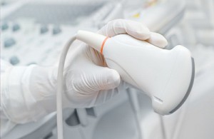
Qualitative diagnosis of diseases of the arteries and veins in the brain and cervical spine spine makes it possible to detect dangerous pathologies, life threatening patient, long before the manifestation of their clinical signs and begin timely adequate treatment. Duplex scanning vessels of the head and neck - a non-invasive modern technique, which is a type of ultrasound, with which you can visualize each artery and assess the speed of blood flow in them. In this case, the tissues that surround the vessels are visible.
USDS (ultrasonic duplex scanning) is based on the possibilities of practical use of the properties of ultra sound wave, which, reflected from red blood cells moving in the vessels, makes it possible to obtain an image of the vessel under study.
This technique allows you to effectively examine both extracranial (cervical) and intracranial (located inside the brain) veins and arteries.
The doctor talks about the diagnosis ultrasound diagnostics Irina Gennadievna Maslova:
Duplex examination is based on two diagnostic technologies. Firstly, it is ultrasonic radiation configured in pulse mode, due to which an image is displayed on the monitor. Studying it, the specialist evaluates the structure of the tissues and examines the vessels. Secondly, the Doppler effect, which involves assessing a moving object, which is the blood flow, by reflected ultrasound on different areas veins.
When an image is fed to the monitor, a two-dimensional image is formed along and across the arteries.
The use of such non-invasive diagnostic method necessary to obtain information reflecting changes in the condition of blood vessels, as well as their effect on blood flow. Transcranial duplex vascular scanning (TCDS) allows for valuable research to detect changes that occur in the tissues and arteries of the brain, making it possible to diagnose brain lesions and establish control over the course of the brain. pathological process.
Using this method, the following information is obtained:
- Condition of the walls of blood vessels in the neck and head;
- State vascular tissue;
- Presence of stenosis and assessment of its severity;
- Disturbances in the flow of the bloodstream;

- Anatomical features circulatory system in the head and neck area;
- The presence of formations inside the vascular canals;
- Deviations associated with changes in the course of the arteries.
To obtain the most reliable information, experts use modern technology, with which you can perform a so-called color scan (CDS) - the ability to display a color sketch of blood flow on the monitor, which facilitates the process of determining its speed at various areas.
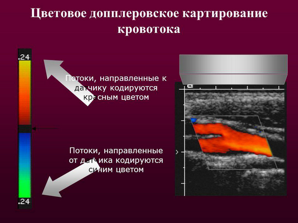
Color Doppler mapping (CDC) is a subtype of manipulation based on the Doppler effect. In this case, data on the speed of movement of structures is displayed in different colors. In particular, the color red determines the speed of blood flow towards the sensor, and the lighter the shade, the slower the speed. The blue color in the study indicates the speed of blood flow in the direction from the sensor. Using this technique, not only a specific vascular pathology is determined, but it is also possible to distinguish a benign process from a malignant one, and to identify the tumor’s tendency to further growth and spread.
Indications for use. What pathologies does the procedure reveal?
Intracranial duplex, which involves the study of veins and arteries located in the cranial cavity, is indicated for patients who have the following complaints:
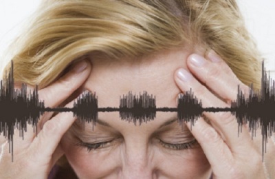
- Regular headaches;
- Dizziness;
- Noise in the head or ears;
- Fainting;
Ultrasound diagnostics doctor Murat Medzhidovich Nagaplev talks about the indications for the examination:
- Manifestations inappropriate behavior;
- Weakness and numbness of the limbs;
- Violation visual functions;
- Poor coordination combined with unsteadiness, with an uncertain gait;
- Deviations in speech production or understanding.
TKDS reveals circulatory disorders in the head area. It is prescribed upon detection the following pathologies:
- Intracranial hypertension;
- Lesions of intracranial vascular canals.
Scanning of extracranial vessels located outside the skull and supplying blood to the brain is carried out in the following cases: pathological signs appearing in the patient:
- Impaired ability to remember information;
- Inability to concentrate on anything;
- Dizziness, intense headaches;
- Coordination problems.
The arteries and veins of the cervical spine should also be examined if it is necessary to perform an operation involving intervention in the vessels of the heart or directly in the muscle structure, as well as when identifying pathologies of the neck organs that can lead to compression of the vessels located in this area.
Duplex scanning of the vessels of the brain and cervical spine is a procedure that should be performed routinely (once a year), even in the absence of any complaints, in following cases:
- If the patient is over 40 years old (men) or 45 years old (women);
- If you have close relatives who have diseases such as diabetes, hypertension, ischemic disease;
- If the patient is an experienced smoker;
- With previously conducted surgical interventions on the spinal cord or brain;
- In case of stroke or disorder cerebral circulation in the anamnesis;
- If available.
Duplex ultrasound scanning is used to diagnose the following pathologies:
- Venous thrombosis, thrombophlebitis, thromboembolism pulmonary artery;
- Anomalies and injuries of veins;
- Varicose veins;
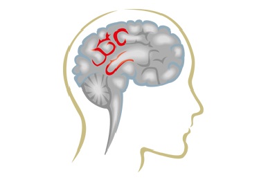
- Aneurysm;
Rehabilitation doctor Sergei Nikolaevich Agapkin tells more about cerebral aneurysm:
- The degree of blood flow deficiency;
- Atherosclerosis;
- Ischemia;
- Angiopathy;
- Vasculitis.
A method such as ultrasound is characterized by high information content, painlessness, and the absence of harmful effects on the body, since the manipulation does not require equipment with radiation.
How is diagnosis carried out?
Diagnostics main arteries head is a safe measure that, if necessary, can be prescribed to pregnant women or a child. A relative contraindication to the procedure is the general serious condition of the patient or the presence of diseases that make it impossible for the patient to accept a horizontal position in which the procedure is performed.
Survey of large cervical arteries
The examination lasts no more than half an hour. Any preliminary preparation no scanning required. Only one thing is required from the subject - not to take substances or drugs the day before that affect vascular tone and distort big picture their condition. These include caffeine, nicotine-containing substances, energy and alcoholic drinks. You should not visit the sauna or bathhouse before duplex scanning.
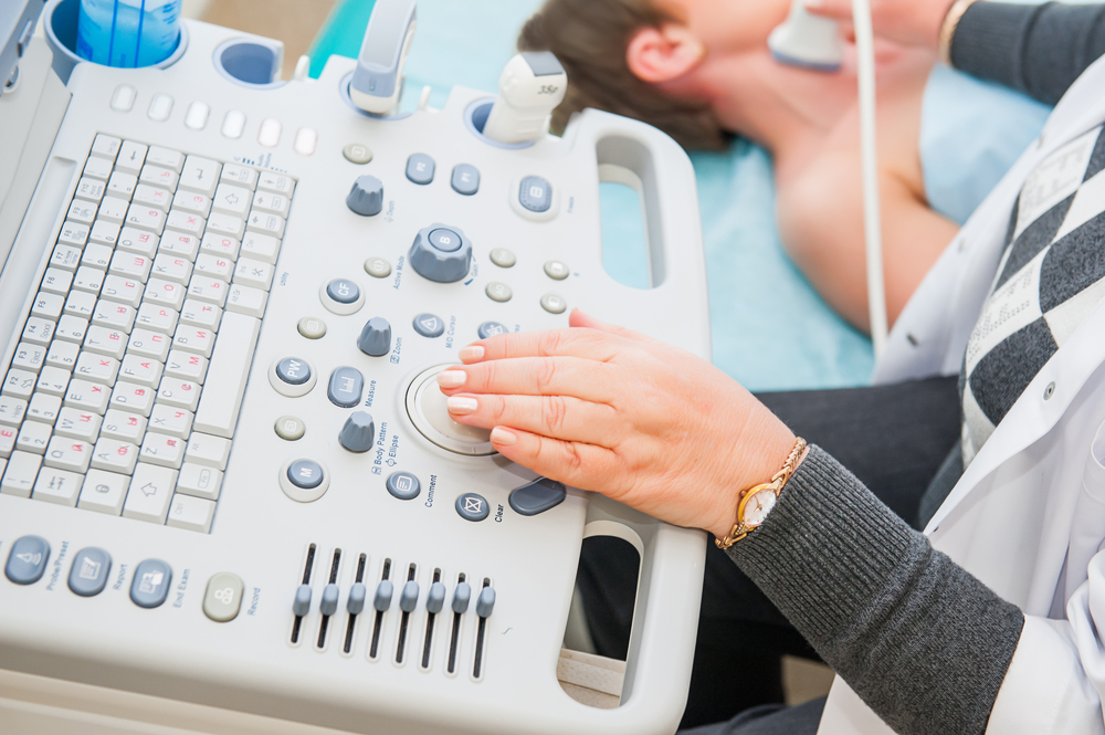
Duplex scanning in progress as follows:
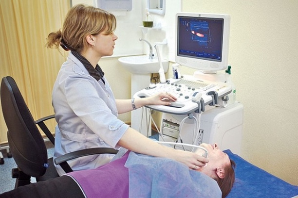
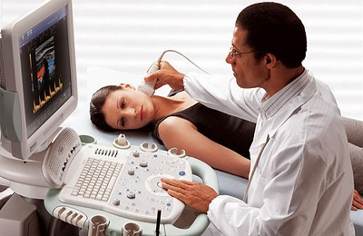
- The patient is in a supine position. The specialist fixes the head so that it is in an elevated position, for which a cushion is placed under the neck. The head is turned in the direction opposite to the area in which the study is being carried out;
- The specialist uses a sensor to trace the area where the vessels are located. First, to facilitate the examination, a gel with a special composition is applied to the surface of the skin. The resulting image is fed to the monitor;
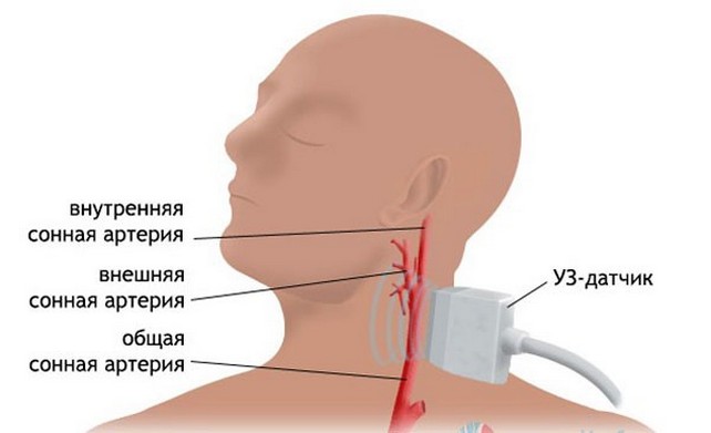
- The examination begins with the diagnosis of the carotid artery before the entrance to the skull, examining it in different planes;
- When examining the brain, areas such as the occipital bone, temporal and supraorbital regions, and the junction are examined. occipital bone with the spine.
During the duplex of the vessels of the head and neck, functional tests to study autonomic regulation. For these purposes, the specialist may ask the subject to hold his breath, cough, and slightly change his body position.
As for the cost of the procedure, it is not too high and, depending on the level medical institution and the city in which it is located ranges from 2300 to 4000 rubles.
Results and their interpretation
The condition of the arteries and veins of the head and cervical region is assessed using such indicators as the thickness of the vessel wall, the nature and speed of blood flow, the ratio between the minimum and maximum speeds.
Level of Doppler status indicators arterial vessels expressed in numbers. Thus, the norm for artery wall thickness is from 0.9 to 1.1. Normal indicator maximum systolic speed should not exceed 0.9, peak velocity in diastole - less than 0.5.
Decoding the results allows you to obtain the following signs of vascular pathology:
- An increase in wall thickness and a narrowing of the artery by less than 20% indicates atherosclerosis;
- Diffuse change wall thickness indicates vasculitis;
- The presence of a fistula between veins and arteries is a sign of malformation.
A lecture on the symptoms and treatment of vascular atherosclerosis is given by general practitioner Yuzef Viktorovich Krinitsky:
The examination protocol allows the specialist to determine the most early signs disease, even before manifestation clinical symptoms.
The advantages of duplex scanning of the vessels of the head and neck are that it is a reliable method that does not require the introduction of any substances into the patient’s blood, as well as exposure to x-rays which may adversely affect your health. The diagnostic method has no contraindications. In addition, the price of the procedure is not too high, which will allow any patient to quickly and accurately determine the condition of the vessels of the neck and head.
Duplex scanning of the brachiocephalic arteries (BCA) is a comprehensive ultrasound examination of the vessels of the head and neck. Non-invasive, safe, no strict contraindications And age restrictions combined with a fairly high information content make it one of the main diagnostic techniques in angioneurology.
The essence of the method
Duplex scanning is based on reflection ultrasonic waves from various fabrics human body. This study includes two components: vascular scanning (B-mode) and Doppler ultrasound. They can be used simultaneously or alternately, depending on the device and the skills of the ultrasound specialist.
B-mode duplex scanning is a two-dimensional gray scale echography. This is what is called a “regular” ultrasound. The piezocrystals located in the device sensor under the influence of alternating electric current generate ultrasonic waves. They are focused and directed to the area of the human body being examined. Such radiation does not cause discomfort or harm. Fabrics and anatomical formations have different densities and abilities to absorb and reflect ultrasonic rays. The sensor perceives the reflected signal, and based on the difference between the emitted and reflected waves, the device program constructs a two-dimensional (planar) black and white image.
B-mode allows you to visualize vessels of different sizes and surrounding tissues. In this case, the doctor evaluates the structure of their walls, identifies the presence of blood clots and plaques, measures the diameter of the arteries and veins, determines their course and the presence of pathological expansions or narrowings.
The Doppler mode is a dynamic study that helps assess blood flow parameters in real time. The method is based on the Doppler effect. This is the change in perceived frequency and wavelength when a signal is reflected from a moving object. The ultrasound generated by the sensor is reflected from shaped elements blood (red blood cells and other cells) and is captured by the device. This allows you to evaluate the direction and speed of blood flow, its linearity and uniformity. Slowing down, the appearance of turbulence (turbulence) or retrograde blood movement indicate the presence of some structural changes.
Thanks to the combination of two modes, duplex scanning provides the doctor with information about the causes of impaired blood supply to the brain and the extent of the existing blood flow deficiency.
What vessels are examined?
Extracranial and some intracranial vessels are subject to examination. From the vessels of the neck, part of the brachiocephalic trunk, the common carotid arteries and the area of their bifurcation, the extracranial part of the internal carotid artery, and the external carotid arteries are examined. The temporal and supratrochlear arteries can also be examined, large veins after they leave the skull, the venous vertebral plexus.
With transcranial duplex scanning, the doctor examines the vessels located in the projection of the acoustic windows (temples, orbits, foramen magnum). It is these areas of the skull in an adult that transmit enough ultrasonic waves for examination. In a child, scanning is also carried out through open fontanelles, which significantly expands the diagnostic capabilities of the method.
Transcranial duplex scanning allows you to examine the intracranial part of the internal carotid and vertebral arteries, cerebral arteries(anterior, middle, posterior), main artery.
What does a BCA duplex scan show?
Using duplex scanning of the vessels of the head and neck, you can identify:
- atherosclerotic damage to arteries with narrowing of their lumen, formation of a parietal thrombus;
- the presence of pathological tortuosity (kinging) of blood vessels;
- various shapes and origin;
- anomalies in the structure, branching and course of blood vessels;
- vasculitis of various etiologies;
- thrombosis;
- violation of the integrity of the vascular wall;
- change in the elasticity of vascular walls;
- the presence of pathological arteriovenous or arterio-arterial shunts (places of abnormal discharge of blood into another vessel);
- violations of the compensatory-regulatory mechanism for maintaining the stability of the blood supply to the brain;
- the degree of blood flow deficiency in narrowed areas of blood vessels, the presence of pathological blood flow and changes in its uniformity and direction;
- state of the collateral network, its functionality and the degree of compensation for insufficient blood flow through the main arteries.
Duplex scanning makes it possible to identify the causes of decreased patency of the great vessels of the head and neck and at the same time assess the nature and extent of the resulting functional disorders.
When is BCA duplex scanning prescribed?

Examination of the vessels of the head and neck is indicated for clinical signs or chronic failure cerebral circulation, to monitor the effectiveness of therapy during systemic thrombolysis. The basis for prescribing duplex scanning may be the patient’s complaints of headaches, memory loss, sudden deterioration of vision, fainting states, noise in the head and, weakness in the arm and leg, difficulty swallowing. Identified during inspection central paresis facial and oculomotor muscles, limb muscles, increasing cognitive disorders, vestibuloatactic syndrome, bulbar palsy are also the basis for diagnostic search with examination of the main vessels of the head and neck.
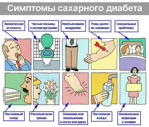
Often, duplex scanning is included in the program of routine examination of people at risk for the development of cerebrovascular diseases. This includes smokers, patients with diabetes mellitus, arterial hypertension, obesity, identified dyslipidemia, systemic diseases. Duplex scanning of the veins of the brachiocephalic system is indicated for suspected thrombosis and thromboembolism.
Research procedure
No special preparation is required before duplex scanning. It is advisable to stop taking medications that may affect the tone and patency of blood vessels for several days. This is agreed with the attending physician, because many medicines require regular use and cannot be cancelled. The patient must inform the ultrasound physician about all medications used. It is also advisable to refrain from drinking alcohol, intensive physical activity, visits to the bathhouse and sauna.
During duplex scanning of the vessels of the head and neck, the patient is lying on his back; if necessary, the doctor asks him to turn on his side. It is undesirable to talk and change body position without permission. If you experience discomfort in the heart area, dizziness or other complaints, you must inform the doctor conducting the study.
During duplex scanning, the doctor presses a sensor with applied contact gel to the skin on the lateral surfaces of the neck, supra- and suboccipital, supraclavicular and temporal areas. And when examining the supratrochlear artery and orbital vessels, the area above the inner corner of the eye is examined. When using the Doppler ultrasound mode, the doctor can use functional tests to assess the state of autoregulation of blood circulation. To do this, short-term compression (compression) of the carotid arteries is performed with fingers or a sensor, and the head end of the couch is lowered. The doctor may also ask the patient to sit up, turn his head, breathe quickly, hold his breath, and strain.
In conclusion, the doctor indicates the diameter of the examined vessels, the speed and nature of blood flow in them, the presence of narrowings (stenoses) and blood clots, pathological changes vascular wall. The condition and thickness of the intima-media complex (IMC) is also described, and the identified blood flow deficiency is additionally indicated as a percentage.
The interpretation of the results of duplex scanning of the vessels of the head and neck is carried out by the attending physician, who also decides on tactics further treatment patient. An ultrasound specialist cannot give recommendations on admission certain drugs or the need for surgical intervention.
Doctor functional diagnostics Murat Nagaplev talks about duplex scanning of the vessels of the head and neck.
Medicine constantly introduces new diagnostic methods to society serious pathologies. Treatment success various diseases depends on their timely detection, purpose necessary therapy. Duplex scanning of head and neck vessels – innovative way research that allows you to see the smallest tubular hollow formations in a two-dimensional projection human body. The non-invasive nature of the technique makes the procedure easier and does not require recovery after the manipulation.
What is duplex scanning of blood vessels
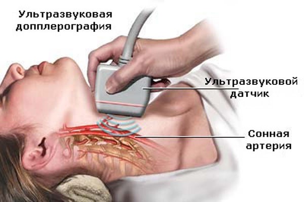
How to check the head non-invasively? Unique properties Ultrasound helps it pass through the tissues of the human body and, reflecting from blood cells, send a signal in the form of an image of the area under study to the diagnostician’s monitor screen. Using duplex scanning of the vessels of the head and neck, a specialist can assess blood hemodynamics and obtain information about anatomical features veins and arteries. Different Doppler technologies use the properties of the sound wave in the same way, but have different functionality:
- Ultrasound Dopplerography (Doppler ultrasound). This study helps to assess the patency of blood vessels in the brain, neck, and other organs. Doppler ultrasound carries only one functional load– determination of hemodynamics.
- Duplex ultrasound scanning. Using this method, it is possible to diagnose the presence of atherosclerotic plaques and blood clots in the arteries and veins, which contribute to the narrowing of the lumen of blood vessels. During monitoring, a tubular formation with surrounding tissues is visualized. Duplex scanning is divided into the following subtypes:
- extracranial – explores great vessels;
- intracranial – checks intracerebral “pools”;
- transcranial – provides color duplex scanning of the brain.
- Triplex scanning. Dopplerography of the vessels of the head and neck, during which, in addition to information about the intensity of blood movement, the diagnostician receives a color image of the tubular formation with the surrounding tissues.
- Ultrasound examination. Shows the “big picture” of the structure of arteries and veins. Doppler ultrasound helps to determine the characteristics of blood flow and conduct an examination for the presence of pathologies.
Indications for the purpose of the study

Vascular examinations of a planned nature should be carried out in mandatory once a year. Anomaly detection on early stage development helps to avoid negative consequences associated with the progressive form of the disease, and take measures to prescribe the necessary therapy. Duplex scanning of the patency of blood vessels in the head and neck is often prescribed to verify the results obtained from MRI, Doppler ultrasound of blood vessels neck and head. Indications for duplex are following symptoms:
- headache;
- dizziness;
- fainting;
- numbness of hands;
- lack of coordination;
- memory loss;
- smoking;
- a history of strokes;
- cervical osteochondrosis;
- arterial hypertension;
- previously identified vascular dystonia;
- family ties with a hypertensive or diabetic;
- vasculitis (inflammation of blood vessels).
How to prepare
The head and neck examination does not require the patient to special training. On the day of the procedure, you must stop using drugs that increase vascular tone: coffee, nicotine, tea, energy drinks. Discontinuation of drugs that can distort ultrasound results– “Betaserc”, “Cinnazirin” - requires consultation with a neurologist. Before scanning, the patient will need to remove anything from the area being examined. foreign objects in the form of chains, hairpins, etc.
How is the procedure performed?
Duplex scanning can be done upon the referral of your attending physician in the neurological departments of large city hospitals or you can go to a clinic according to your area of residence. The manipulation takes place according to general rule. The patient is placed on the couch, a hard pillow or cushion is placed under the head, and the head is moved to the side opposite to the sensor.
Before starting the procedure, the doctor applies a little special gel to the area under study, with which you can easily “move” the transducer over the surface of the skin, analyzing the arterial and venous beds. The blood vessels of the brain are checked through the bones of the skull. Previously skin are treated with a water-soluble gel, then the doctor places sensors on the following areas:
- temples;
- above the eye sockets;
- alignment of the occipital bone with the spine;
- occipital bone.
Decoding the results
![]()
Upon completion of the examination, the doctor receives comprehensive information about the condition of arteries and veins. Analysis of the venous bed contains practically no digital data, but includes the following parameters:
- anatomy;
- cross-country ability;
- blood flow speed;
- the presence of abnormal formations inside the lumen.
Dopplerography of arterial vessels collects digital data that is compared with normal values. Satisfactory condition of general and carotid arteries the presence of the following indicators can be considered:
- the maximum speed of blood movement in the artery is less than 0.9;
- percentage of stenosis – 0;
- peak velocity in diastole – less than 0.5;
- absence of formations inside the lumen;
- wall thickness – 0.9-1.1.
Are there any contraindications

The advantage of duplex scanning is the absence negative influence on the human body. The non-invasive nature of the study helps to diagnose blood vessels in adults and children without any restrictions. Relative contraindications can be considered serious condition patient or the presence of diseases that prevent the patient from transitioning to horizontal position.
Video: what ultrasound of the vessels of the head and neck shows
Duplex scanning of the vessels of the head and neck is an ultrasonic examination of vessels that are located extracranial (vessels of the neck) and intracranial (vessels of the head). This modern method diagnostics, which allows you to visualize the lumen, its walls and identify functional indicators blood flow Widely used in neurology, neurosurgery, cardiology, and other branches of medicine.
What is duplex scanning?
First of all, this is a non-invasive diagnostic method that is available in almost all medical centers. The basis of the diagnostic manipulation is the Doppler effect, which helps to calculate the speed of blood flow and determine its location. possible violation. In addition, the ability to examine the anatomical structure of blood vessels, the presence of congenital or postoperative anomalies of the vascular bed, and the identification of atherosclerotic plaques and blood clots provide the basis for making a diagnosis.
Ultrasound of neck vessels
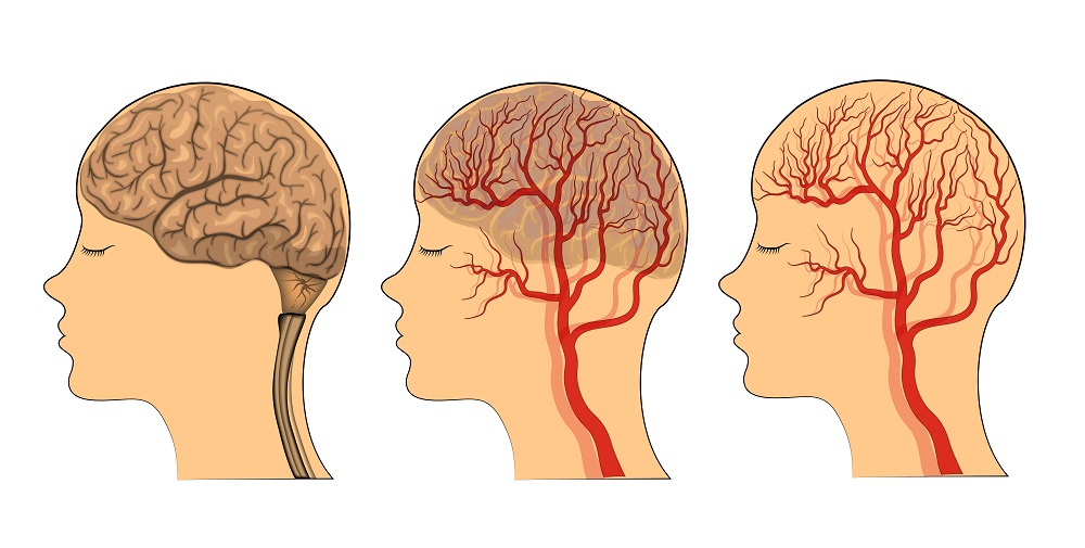
Some of the main vessels that supply blood to the brain are located in the neck. Examination using ultrasound waves allows you to examine their structure, evaluate the lumen, and identify the presence of pathological structures (plaques, blood clots).
The atherosclerotic process occurs in the body simultaneously throughout the entire body, leading to damage, which becomes the cause of vascular accidents (heart attacks).
Estimate prevalence vascular changes possible in areas of the body where the vessels are close and accessible to duplex scanning. Carrying out an ultrasound examination of the vessels of the neck makes it clear what they look like vascular lesions throughout the body. In addition, you can evaluate the influence of nearby bone structures directly on the vessels of the neck, and therefore on the blood supply to the brain.
Duplex scanning of cerebral vessels can visualize the following parameters:
- evaluate anatomical structure, abnormalities, or changes;
- determine the presence of atherosclerotic lesions: plaques, thickening of the intima-media complex (IMC);
- identify venous stasis in the veins of the neck;
- assess the effect of the cervical spine on blood flow;
- identify vascular patency, occlusion or the presence of a thrombus.
The attending physician can prescribe and evaluate the results of duplex scanning of extracranial vessels. Depending on the identified changes, treatment tactics are selected, drugs are selected taking into account the course of the disease and individual characteristics body. In the future, the effectiveness of therapy can also be assessed by performing an ultrasound examination.
Vascular ultrasound has the following indications:
- headaches;
- dizziness, ;
- decreased memory and ability to work;
- head injuries;
- attacks of numbness in the fingers;
- high blood cholesterol;
- , ears;
- presence of symptoms of developing acute disorder cerebral circulation;
- cervical osteochondrosis.
How the research is carried out
On the eve of the examination, it is not recommended to take caffeine-containing medications, drink alcohol, or smoke. The procedure is also not carried out after intravenous injections, as this may affect the result. Any drugs that affect blood flow and blood pressure, except in cases where taking these medications is vital.
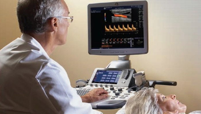
 The patient is positioned on the couch with his head thrown back and to the side opposite to the study. The ultrasound device sensor is installed on one side of the neck first. Next they evaluate vascular bundles neck on the other side. The duration of the study varies individually, but on average it lasts 10–15 minutes.
Ultrasound examination of the vessels of the head
Duplex scanning of the vessels of the neck and brain visualizes the vessels not only located in the neck, but also those supplying the brain, determining blood flow through the intracranial vessels. Since bone tissue(skull) serves as an obstacle to the penetration of ultrasonic waves, then for transcranial Doppler ultrasound, so-called acoustic windows are used: temporal region, orbits of the eyes, areas of the occipital bone and vertebra.
Duplex examination of blood vessels is carried out when:
- chronic headache;
- neurocirculatory dystonia;
- blood flow disturbances;
- dizziness and tinnitus;
- periodic fainting states;
- suspicion of abnormally located cerebral vessels;
- assessing the degree of atherosclerotic damage to the vessels of the head;
How is duplex examination of the vessels of the head and neck performed?
The day before the study is excluded vascular drugs, which affect the results of the procedure, alcohol and nicotine. The examination is carried out lying on the couch. An ultrasound sensor is installed on the back of the head, in the area of the orbit and temple. In the process diagnostic measure It will be necessary, at the request of the doctor, to turn your head to the side, breathe quickly or hold your breath. The duration of the procedure is no more than 15 minutes.
It is possible to evaluate the results by the following indicators: pulsation and resistive indices, linear blood flow velocity and its nature, vessel diameter and percentage of occlusion by thrombus, systole-diastolic ratio.
Ultrasound duplex scanning of head and neck vessels is a modern diagnostic method, allowing not only to install accurate diagnosis, but also to evaluate the effectiveness of treatment. The examination is prescribed by the attending physician. After receiving the results, he determines the further direction of therapy and selects medical supplies taking into account the characteristics of the course of the disease.
To date ultrasonic methods tests that can be used for diagnosis vascular pathology brain are represented by:
Duplex scanning of the vessels of the head and neck is recognized as the gold standard for diagnosing angiological pathology in this area. What is the essence of this method? How does it differ from ultrasound and ultrasound? Who is indicated for this study and how to prepare for the procedure? We will answer these and many other questions in the article.
Review from our reader Victoria Mirnova
I’m not used to trusting any information, but I decided to check and ordered a package. I noticed changes within a week: constant pain in my heart, heaviness, pressure surges that tormented me before receded, and after 2 weeks disappeared completely. Try it too, and if anyone is interested, below is the link to the article.
What is a duplex?
Duplex scanning combines the advantages of classic ultrasound and Doppler.
 This research makes it possible to:
- obtain an image of the vessel, evaluate its morphology (something that can be done with regular ultrasound);
- visualize the blood flow in the lumen of the vessel, evaluate its direction, intensity, as well as a number of other characteristics (what ultrasound scanning allows).
Thus, it is possible to evaluate two basic parameters – structure and function. In addition, duplex allows you to visualize atherosclerotic plaques, blood clots and emboli of various origins, pathological tortuosity of blood vessels, thickening or thinning of vascular walls.
Modern ultrasound machines, which provide duplex scanning, produce color images.
The resulting picture is displayed in the form of a cartogram, which contains information about the speed, direction, and intensity of blood flow. This option The technique is called color Doppler scanning (CDS).
 The extensive possibilities revealed by this method in the diagnosis of angiological pathology determine wide range indications for the study.
Using this method ultrasound diagnostics, you can examine almost any blood vessels of the human body. Since this article is about the head and neck duplex, below we will provide a list of vessels in this area that are most often examined using this technique.

These are the main groups of vessels that are available for duplex scanning of the corresponding area.
The method of transcranial Doppler scanning (TCDS) does not lose its relevance. This study allows you to evaluate blood flow in the same vessels that are accessible to the duplex. The disadvantage of the method is the inability to assess the morphology of the vascular bed. Therefore, transcranial Doppler is used only when duplex is not possible.
To clean VESSELS, prevent blood clots and get rid of CHOLESTEROL - our readers use the new natural preparation, which is recommended by Elena Malysheva. The product contains blueberry juice, clover flowers, native garlic concentrate, rock oil, and wild garlic juice.
Indications for the study
Let's start with the cases in which routine duplex scanning of the head and neck area is necessary. Duplex scanning of cerebral vessels is indicated for:

Separately, cases should be analyzed when the patient is bothered by certain symptoms, the diagnosis has not been established and an ultrasound examination is recommended. Symptoms that indicate the need for a duplex of the head and neck vessels include:
In the above cases, duplex scanning will reveal pathology and establish a diagnosis. The efficiency of the study increases significantly when using the CDS mode, which was already mentioned above.
Several nosological units can be identified, in the diagnosis of which the method of duplex scanning of cerebral vessels plays a decisive role. This:
Thus, modern techniques Ultrasound allows you to diagnose many different pathological processes. And an accurate diagnosis is the key the right choice patient management tactics.
How is the research conducted?
Carrying out this type of ultrasound (including with CD) does not require special preparation of the patient. The only thing the attending physician can ask is a refusal to use substances that can distort the real picture. It's about about substances that significantly affect vascular tone.
Many of our readers actively use the well-known method based on Amaranth seeds and juice, discovered by Elena Malysheva, to CLEAN VESSELS and reduce the level of CHOLESTEROL in the body. We recommend that you familiarize yourself with this technique.
 Therefore, on the eve of the duplex, the head and neck area should be avoided:
- nicotine-containing substances;
- caffeine-containing substances;
- alcohol and energy drinks.
If the patient regularly takes medications that affect vascular tone, then before performing this type of ultrasound, he should discuss the issue of medication regimen with his doctor.
All types of ultrasound of the vessels of the head (including duplex with CDS) are carried out according to a single scheme. The patient is in a lying position, with a special cushion (or pillow) under his head.
The neck should be free and the head should be turned in the direction opposite to the side of the study. A special gel is applied to the skin, along which the sensor moves. The doctor places the sensor at certain points, which makes it possible to visualize the required part of the vascular bed.
Duplex is a simple, non-invasive and absolutely painless method. At the same time this study is extremely informative and effective.
About the information content of this method
In some cases, the duplex of cerebral vessels allows solving diagnostically complex problems. Below we give several examples in which cases we can resolve the issues of diagnosis and choice. therapeutic tactics is not possible without using duplex scanning:
From all of the above, it becomes clear that today duplex scanning of cerebral vessels plays an important role in diagnosing diseases of neurological and angiosurgical patients. Duplex provides high quality diagnostics, and therefore subsequent treatment.
Do you still think that it is completely impossible to RESTORE blood vessels and the BODY!?
Have you ever tried to restore the functioning of your heart, brain or other organs after suffering pathologies and injuries? Judging by the fact that you are reading this article, you know firsthand what it is:
- often occur discomfort in the head area (pain, dizziness)?
- You may suddenly feel weak and tired...
- is constantly felt high blood pressure…
- about shortness of breath after the slightest physical stress and there is nothing to say...
Did you know that all these symptoms indicate INCREASED CHOLESTEROL levels in your body? And all that is necessary is to bring cholesterol back to normal. Now answer the question: are you satisfied with this? Can ALL THESE SYMPTOMS be tolerated? How much time have you already spent on ineffective treatment? After all, sooner or later the SITUATION WILL GET WORSE.
That's right - it's time to start putting an end to this problem! Do you agree? That is why we decided to publish an exclusive interview with the head of the Institute of Cardiology of the Ministry of Health of Russia, Renat Suleymanovich Akchurin, in which he revealed the secret of TREATING high cholesterol.
