What is perinatal damage to the central nervous system of hypoxic origin. Perinatal brain pathology and its consequences
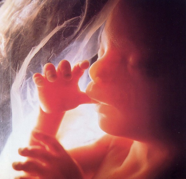
Perinatal pathology NS is general definition functional or structural damage hemispheres of the brain, the source of which was various phenomena during prenatal development. Actually, it includes antenatal, intranatal and early neonatal development, which begins at 28 weeks.
In order to protect your child as much as possible from pathologies, it is important to answer the question “What is PCPNS?” It is the answers to these questions that will allow us to understand how to prevent an undesirable future for a child.
In modern medical practice diseases of perinatal encephalopathies do not exist, however, due to the complexity of diagnostic and treatment measures, domestic specialists continue to use this term to determine the disease.
Hypoxic ischemic lesion The CNS is a common source of various neurological abnormalities in children. Suspicious symptoms appear from the first days of life, but by the end of the 12th month they become more pronounced.
After this period, the neurologist is obliged to identify damage to the central nervous system, as well as develop a treatment strategy for the child. Brain little patient extremely flexible, which allows you to achieve high efficiency treatment.
Remember that the consequences of perinatal damage to the central nervous system will manifest themselves at all stages of life, so it is important to carry out therapy to improve future functioning.
Classification of PPCNS today
IN medical literature Two ways of causing CNS damage have been described:
- Hypoxic ischemic damage to the central nervous system during pregnancy – intrauterine;
- Acute fetal hypoxic syndrome that occurs during pregnancy;
If the first class of pathologies arises due to the anatomical and morological characteristics of a woman during pregnancy, then acute labor hypoxia is most often of traumatic origin.
Perinatal damage to the nervous system is caused by multiple sources that significantly affect the health of the baby. Sometimes such disorders do not in any way manifest themselves in the baby’s functionality, but develop in the future in serious illnesses of a different origin.
The combination of two factors can sometimes lead to catastrophic consequences. This condition is called perinatal lesion CNS mixed origin. Perhaps, in some cases, a single manifestation of each cause would not lead to the development of pathology, but their simultaneous appearance leads to significant complications.
Intrauterine disorders of the central nervous system largely depend on the mother, her health and lifestyle, and responsibility for postnatal disorders lies on the shoulders of doctors delivering babies.
Common causes of pathologies
As with any other pathology, it is important to understand the causes of the disease so that it is possible to develop effective measures treatment. Perinatal pathology of the nervous system can be caused by the following reasons:
- Somatic disorders in the maternal body, which are accompanied by chronic intoxication;
- The presence of an acute infectious disease or aggravated chronic processes during pregnancy;
- Poor nutrition or physiological immaturity of the maternal body;
- Tendency to hereditary pregnancy disorders;
- Unfavorable environment;
- Pathological situations during delivery;
As you can see, there are many various reasons, which could potentially ruin the health of your unborn child. Hypoxic ischemic damage to the central nervous system is an extremely difficult prognostic pathology, the development of which is almost impossible to predict or prevent.
Early delivery can also lead to adverse consequences . Exchange processes immature babies are not adapted to independent work organism, which is difficult when artificially “gestating” them. That is why hypoxic ischemic damage to the central nervous system can appear after childbirth.
Prognostic course of the disease
Ischemic damage to the central nervous system in newborns can be accurately diagnosed after the first months of their life. Experienced doctor is able to assess not only the degree of brain damage, but also make a relatively accurate prognosis of its condition.
The outcome of PPCNSL can be of two types: complete recovery with minimal impairment of the central nervous system or severe manifestations that will require long-term or lifelong treatment by appropriate medical specialists. Every clinical case requires individual approach so that the effectiveness of treatment is maximized.
In general, manifestations of cerebral ischemia in newborns have various consequences, characterized by:
- Full restoration of health;
- Inhibition of mental, motor or speech activity;
- Neurotic deviations;
- Post-traumatic abnormalities;
- Autonomic-visceral dysfunctions;
- Hydrocephalic syndrome;
Some deviations can ruin the patient’s future for the rest of his life, but some (for example, motor disorders) can only slightly limit the level and quality of the baby’s vital activity with proper treatment.
Remember that perinatal hypoxic damage to the brain can often be complicated in late childhood and adolescence neurotic syndromes and inability to adapt to the surrounding society. Children will have a negative attitude towards peers with central nervous system disorders of hypoxic origin. Such actions will negatively affect internal state the latter.
Diagnostic measures
To make a diagnosis of perinatal CNS lesions, irrefutable data are required clinical examination, and all other examinations are only auxiliary, which do not play a major role.
In addition, additional methodology in the study of the central nervous system has only clarifying properties to determine a more accurate source of pathology of ischemic origin, as this will allow the selection or development of organ and regionally specific therapy.
The following methodology is used as diagnostic measures to determine the origin of the source of the problem:
- Neurophysiological procedures;
- X-ray diagnostic procedures;
Unfortunately, today there is no single unified method that will accurately determine the source of the problem. Each method is important and unique in its own way. It is based on certain ones, which allows a comprehensive study of the pathological processes in it.
It is unacceptable to independently prescribe and carry out any diagnostic measures on one's own. Although many methods are relatively safe for your baby, they can make him feel uncomfortable or anxious, which can adversely affect his mental health.
Diagnostic methods are aimed at identifying excitation in various departments and his assessments. It is important to identify the pathological origin nerve impulses so that the treatment is as accurate and effective as possible.
Therapeutic measures

Brain damage most often leads to disability of a small patient, which makes him unfit for life in modern society. Fortunately, there are modern therapeutic measures, which are able to compensate for the pathological condition of the baby.
General complex medical procedures consists of several stages:
- Drug therapy;
- Massage treatments;
- Physical therapy exercises;
- Physiotherapy;
Relatively non-standard methods of assistance are often used in the form of acupuncture and intensive pedagogical work. Extremely high demands are placed on treatment, since doctors often do not have enough time for treatment, so it is unacceptable to waste it in vain.
Shows the greatest efficiency physical therapy, massages and other methods physical impact. Pharmacological therapy is used for symptomatic treatment seizures, hydrocephalus, etc.
Treatment tactics huge variety, and only an experienced pediatric neurologist can choose the best one. Often, the doctor may change the treatment plan to identify only the most effective techniques, which will be actively included in further therapy.
Common syndromes
CNS disorders may have general character, but often they manifest themselves in the form of a set of symptoms (syndromic complexes):
- Increased ICP;
- Disorders of nerve-reflex conductivity;
- Epileptic seizures;
- Minimizing brain activity;
Despite the fact that these syndromes have quite unpleasant manifestations, modern medicine is able to effectively hide them and expose at least minimal treatment. Pharmacological drugs are able to stabilize the patient's condition, allowing him to lead a relatively normal life.
So, although pregnancy and childbirth are physiological processes, exists a whole series various complications that can ruin the life of your heir.
Perinatal pathologies of the nervous system are rare, but it is impossible to calculate and predict their occurrence. Even if you are faced with a similar pathology, do not despair!
Literate medical specialist, using all the achievements of modern medicine, is able to stabilize the baby’s condition so that he can lead normal image life. Remember that only together with your child will you be able to overcome all the difficulties encountered on your common path in life.
The clinical consequences of perinatal CNS lesions have been a topic of heated debate among pediatricians, neonatologists and neurologists for many decades. It is widely believed that the human central nervous system after damage is not capable of regeneration.
However, modern literature data and experience practical work convince us that children with cerebral damage experience partial or full recovery neurological functions. This is explained by the fact that in the nervous system, in response to the influence of a traumatic agent, compensatory-adaptive mechanisms are activated, ensuring the restoration of lost nerve connections and maintaining the functional unity of the nervous system. However, it is difficult to underestimate the role of perinatal lesions of the central nervous system in the formation of childhood pathology: in the structure of childhood disability, lesions of the nervous system account for about 50%, with 70-80% of cases accounting for perinatal lesions.
Currently it is customary to distinguish the following types perinatal brain lesions: 1) traumatic injuries; 2) hypoxic-ischemic encephalopathy (HIE); 3) infectious lesions of the brain and/or its membranes; 4) congenital anomalies brain development; 5) dysmetabolic lesions of the central nervous system. Hemorrhagic lesions of the central nervous system are related to several groups at once, since the main cause of intracranial hemorrhage is hypoxia, and as a component of injury they are always present in traumatic hemorrhages.
Most common cause perinatal damage to the central nervous system are hypoxic-ischemic brain damage (HIP) - 47%, the consequences of which occupy a leading place in the structure of morbidity and mortality in children of the neonatal period and early age. Further, it is advisable to distribute the causes of perinatal brain damage depending on the frequency of occurrence: as follows: brain anomalies and dysplasia - 28%; TORCH infections - 19%; birth trauma — 4 %; hereditary diseases exchange - 2%. The frequency of brain damage with hyperbilirubinemia depends on the level of bilirubin and gestational age: with a bilirubin level in the blood of 428-496 µmol/l, kernicterus develops in 30% of newborns, and with a level of 513-684 µmol/l - in 70%, in premature infants it develops with hyperbilirubinemia 171-205 µmol/l.
At birth, the child's brain is immature, especially the cerebral hemispheres. It is the immature brain, which is in the stage of rapid development, that has the highest compensatory capabilities. The main damaging factor in this category of children is hypoxia, which leads to both hypoxemia and cerebral ischemia and is the main factor predisposing to the development of hypoxic-ischemic encephalopathy. Acute severe asphyxia mainly causes changes in the stem structures, less pronounced long-term asphyxia causes diffuse cortical disorders. However, not all children who have suffered severe hypoxia experience severe neurological consequences. Their brain, exposed to hypoxic effects, has a number of features assessed as self-defense phenomena.
Such phenomena include increased tolerance of the developing brain to hypoxia (fewer neurons and processes, fewer synapses and, ultimately, less dependence on the energy-consuming ion pump), its neuroplasticity (modern researchers argue that the brain, in response to damage, can form new neurons and carry out transplantation of immature neurons into certain sections, thereby promoting the formation of stable nerve connections, and denervated neurons are capable of reinnervation of the structure), minimizing the source of damage due to neurotrophic factors (when neurons are damaged, neurotrophic factors enter the extracellular space, which promotes not only preservation of functions, but also active restoration of brain tissue), autoregulation cerebral blood flow and redistribution of blood in the brain (during hypoxia, redistribution of blood flow in the brain occurs, with blood flow increasing in the brainstem and spinal cord and weakening in the white matter and cerebral cortex).
Newborn babies, revived after severe perinatal asphyxia and a long period of oxygen deficiency, can retain their cerebral functions in 50-75% of cases.
Clinical syndromes associated with perinatal hypoxia depend on the period of HIE: to syndromes acute period include increased neuroreflex excitability, syndromes of general depression of the central nervous system, vegetovisceral dysfunction, hydrocephalic-hypertensive, convulsive, coma; The structure of the recovery period of HIE includes syndromes of delayed speech, mental, motor development, hypertensive-hydrocephalic, vegetovisceral dysfunction, hyperkinetic, epileptic, cerebroasthenic. Some authors in recovery period syndromes of motor disorders and increased neuro-reflex excitability are distinguished.
In the structure of the main prognostic factors, three main groups of signs should be considered: Apgar score in the first 20 minutes of life; neurological disorders during the neonatal period; data modern methods brain imaging during the acute period of the disease.
K. Nelson et al. noted in their works that children with an Apgar score of less than 3 at 10, 15, 20 minutes and survivors were more likely than children with a higher score to have children's cerebral palsy, delayed psychomotor development, convulsions. Prognostic signs depend on severity clinical manifestations. The mortality rate of newborns with perinatal damage to the central nervous system of a hypoxic nature is 11.5% (among children with moderate cerebral disorders - 2.5%, severe - 50%). In children with light current Hypoxic-ischemic encephalopathy does not cause complications in the neonatal period. According to M.I. Levene, in 80% of full-term newborns, severe CNS HIP leads to death or severe neurological impairment.
Of the clinical manifestations, the most unfavorable in terms of prognosis and long-term neurological consequences are the appearance of seizures in the first 8 hours of life, recurrent seizures, persistent muscle hypotonia and the transition of the phase of lethargy and hypotension into a state of pronounced hyperexcitability and hypertension of the extensor muscles. It was noted that in children who suffered asphyxia with the subsequent clinical picture of DIE, and also had neurological symptoms along with asphyxia, the development of cerebral palsy occurred more often.
Symmetry in the motor sphere is of particular importance: an unfavorable prognostic sign for cerebral palsy is asymmetry of movements in the neonatal period. Data from brain imaging methods are also important, although the diagnosis of perinatal CNS lesions in newborns is difficult due to unclear clinical picture, extremely rapid dynamics of liquorological parameters and neurological symptoms, especially in the first hours and days of life.
One of the most available methods brain imaging is neurosonography, with which it is possible to assess the macrostructure and echogenicity medulla, size and shape of liquor spaces. The method allows you to objectify morphological changes brain in newborns, in whom routine anamnestic and clinical neurological methods may not be enough to make a diagnosis, it allows one to suspect periventricular leukomalacia on the first day, assume the presence of peri- or intraventricular hemorrhage and clarify its degree. Data from neurosonographic studies at various stages of the pathological process make it possible to evaluate the results of the therapy and determine tactics further treatment, and are also used for clinical monitoring of children in the first year of life with perinatal damage to the central nervous system.
However, the connection between neurosonography data and clinical outcomes not always natural: comparative study brain imaging methods and clinical outcomes showed that in the presence of changes in neurosonograms ( ultrasound signs hemorrhages, leukomalacia) normal neurological outcomes are possible. Currently, neurosonography is considered mainly as a screening method, with the help of which a group of children is identified, which is subject to a more in-depth computed tomography, magnetic resonance, proton spectroscopic study. However this method remains indispensable in the diagnosis of subependymal and intraventricular hemorrhages.
Dopplerography, which allows one to assess the amount of blood flow in intra- and extracerebral vessels, is used in HIE to assess the intensity of cerebral blood flow in different phases vascular reactions to hypoxia. However, any relationship between the intensity of cerebral blood flow during the neonatal period and neurological outcome absent at 6 and 12 months.
Computed tomography makes it possible to diagnose selective neuronal necrosis, lesions of the thalamus and subcortical ganglia, parasagittal lesions of the ganglia, periventricular leukomalacia, focal and multifocal necrosis. Magnetic resonance imaging makes it possible to assess not only disturbances in the macrostructure of the brain matter, the localization and volume of intracranial hemorrhage, the size of the cerebrospinal fluid ducts, but also to identify foci of decreased and increased density brain matter, in particular white matter. Thus, this method is indispensable in the diagnosis of periventricular and subcortical leukomalacia.
The positron emission tomography method makes it possible to determine at various levels and in various structures brain intensity of regional metabolism, intensity of cerebral blood flow. Magnetic resonance spectroscopy makes it possible to reflect delayed brain death and is informative not only for the diagnosis of DIE in the acute period, but also for the prognosis of the disease.
An important prognostic criterion is electroencephalography (EEG) data. Normal indicators EEGs are highly correlated with favorable outcomes. On the contrary, indicators such as low voltage, bursts, suppression or absence of electrocerebral activity, and paroxysmal EEG are highly associated with adverse outcomes.
One of the most severe and frequent (after peri- and intraventricular hemorrhages) forms of brain damage of hypoxic-ischemic origin is periventricular leukomalacia (PVL). The incidence of PVL in the group of surviving preterm newborns of gestational age up to 33 weeks is 4.8% with ultrasound and 7.7% with magnetic resonance imaging or computed tomography.
The neurological consequences of PVL are caused by the formation of focal coagulative necrosis of the periventricular white matter between the optical emissivity level and the triangle lateral ventricle, occipital periventricular region and frontal medullary white matter at the foramen of Monro. The lesions are mostly bilateral, with dilatation of the lateral ventricles, often due to atrophy of the white matter of the brain. An objective sign of periventricular leukomalacia is the formation of cysts in areas of ischemic necrosis. However, their presence does not always cause severe neurological disorders.
Prognosis depends on prevalence cystic degeneration. Large cystic PVL in 100% of cases is accompanied by severe motor disorders (spastic di-, hemi-, quadriplegia), in 65-100% - delays mental development to varying degrees, in 30-100% - visual impairment (strabismus, hemianopsia, blindness). Possible hearing impairment, microcephaly, convulsions.
In addition to the prevalence clinical variant the consequences depend on the affected area and the size of the cysts. The development of cerebral palsy is associated with damage to the central part of the internal capsule, medial middle and posterior frontal segments of white matter cerebral hemispheres brain Strabismus is caused by damage to the projection and commissural connections of the posterior adversive field. Delay mental development observed with damage to the lateral frontal and parietal segments of the cerebral hemispheres, with changes in the system of the upper longitudinal beam. Periventricular leukomalacia leads to minor neurological disorders in the form of dyspraxia, transient changes muscle tone or does not cause any neurological abnormalities in children with isolated unilateral brain damage in the medial posterior frontal and parietal segments of the cerebral hemispheres, as well as in the presence of single small pseudocysts of any location. Small cysts (diameter< 3 мм) не вызывают каких-либо последствий .
Of the hemorrhagic cerebral injuries that occur during the perinatal period, the most commonly observed are subependymal hemorrhages (SEH) and intraventricular hemorrhages (IVH), and their frequency increases as the degree of maturity of the newborn decreases.
The outcome of SEC and IVH depends on the degree of hemorrhage and the nature of its complications. With IVH of grade I, complete compensation of neurological abnormalities is observed in the first year of life, with IVH of grade II and IIIA, a favorable prognosis is observed in 80% of cases; in grades IIIB and IV, an unfavorable prognosis is typical in 90% of cases.
Some authors do not divide stage III IVH into A and B; according to them, the survival rate of such children is about 50-70%; according to other authors, in 40% of patients with IVH III degree there are neuropsychological problems of varying severity both in early and school age, and 10% of children with IVH I-II degrees there are motor disorders (mainly spastic diplegia).
The criteria for an unfavorable prognosis are: spread of hemorrhage to the brain parenchyma; catastrophic onset of clinical manifestations with bulging fontanel, convulsions, respiratory arrest; posthemorrhagic hydrocephalus that does not stabilize spontaneously; signs of increase intracranial pressure which indicate posthemorrhagic hydrocephalus.
The outcome of PVL and IVH also depends on the timeliness and completeness of the procedure. resuscitation measures aimed at combating the main pathogenetic mechanisms leading to their development, and this is primarily adequate ventilation of the lungs, elimination of hypovolemia, maintenance of adequate brain perfusion, protective regime, systematic delivery of energy to the brain, prevention of hemorrhagic complications, neuroprotection and treatment of cerebral edema. Since the course of hypoxic-ischemic lesions of the central nervous system is progressive, using the above measures, it is possible to prevent the development severe consequences, which influence both the immediate and long-term prognosis. It is necessary to take full advantage of the high neuroplasticity of the developing brain and actively promote the restoration of damaged structures and functions of the central nervous system.
The prognosis of traumatic hemorrhages, most often represented by subdural and epidural hematomas, depends on the timeliness of diagnosis and treatment. Favorable long-term consequences are timely removed epidural and supratentorial hematomas (50-80%); with subtentorial hematomas without damage to the cerebellum is possible favorable outcome, however, there is a high risk of developing hydrocephalus as a result of obstruction of the cerebrospinal fluid pathways. When an unrecognized subdural hematoma occurs, its encapsulation occurs, which causes atrophy of brain tissue due to compression and ischemia, which determines the prognosis. Long-term neurological consequences in isolated subarachnoid hemorrhages, which can be of either traumatic or hypoxic origin, are usually absent.
The course and prognosis of spinal injuries depend on the severity, localization of the pathological process and the nature of the anatomical and morphological changes. When the upper cervical segments are affected, a picture of spinal shock, Cofferat syndrome, is observed; with damage to the lower cervical segments and brachial plexus paresis or paralysis of the arms develops; in case of defeat thoracic clinic prevails respiratory disorders; injury in the lumbosacral region is accompanied by lower flaccid paraparesis.
At minor injury, as a rule, spontaneous recovery occurs, with moderate and severe damage, when there are organic changes, restoration of impaired functions is slow and requires a long rehabilitation treatment, in some cases operational.
Long-term consequences of birth spinal injury may include peripheral cervical insufficiency(hypotrophy of the muscles of the shoulder girdle, protruding shoulder blades, general myopathic syndrome with hyperflexibility of the child), acute disorders cerebral and spinal circulation, myopia, hearing impairment, nocturnal enuresis, convulsive states, hypertension, vomiting and regurgitation syndrome.
Consequences of perinatal CNS damage infectious nature always serious. Among intrauterine infections, accompanied by lesions of the central nervous system, a number of pathological conditions, at which detectable brain disorders are of a specific nature. These include embryo- and fetopathies with TORCH infections. In such newborns, against the background common symptoms characteristic signs of multiple organ lesions with a predominance of one or another system, depending on the tropism of the pathogen.
The pathogens that exhibit the greatest affinity for the central nervous system are rubella, cytomegaly, toxoplasmosis, and herpes. Antenatal damage by representatives of this group entails severe, often irreversible organic and functional damage to the central nervous system (cerebral palsy, deafness, blindness, microcephaly, oligophrenia, hydrocephalic, convulsive syndrome s, impaired thermoregulation, intracerebral foci of calcification, severe meningoencephalitis). With early diagnosis and active treatment The prognosis for life is usually favorable for full recovery unclear because after past infection the pathogen is capable of persisting for months and sometimes years, predisposing to a number of diseases.
A separate group consists bacterial infections CNS, in which either the CNS is involved in pathological process in the form of a general nonspecific reaction, as a manifestation of infectious toxicosis, or a generalized infection leads to secondary brain damage, affecting cerebral vessels and breaking cerebral blood supply with the development of hypoxic-ischemic or hypoxic-hemorrhagic damage to brain tissue with a favorite localization in the area of passage of certain vessels (periventricular zone).
Most common early complications are edema and swelling of the brain, convulsive syndrome, bacterial (septic) shock. After neonatal meningitis, hydrocephalus, multicystic encephalomalacia, atrophy of the white matter of the cortex, blindness, deafness, spastic paresis and paralysis, mental retardation, and epilepsy may develop. The presence of these changes in to a large extent affects the prognosis.
The mortality rate of newborns from purulent meningitis ranges from 6.5 to 37.5%. In 40-50% of surviving children, neurological defects persist or develop at follow-up (half have mild or moderate severity), including blindness and deafness. The outcome depends on timely diagnosis and started intensive treatment.
When predicting the consequences of purulent meningitis, laboratory and instrumental methods examinations. Prognostically unfavorable factors In terms of both death and the development of complications, high numbers of proteinorrhachia (more than 3-5 g/l), cytosis (more than 1000 in 1 μl of cerebrospinal fluid) are considered. Ultrasound scanning brain, CT, MRI make it possible to diagnose the development of complications of purulent meningitis in the form of ventriculitis, various forms of hydrocephalus, brain abscess, hemorrhagic complications, which largely determine the possible short-term prognosis. For forecasting long-term consequences EEG data is more informative: pronounced EEG changes at the end of the acute period are an unfavorable prognostic factor for long-term consequences.
Adequate and timely treatment directly correlates with the outcome and prognosis of the disease, preventing its progression and the occurrence of complications.
A special place in the structure of perintal pathology is occupied by toxic and dysmetabolic lesions of the central nervous system. Metabolic products (for example, indirect bilirubin), alcohol, tobacco, narcotic drugs, some medications. Hyperbilirubinemia of any origin carries the risk of damage to the central nervous system.
There are 4 phases of bilirubin encephalopathy: dominance of bilirubin intoxication, the appearance of classic signs of kernicterus, a period of false well-being, and the period of formation of a clinical picture of neurological complications. In the first phase, brain damage is reversible and does not lead to long-term neurological consequences. After breaking the blood-brain barrier and staining of the nuclei, irreversible changes CNS. At kernicterus the basal ganglia are primarily stained; the cerebral cortex, cerebellum, subtubercular region, and nuclei may also be damaged medulla oblongata, area of the cochlear and vestibular nuclei. The pyramidal cells of the 3rd layer of the cortex, the motor area of the spinal cord and the stem part of the brain are especially reduced. The syndromes of bilirubin encephalopathy include the syndrome of vegetative visceral disorders with liquor hypertension, convulsive syndrome, syndrome of motor disorders and mental retardation. As a rule, each patient has a combination of several syndromes, but all patients without exception have a syndrome of motor disorders, which is due to the involvement of the pyramidal and extrapyramidal systems in the process. Concomitant manifestations of the disease include limitation of upward gaze, icteric staining and defect of tooth enamel, dysarthria.
The severity of both immediate and long-term consequences can be adjusted timely appointment adequate treatment, aimed primarily at preventing the development of bilirubin encephalopathy. Timely and correctly performed phototherapy reduces the likelihood of developing complications of neonatal jaundice.
Central problem drug therapy pregnant is possible impact drugs for the fetus. There are a number of medications that disrupt the normal morphogenesis of the central nervous system and cause the formation birth defects development of this system. However, the current practice of testing drugs involves identifying embryotoxicity in animals, therefore, taking into account species sensitivity, as well as the hereditarily determined sensitivity of the body to the effects of drugs, difficulties arise in unambiguously predicting the use of a particular drug by a pregnant woman.
Nicotine has a wide range of effects on the fetus. Women who smoke are more likely to have spontaneous abortions and premature birth, which is associated with inhibition of progesterone and prolactin production and the development of circulatory disorders in the placenta, uterus, and umbilical cord. Decreased uterine circulation leads to chronic hypoxia fetus, and as a result intrauterine hypoxia and hypovitaminosis, accumulation of carboxyhemoglobin, nicotine, thiocyanate in the blood, 25% of children are born with asphyxia with all its consequences. Smoking during pregnancy is a risk factor for the development of hypoxic lesions of the nervous system in the newborn. In addition, mothers who smoke are 2-3 times more likely to give birth to children with central nervous system defects.
The teratogenic effect of alcohol manifests itself as alcohol syndrome fruit - a special combination birth defects, disorders of physical and mental development. The main clinical manifestations: discrepancy between height and body weight and gestational age, underdevelopment of the brain, tendency to seizures, cerebral edema, incoordination of movements, decreased intelligence.
In the absence of severe congenital malformations, the prognosis for life is favorable. The neonatal period is characterized by changes circadian rhythm, chin tremor, difficulty sucking and swallowing, possible convulsions, hydrocephalic syndrome; in the future - a decrease in intelligence up to oligophrenia, aggressiveness, speech disorders, neuroses, epilepsy, enuresis, hearing and vision impairment, hypotension.
Thus, despite the significant relevance of the problem of the consequences of perinatal lesions of the central nervous system and the fact that sufficient attention is paid to it, the true frequency of perinatal brain lesions cannot be considered established, which is due to the vagueness of the criteria that make it possible to differentiate neurological pathology in newborns from normal, transitional states from normal to pathological. The expansion of technical capabilities for assessing the state of the brain during the newborn period (neurosonography, electrophysiological examination methods, computed and magnetic resonance imaging, assessment of the level of neurospecific proteins in the blood, etc.) inevitably entailed an increase in the frequency of detection of neonatal brain lesions.
It should be noted that such a high frequency of diagnosis of neurological pathology in newborns in some cases is a consequence of overdiagnosis, since the follow-up consequences of certain events of the perinatal period are not always clear: often severe neurological defects are found in follow-up in children with mild neurological symptoms, and vice versa , normal neuropsychological development occurs in children with clinically very severe disorders of the nervous system immediately after birth. However, in any case, children who have suffered perinatal damage to the central nervous system, in mandatory must be under careful dispensary observation pediatrician, neurologist and other medical specialists.
References
1. Avenarius S., Knie K., Ghosh G. et al. Hypoxic and ischemic damage brain in premature newborns - new pathophysiological aspects and diagnostic features// Echography in perinatology, gynecology and pediatrics: Shchorichn. zb. Sci. pratz Ukr. asoc. Ultrasound diagnostic medicine in perinatology and gynecology. - Krivy Pig, 1997. - P. 139.
2. Barashnev Yu.I. Perinatal neurology. - Moscow: Triada-X, 2001. - 640 p.
3. Barashnev Yu.I., Burkova A.S. // Journal of neuropathology and psychiatry. - 1990. - T. 90, No. 8. - P. 3-5.
4. Barashnev Yu.I. The influence of drug therapy on compensation processes in the brain (experimental study) // Neuropath. and psychiatrist. - 1970. - No. 12. - P. 1815-1819.
5. Barashnev Yu.I., Ozerova O.E., Vyaskova M.G., Sorokina Z.Kh. Compensatory capabilities of the central nervous system in premature infants // Obstetrics. and gynek. - 1990. - No. 11. - P. 49-53.
6. Barashnev Yu.I. Compensation for impaired functions of the central nervous system and the importance of stimulating therapy for perinatal brain damage in newborns // Ros. Vest. perinatol and pediatrician. - 1997. - No. 6. - P. 7-13.
7. Belkina A.A. Purulent meningitis newborns // Antibiotics and chemotherapy. - 2000. - No. 7. - P. 22‑36.
8. Botvinyev O., Razumovskaya I., Doronina V., Shalneva A. Purulent meningitis in newborns // Medical newspaper. - 2003. - No. 49.
9. Burtsev E.M., Dyakonova E.N. // Journal. neuropath. and a psychiatrist. - 1997. - No. 8. - P. 4-7.
10. Vatolin K.V. Ultrasound diagnostics brain diseases in children. - M.: Vidar, 1995. - 120 p.
11. Veltishchev Yu.E. The state of children's health and the general strategy for disease prevention // Russian Bulletin of Perinatology and Pediatrics. - M., 1994. - 67 p.
12. Golovchenko O.V., Luk Yanova I.S., Dzyuba O.M., Medvedenko G.F. Peculiarities of cerebral hemodynamics in newborns with acute and chronic hypoxia // Perinatology and Pediatrics. - 2003. - No. 1 . - pp. 8-11.
13. Evtushenko S.K., Shestova O.P., Morozova T.M. Hypoxic damage to the brain in newborns. - K.: Intermed, 2003. - 101 p.
14. Zhovtyanitsa of newlyweds. Clinical protocol for providing neonatological care to children. Approved by order of the Ministry of Health of Ukraine dated April 27, 2006. No. 255.
15. Zaporozhan V.M., Aryaev M.L. Perinatology: Podruchnik. - Odessa, 2000. - 302 p.
16. Znamenska T.K., Zadorozhna T.D., Zakrevsky A.O. ta in. The cause of thrombo-hemorrhagic syndrome among the causes of perinatal mortality // Perinatology and Pediatrics. - 2003. - No. 3. - P. 19-20.
17. Katonina S.L., Sulima E.G., Makarova E.A. Treatment and diagnostic technologies and methods for predicting the outcomes of perinatal lesions of the central nervous system: Methodical recommendations- K., 1995. - 30 p.
18. Klimenko T.M. Sexual and biorhythmological aspects of the clinic, diagnosis and treatment of newborns with asphyxia: Diss... Dr. med. Sci. - Kharkov, 1999. - 309 p.
19. Klimenko T.M., Vodyanitskaya S.V., Koroleva G.A., Serdtseva E.A., Karatay O.S., Tomchuk A.I., Gritsenko S.M., Zakrevsky A.N. Status marmoratus and brain leukomalacia in newborns: features of the course and prospects for therapy // Planet of Health. - 2005. - T. 6, No. 3.
20. Makarova E.A., Zdvizhkova V.Yu., Martynyuk V.Yu. Periventricular leukomalacia: risk factors and prognosis // Modern Pediatrics. - 2007. - No. 1(14). — pp. 195-197.
21. Marushchenko L.L. Dynamic neurosonographic studies of birth injuries of the brain // Bulletin of the Ukrainian Association of Neurosurgeons. - 1998. - No. 6.
22. Bear V. Theory and practice of drying the face during the hour of pregnancy: alarming disharmony // Spring. Pharmacol. and pharmacy. - 2001. - No. 7-8. — P. 27-31.
23. Montgomery T.R. Early diagnosis cerebral palsy // Pediatrics. - 1993. - No. 5. - P. 89-91.
24. Moshchich P.S., Sulima O.G. Neonatology: Head. Pos_bnik. - K.: Vishcha School, 2004. - 407 p.
25. Palchik A.B., Shabalov N.P. Hypoxic-ischemic encephalopathy of newborns: A guide for doctors. - St. Petersburg: Peter, 2000. - 224 p.
26. Ratner A.Yu. Neurology of newborns. - Kazan, 1995. - 367 p.
27. Sapozhnikov V., Nazarova E. Periventricular leukomalacia in premature infants // Medical newspaper. - 2000. - No. 43.
28. Sugak A.B. The state of cerebral hemodynamics in perinatal encephalopathy in children: Dis... cand. honey. Sci. - M., 1999.
29. Timofeeva L. Hemolytic disease newborns // Medical newspaper. - 2001. - No. 34.
30. Titova N.S. Perinatal pathology of the central nervous system in newborns: Tutorial for students and medical interns. - Kharkov: KhSMU, 2002. - 86 p.
31. Uchaikin V.F. Guide to infectious diseases in children. - M.: GEOTAR-MED, 2004. - 824 p.
32. Friese K., Kachel V. Infectious diseases pregnant women and newborns: Trans. with him. - M.: Medicine, 2003. - 422 p.
33. Kharchenko O., Gavrish L., Ostapenko L. Toxic effect of ethanol and its products on the body // Bulletin of the National Academy of Sciences of Ukraine. - 2006. - No. 3.
34. Tsinzerling V.A., Melnikova V.F. Perinatal infections. - St. Petersburg: Elbi St. Petersburg, 2002. - 351 p.
35. Tsypkun A. Assessment of the effect of drugs on reproductive functions person // Visn. Pharmacol. and pharmacy. - 2004. - No. 6. - P. 4-10.
36. Shabalov N.P. Neonatology: Textbook: In 2 volumes - 4th ed., revised. and additional - T. 1. - M: MEDpress-inform, 2006. - 608 p.
37. Shabalov N.P. Neonatology: Textbook: In 2 volumes - 4th ed., revised. and additional - T. 2. - M. MEDpress-inform, 2006. - 656 p.
38. Shunko E.E., Konchakovska T.V. The role of TNFa, IL-1b and IL-6 in hypoxic-ischemic disorders of the central nervous system of newborns // Pediatrics, obstetrics and gynecology. - 2002. - No. 1. - P. 15-19.
39. Yakunin Yu.A., Yampolskaya E.I., Kipnis S.L., Sysoeva I.M. Diseases of the nervous system in newborns and young children. - M.: Medicine, 1979. - 280 p.
40. Fujimoto S. et al. National survey of periventricular leucomalacia in Japan // A cta diatrica Japonica. - 1998. - Vol. 40(3). — P. 239-243.
41. Gaffney G., Flavell K., Johnson A. et al. //Arch. Dis. Child. - 1994. - Vol. 70. - P. 195-200.
42. Guide to A ntimicrobial Therapy. — 23d ed. — 1994.
43. Gunn A., Edwards A.D. Central nervous system response to injury // Pediatrics Perinatology / Ed. by P.D. Gluckman, M.A. Heyman-Arnold. - London, 1996. - R. 443-447.
44. Levene M.L., Kornberg J., Williams T.H.C. The incidence and severity of post-asphyxial encephalopathy in full-term infants // Early Human Dev. - 1985. - Vol. 11. - P. 21-28.
45. Ment L.R., Bada H.S., Barnes P., Grant P.E., Hirtz D., Papile L.A., Pinto-Martin J., Rivkin M., Slovis T.L. Practice parameter: Neuroimaging of the neonate: Report of the Quality Standards Subcommittee of the American Academy of Neurology and the Practice Committee of the Child Neurology Society // Neurology. — 2002, June 25. — 58(12). - R. 1726-1738.
46. Nelson K.B., Ellenberg J.H. Apgar Scores as Predictors of Chronic Neurological Disability // Pediatrics. - 1981. - Vol. 68. - R. 36-44.
47. Nelson K.B., Leviton A. // Am. J. Dis. Child. - 1991. - V o l. 145, No. 11. - R. 1325-1331.
48. Prober C.G. et al. Consensus: Varicella-zoster infections in pregnancy and the perinatal period // Pediatr. Infect. Dis. J. - 1990. - 9. - R. 865.
49. Richardson B.S. Fetal adaptive responses to hypoxemia // Pediatrics and Perinatology / Ed. by P.O. Gluckman, M.A. Heyman-Arnold. - London, 1996. - R. 228-233.
50. Stewart B.W. Mechanisms of apoptosis: integration of genetic, biochemical, and cellular indicators // J. Natl. Cancer Inst. - 1994. - 86. - R. 1286-1289.
51. Volpe J.J. Neurology of the Newborn. — Philadelphia: Saunders, 2001.
52. Yudkin P.L., Johnson A., Clover L.M., Murphy K.W. Clustering of perinatal markers of birth asphyxia and outcome at age five years // Br. J. Obstetr. Gynaecol. - 1994. - Vol. 101, No. 9. - P. 774-781.
Relevance neurological disorders childhood associated with perinatal brain pathology requires the creation of an algorithm for step-by-step observation and treatment of the patient from the first hours after birth and in subsequent periods of growth and development. The authors consider some neurological problems in children school age in connection with perinatal history.
Perinatal pathology of the brain and its consequences
The currency of neurological disorders of childhood, associated with perinatal pathology of the brain, require an algorithm of phased observation and treatment of the patient from the first hours after birth and in subsequent periods of growth and development. The authors look at some of the neurological problems of children of school age in relation to the perinatal history.
Medical science convinces us that famous phrase“It was and passed, does not mean it did not exist” has a direct bearing on it. Every day, a practicing doctor has to remember another famous saying - “We all come from childhood,” the comprehensive meaning of which is fully applicable to numerous health problems. When collecting anamnesis, a practicing doctor often has to return to diseases not only of childhood, but also of the perinatal period.
Perinatal pathology of the central nervous system is one of the most “hackneyed” and generalizing diagnoses in pediatrics and child neurology. It's surprising that the analysis medical card child, the first year of his life, shows that the abbreviation “PPTSNS”, having sounded once, is repeated in the conclusions of almost every specialist in the future. This diagnosis may hide a pathology of the brain and spinal cord varying in severity and clinical manifestations. IN perinatal period The nervous system is still in a state of maturation, so damaging factors disrupt brain embryogenesis, which is clinically manifested by non-classical neurological syndromes. Observation of such patients by clinical neurologists of the 19th-20th centuries made it possible to identify special group diseases with the illogical neurological term “cerebral palsy,” which is still widely used in pediatric neurology.
The history of the study of birth injuries to the nervous system begins in 1746, when Stelly first described paralysis of the arm in newborns and associated their occurrence with birth trauma. It would be 130 years later that scientists would look again at birth brachial plexitis, taking the next step toward understanding a variety of perinatal problems. Today, after another 130 years, we can sadly state that perinatal neurology has not begun to play a worthy role either in child neurology in general or in pediatrics.
This can be explained by several reasons. First of all, the lack of recognition of its leading role in the formation of a significant number of problems in childhood and adolescence. Therefore, evidence-based studies in perinatal neurology are few and far between. As many years ago, there are no perinatologist specialists who early stages in a child’s life, they would be able to identify even mild neurological pathology and take the first steps in its treatment. This is a chance for the newborn to avoid subsequent complications, and in more severe cases, disability. Child neurology is taught at medical universities within two weeks. There is no perinatal neurology in the institute's program at all.
Next most important factor- unreliable statistics. The percentage of perinatal damage to the nervous system, judging by the reports of leading experts, varies widely. In most cases the numbers are very small. The subtle symptoms of the first days of life remain underestimated, which, being not manifest and therefore unnoticed, manifests itself in many neurological disorders, both in the first year of a child’s life and at school age.
In the majority foreign countries There are no pediatric neurologists at all, and there is no talk of systematic monitoring of newborns - this mission is taken on by pediatricians. At the same time, neonatal neurology requires both experience and knowledge that a pediatrician does not have. In our country today the situation is not much better. Pediatric neurology as a specialty has ceased to exist, and examinations of newborns by a neurologist in maternity hospitals are very rare. Thus, the unreliable figures of birth damage to the nervous system become clear - in the absence of specialists, they cannot be adequately assessed. Domestic obstetrician M.D. Gütner called perinatal damage “the most common disease” that we are unlikely to overcome in the absence of a clearly developed strategy that combines the efforts of many specialists.
Only in pediatric neurology can one find such diagnoses as “hyperexcitability syndrome”, “syndrome movement disorders", "delayed psychomotor development." Classical neurology has always required and still requires a topical diagnosis, and there can be no age limit in this matter. In the neurology of adults you will not find the diagnosis “hypoxic-ischemic encephalopathy” and not because such processes do not occur in the adult brain, but because they are a consequence of the main process that triggered the mechanism of its development. A newborn, just like an adult patient, requires an answer to the questions: is the head or spinal cord, the damage occurred in the ante-, intra- or postnatal periods what nature of the lesion - hemorrhage, ischemia, metabolic disorders or genetic pathology. Modern medicine has all the capabilities to answer the above questions. It is important that the doctor tries to understand them. Unfortunately, for many novice neurologists, existing diagnoses are life-saving, but losses are borne by perinatal neurology and an army of patients requiring rational, “causal” therapy, especially in the first days of life, when much can still be corrected.
If you trust statistical data about low numbers of perinatal and, especially, natal pathologies, then there is no point in studying their long-term consequences - the data obtained should not be significant. There is no mention of such studies in any medical publication. Consequently, either such a problem does not exist, or no one has dealt with it. The only monograph on this topic was published in 1990 - “ Late complications birth damage to the nervous system” edited by Professor A.Yu. Ratner. For recent years serious research, devoted to this problem, did not appear.
At the same time, many practitioners are sounding the alarm - children with neurological problems in the first year of life have similar diseases and in the future. This can hardly be considered an accident. Many scientific research recent years have shown that perinatal damage to the nervous system is extremely frequent and in most cases does not go away without leaving a trace. In some children, the resulting neurological disorders are very severe, practically do not regress and lead to permanent disability. In another group of patients, the symptoms of perinatal damage gradually decrease, remaining minimal focal neurological deficit, and then we talk about residual effects. Those children who have neurological manifestations were transient or minimal. Years later, it is in such patients, as the organs and systems of the body develop, as well as increasing loads, that neurological and somatic disorders appear, which force the doctor to return to the perinatal history.
Perinatal neurology is a special field of medicine, formed at the intersection of obstetrics, pediatrics and neurology. The discipline is neuroscience, and the subject of research is developing brain. Etiological factors, causing damage to the nervous system of the fetus and newborns, can affect the prenatal, intrapartum and neonatal periods, and infectious and genetic factors have a predetermining significance even before conception. When analyzing modern classification It becomes obvious that the leading role in the structure of perinatal brain damage belongs to hypoxia-ischemia, while at the same time it remains obviously underestimated birth trauma as one of its main reasons - an insignificant 4%. The same insignificant figures apply to spinal natal injuries.
Yet the number of scientific studies in perinatal neurology has increased significantly in recent years. Russian Association Perinatal medicine has developed a classification of nervous system damage in newborns. It is important to comply with the two classifications - International and Russian. This means a convergence of views among neurologists around the world and makes it easier for practitioners to understand the problem. Classification requires assessment of both the leading damaging factor and nosological form, as well as the severity of damage to the newborn’s brain. In addition, it highlights the main neurological syndromes. For the first time, the mechanisms of injury, namely ischemia and hemorrhage, were separated. The disappearance in new classification outdated and far from the principles of classical neurology term “ perinatal encephalopathy" Today, perinatal brain pathology is divided into 4 main groups depending on the leading mechanism of damage:
1) hypoxic, 2) traumatic, 3) toxic-metabolic, 4) infectious.
The positive attitude in perinatology is also due to the growing number of scientific publications concerning the neurology of premature infants. Data from numerous studies have been obtained on the pathogenesis and morphology of the most common and disabling brain injury premature newborn- periventricular leukomalacia. It has been proven that it is based on vascular disorders, associated with the immaturity of the vascularization system and traumatization of premature infants during the birth process. Periventricular leukomalacia is an outcome of cerebral ischemia or hemorrhage.
It is necessary to draw the attention of doctors to the significance of even mild neurological symptoms identified in the first hours and days of life and its relationship with numerous disorders in school-age children and adolescents.
Couldn't be more actual problem in modern healthcare than the health of the younger generation, which shapes the health of the nation. Despite this, it is teenagers who are deprived of the attention of doctors. Not yet adults and no longer children, they are formally under the supervision of pediatricians, without actually receiving the due comprehensive examination. Medical and social research shows that the complaints of adolescent children remain underestimated even by their parents.
We have to admit with bitterness that over the past 30 years the health of schoolchildren has significantly deteriorated. The number of healthy children in first grade decreased from 38.7% to 5.2%. The incidence of chronic diseases of the digestive, nervous and immune systems. Hippocrates warned in 460 BC that diseases in boys that do not go away during puberty become chronic course. The socio-economic development of society is largely determined by the level of development of youth, which forms the future labor resources, the health of the nation, and ensures the country's defense capability.
By the time of initial military registration, it is revealed significant amount neglected chronically ill adolescents. In recent years, the health level of school graduates has decreased by 4 times! Only 10% of schoolchildren can be considered healthy, 50% have morphological deviations from the norm, and another 40% are found to have chronic diseases. Despite such frightening figures, works devoted to a comprehensive study of the health of young men are few in number. Main problem of a growing organism is its ability to adapt. According to the intensity of processes occurring in the body, adolescence occupies second place in ontogenesis after newborns. It has been proven that the state of health and development of a teenager determines the health of the individual in subsequent years. age periods. The slightest disruption of adaptation mechanisms leads to the development of the disease, and in the absence of timely therapeutic activities- to its chronicity. Doctors who study adolescent pathology are sounding the alarm about the rise of borderline psychiatric conditions during puberty, venereal diseases, diseases genitourinary system, musculoskeletal system, alcoholism and substance abuse.
Psychiatrists who deal with issues mental disorders in children's and adolescence, believe that approximately 20% of school-age children need consultation with a neuropsychiatrist regarding neurotic conditions due to difficulties with upbringing or poor performance at school. The fewest psychoneurological hospitals have been created for adolescents (age 15-18 years). At the same time, it is known that in a considerable number of adolescents, along with pubertal mental disharmony, signs of distinct deviations in personality development appear. Often these features in personality development can be observed from childhood. Part similar violations associated with mild perinatal brain pathology. The changes are considered mild, since they do not reveal distinct focal symptoms. They are based on small diffuse damage to brain tissue that occurs perinatally. Mental abilities such children remain average or below average. At the same time, disorders of perception, thinking, behavior, fine motor skills, and often motor awkwardness in combination with coordination disorders are detected.
Recent scientific studies have shown that transient ischemic attacks, cephalgia, cervicalgia and other serious neurological disorders detected in adolescence threaten irreversible complications without proper treatment. Borderline neuropsychic conditions in adolescents are underestimated and, most often, referred to psychiatrists.
One of these diseases is attention deficit hyperactivity disorder (ADHD), which is increasingly attracting the attention of doctors of various specialties. The search continues for the causes and pathogenetic aspects of the formation of the main clinical symptoms. ADHD is recognized as a neurobiological disease, and its neurochemical and neurohumoral mechanisms are being studied. By the end of the 20th century, ADHD became not only medical diagnosis, corresponding to all components of the definition of “disease” as defined by WHO, but has also become relevant medical and social problem, which is addressed by pediatricians, neurologists, psychiatrists, psychotherapists, psychologists, and teachers. It is believed that in Russia the number of children with ADHD under the most optimistic forecast under the age of 14 is at least 400 thousand people. Considering that up to 1 million members of their families are involved in the sphere of influence of children with ADHD, the figure becomes frightening.
Not so long ago, it was believed that symptoms of attention deficit hyperactivity disorder were characteristic only of children, primarily of primary school age. Today we can say with confidence that ADHD manifestations obvious already in infancy, distinct in preschoolers, reaching maximum manifestations in elementary school and, evolving, do not disappear, but change in their manifestations in adolescents and adults. If in children of preschool and school age manifestations of hyperactivity predominate, then in adolescents and adults attention deficit and borderline disorders are more obvious. mental disorders such as anxiety and depressive disorders. If in young children it is aggressiveness in games with peers and the inability to find a common language, then in adults the problems become more multifaceted and interfere with adaptation in a team of employees, contribute to more frequent divorces, increased accident rates when driving.
The contradictory views of scientists on the origin of ADHD still clearly move away from categorical conclusions about exclusively genetic origin diseases in favor of organic brain damage.
Hyperactive and absent-minded children worried doctors back in the 19th century. This child became a character known history called "Der Sturvel Peter", in which a certain spinner nicknamed Tsapel-Philipp constantly drops dishes on the floor. WITH light hand German doctor and father of the family G. Hoffmann already in the 19th century the name Tsapel-Philipp (German: Zappeln - to twirl restlessly, twitch, make nervous movements, rush back and forth) became a household name. The author described the restless boy in poetic form in 1845. In scientific parlance, such children began to be called hyperactive or children with “hyperkinetic syndrome.” For the first time biological basis“hyperactivity” was noted in his work by G. Still at the beginning of the twentieth century, referring to hereditary pathology or birth trauma. In 1938, P. Lewin as a result experimental research conducted on primates, concluded that severe forms motor restlessness caused by organic damage frontal lobes brain
In 1934, E. Kahn proposed the term “minimal brain damage” for children with inadequate motor activity, emotional instability, increased excitability and distractibility, the cause of which is brain damage unknown etiology. In the 1950s, manifestations of “minimal brain injury” in children were correlated with disorders associated with traumatic brain injury in adults. This term later gave way to the more flexible concept of “minimal brain dysfunction” (MBD), which applies to children “of average intelligence, with mild to severe behavioral disturbances in combination with minimal abnormalities in the central nervous system that may be characterized by various disorders speech, memory, attention control, motor functions" According to some authors, MMD is a symptom complex without focal damage to the central nervous system. The question then arises: how to explain such associated symptoms such as dyspraxia, dyslexia, dyscalculia, which from the standpoint of classical neurology are focal symptoms violations of higher cortical functions.
The term “attention deficit hyperactivity disorder” was first identified by M. Laufer within the framework of MMD to explain learning difficulties in children who do not have focal neurological symptoms. In 1980, this term was introduced as a separate nosology into the classification of the American Psychiatric Association and is characterized by a triad of symptoms: impaired attention, hyperactivity and impulsivity.
According to modern concepts pathogenesis of ADHD, the development is based on brain damage in the pre- and perinatal period and hereditary predisposition, realized under the influence adverse influences external environment. Unlike genetic factors, perinatal pathology of the nervous system, with timely and correct diagnosis, can be corrected, which may contribute to a more favorable prognosis of the disease.
A study of cerebral blood flow in children with attention deficit hyperactivity disorder revealed impaired arterial inflow and/or difficulty venous outflow, and in children with a history of asphyxia, symptoms of attention deficit and hemodynamic disturbances prevailed of various nature mainly in the vertebral-basilar basin. When conducting spectral tomography and SPECT studies of the brain, a decrease in cerebral blood flow was detected specifically in the prefrontal areas that control processes associated with the level of attention. Positron emission tomography revealed a decrease in metabolic activity in the prefrontal cortex and basal ganglia. MRI studies of the brains of patients with ADHD reveal smaller volumes of white matter in the right frontal lobe, smaller sizes of the caudate nucleus, putamen, corpus callosum and cerebellum.
In the clinic of pediatric neurology in December 2008, a scientific and practical center for children with ADHD. The number of patients examined in a year showed how relevant the problem we are studying is, how common is the overdiagnosis of ADHD and how significant the role of perinatal disorders is in the origin of its main symptoms. The main conclusion is that it is possible to create a modern algorithm for the prevention and step-by-step treatment of these patients.
Epileptology is rightfully considered one of the most developing areas of neurology. The relevance of studying epilepsy can hardly be overestimated, especially when we're talking about about children, since this is one of the most severe and disabling diseases of the brain. It is known that the onset of 75% of epilepsies occurs in childhood, and adult patients have various manifestations evolution epileptic syndromes. The prognosis of epilepsy in children depends on the cause, age of onset, clinical manifestations, timeliness and adequacy of antiepileptic therapy. In most cases of epilepsy, the earlier the onset of the disease, the more severe the prognosis. The second critical age of onset and poor prognosis, according to the International League Against Epilepsy (ILAE) is 12-16 years. The mortality rate of patients with epilepsy is maximum in the first year of life and decreases in older years. age groups. In this regard, it seemed important to us to assess the role of perinatal pathology in the formation of epilepsy.
Classification of perinatal lesions of the nervous system in newborns (1999) indicates a high frequency of seizures in various options damage to the central nervous system. Convulsions develop with cerebral ischemia of II, III degrees, intracranial hemorrhages hypoxic origin, intraventricular and subarachnoid hemorrhages, traumatic lesions CNS, dismetabolic and toxic-metabolic disorders of the central nervous system, infectious lesions CNS perinatal origin. Thus, neonatal seizures (NS) are polyetiological clinical syndrome, reflecting early cerebral disorders. According to ILAE, more than 90% of NS are symptomatic, but about 10% are hereditarily determined (idiopathic). Hypoxic-ischemic pathology of the central nervous system underlies the nervous system in 32-56% of cases. The onset of NS caused by hypoxic damage to the central nervous system is observed in 90% of cases in the first 72 hours of postnatal life. Cerebral hemorrhages are the cause of NS in 23-33% of cases. A particular frequency of NS is noted in premature infants, and the greater the degree of prematurity, the more often intraventricular hemorrhages and periventricular strokes develop, in 80% of cases accompanied by NS. According to A.I. Boldyreva (1990), the earlier epilepsy begins, the more significant specific gravity birth trauma in the etiology of the disease, and vice versa. In children under 5 years of age, birth trauma is the cause of epilepsy 2 times more often than in children 6-10 years of age; and in the latter - 2 times more often than in children aged 11-15 years. The most common reason, according to P.V. Melnichuk (1986), is a birth injury accompanied by anoxia or mechanical trauma to the brain, often combined with hemorrhage. T. Brown and G. Holmes (2006) united the opinions of many researchers that neonatal seizures are the most common and most serious neurological disorder in newborns. Epileptic seizures, as the authors rightly assess, can be the first and sometimes the only symptom of damage to the nervous system. It is difficult to disagree with the fact that their recognition and timely adequate therapy extremely important. At the same time, the further fate of children who have suffered NS is unknown, since their long-term consequences have not been sufficiently studied. The issue of transformation of neonatal seizures into various shapes epilepsy remains open today. According to the literature, 4-20% of children subsequently develop epilepsy, and 9-31% develop cerebral palsy. According to J. Aicardi (1996), children with neonatal seizures have a risk of development of cerebral palsy(55-70 times) and epilepsy (18 times) higher than in the general population.
At the Department of Nervous Diseases of St. Petersburg Medical Academy, a study of the epidemiology of epilepsy in children of Yakutia was conducted. 1309 children from 1 month to 18 years with a clinically reliable diagnosis of epilepsy were examined. The study showed that perinatal brain pathology was the leading factor in the development of epilepsy in 79.75% of patients. In symptomatic epilepsy, perinatal brain damage was observed in the greatest number cases (33%). Temporal lobe epilepsy was detected much more often than other forms (20.4% of cases). This is not surprising. Since it is known that temporal region suffers during childbirth most often due to compression, while possessing the most developed cortex and vulnerable blood flow. In 21.6% of the examined children, MRI showed brain atrophy; in 12% of cases, intracerebral and arachnoid cysts were detected. And these are not congenital anomalies, as is often regarded, but the outcome of a significant, primarily perinatally caused, circulatory disorder. True brain malformations were found in a small percentage of cases - 2.8%. The figures obtained by the authors are extremely important for assessing the role of perinatal pathology in the development of epilepsy. We find it interesting to evaluate neurological status, EEG patterns and the risk of epilepsy in children who experienced seizures during the first month of life as the main symptom of perinatal brain pathology. Already the first studies conducted at our department showed how underestimated the role of perinatally caused epilepsy in the formation of serious neurological problems in the future for this group of children. Most of them do not undergo modern and extremely necessary research for the treatment of the patient - video-EEG monitoring. MRI of the brain as one of the main steps in the algorithm for examining patients with epilepsy, especially symptomatic, is carried out minimum quantity sick. Thus, a large group of patients with resistant, to some extent iatrogenic, epilepsy is formed.
In one article it is impossible to discuss the details of many neurological disorders that are a consequence of such a serious and global pathology as perinatal brain damage. And this, in addition to the already mentioned vascular cephalgia and transient ischemic attacks, is a delay speech development, visual and hearing impairments, posture, early development cervical osteochondrosis, dyskinesia gastrointestinal tract, enuresis and many others. Therefore, we presented the results on the most common and promising problems in child neurology in therapy, which force us to return to the roots - the birth and first days of a child’s life. Perhaps this is what will allow them to change something in their “neurological fate” in the future, including a chance to avoid disability.
V.F. Prusakov, E.A. Morozova, V.I. Marulina, M.A. Utkuzova, M.V. Belousova, F.M. Zaikova
Prusakov Vladimir Fedorovich - doctor medical sciences, Associate Professor, Head of the Department of Child Neurology
Literature:
1. Barashnev Yu.I., Bubnova N.I., Sorokina Z.Kh. and others. Perinatal pathology of the brain: safety limit and long-term prognosis. Ros.vestnik perinat. and pediatrician. 1998; 4: 6-12.
2. Boldyrev A.I. Epilepsy in children and adolescents. M.: Medicine, 1990. 320 p.
3. Brown T., Holmes G. Epilepsy. Clinical guidelines. Moscow, 2006. 288 p.
4. Glerman T.B. Brain dysfunctions in children. Moscow, 1983. P. 239.
5. Guzeva V.I. Guide to Child Neurology. Moscow, 2009. P. 640.
6. Melnichuk P.V. Epilepsy: a guide. M., 1986. pp. 322-341.
7. Morozova E.A., Belousova M.V. Attention deficit hyperactivity disorder: evolution, clinical picture, treatment. “Journal of Neurology and Psychiatry named after. S.S. Korsakova", 2, 2009. pp. 31-34.
8. Morozova E.A., Morozov D.V. Perinatal pathology of the central nervous system in the genesis of attention deficit hyperactivity disorder and its treatment. Journal of Neurology and Psychiatry 2008; 10: 70-72.
9. Mubarakshina A.R. Asphyxia as a risk factor for the development of attention deficit hyperactivity disorder in children. Ros.Vestnik of Perinatology and Pediatrics 2007; 6: 67-72.
10. Mubarakshina A.R., Tukhvatullin M.G., Prusakov V.F., Zaikova F.M. Complex echography in assessing cerebral blood flow in children with ADHD. Child neurology: materials scientific-practical conference. Kazan, 2008. pp. 38-42.
11. Obreimova N.I., Petrukhin A.S. Fundamentals of anatomy, physiology and hygiene of adolescents. Moscow, 2007.
12. Petrukhin A.S., Volodin N.N. Classification of perinatal injuries of the central nervous system. Moscow, 1999.
13. Petrukhin A.S. Neurology of childhood. Moscow, 2004. 784 p.
14. Petrukhin A.S. Epileptology of childhood. Moscow, 2000; 624 pp.
15. Ravich-Shcherbo I.V., Maryutina T.M., Grigorenko E.K. Psychogenetics. Moscow, 1999. 447 p.
16. Attention deficit hyperactivity disorder (ADHD): etiology, pathogenesis, clinical picture, course, prognosis, therapy, organization of care (Report expert commission for ADHD). Russian Journal of Child Neurology 2007; 2:1:3-21.
17. Skoromets A.A., Skoromets A.P., Skoromets T.A. Nervous diseases. Moscow, 2007. 552 p.
18. Smirnov D.N., Suvorova N.D., Asmolova G.A., Medvedev M.I., Volodin N.N. Cerebral palsy and symptomatic epilepsy in a child with neonatal seizures. Russian Bulletin of Perinatology and Pediatrics (issues of motherhood and childhood) 2003; 48:2:38-42.
19. Chutko L.S. Attention deficit hyperactivity disorder and comorbid disorders. St. Petersburg, 2007. 136 p.
20. Shklovsky V.M., Volodin N.N. Features of speech development in early age in children with consequences of perinatal pathology of the nervous system. Early diagnosis of speech disorders and their correction. Methodological recommendations. Moscow, 2008. 45 p.
21. Aicardi J. Epilepsy in children. 1996.
22. Aicardi J. Clinics in Developmental Medicine. Diseases of the Nervous System in Childhood. London: Vac Keith Press 1998; 573-675.
23. Amen D.G., Carmichael B.D. High-resolution brain SPECT imaging in ADHD. Ann Clin Psychiatry, 1997; 9:2:81-86.
24. Belmont L.Handbook of Minimal Brain Dysfunction: A critical View. New York: Eds.H.Rie, 2000; 55-74.
25. Berquin P.C., Giedd J.N., Jacobsen L.K. et al. Cerebellum in attention-deficit hyperactivity disorder: a morphometric MRI study. Neurology 1998; 50:4:1087-1093.
26. Biederman J., Faraone S.V. Current concepts on the neurobiology of attention-deficit hyperactivity disorder. Journal of attention disorders 2002; 6:1:7-16.
27. Biederman J., Faraone S.V. Attention-deficit hyperactivity disorder. Lancet 2005; 366(9481): 237-48.
28. Castellanos F.X., Lee P.P., Sharp W. et al. Developmental trajectories of brain volume abnormalities in children and adolescents with attention-deficit hyperactivity disorder. JAMA 2002; 288: 1740-1748.
29. Clements S.D. Minimal brain dysfunction by children. National J.Neurolog.Bull. 1966. 9 p.
30. Kim B.N., Lee J.S., Shin M.S. et al. Regional cerebral perfusion abnormalities in attention deficit hyperactivity disorder. Statistical parametric mapping analysis. Eur.Arch.Psychiatry Clin. Neurosci. 2002; 252: 219-225.
31. Laufer M., Denhoff E., Solomons G. Hyperkinetic impulse disorder in children’s behavior problems. Psychosomatic Medicine 1957; 19: 38-49.
32. Lewin P.M. Restlessness in children. Arch. Neurol. Psych. 1938; 39; 764-770.
33. Micco Jamie A. et al. Anxiety and depressive disorders in offspring at high risk for anxiety: a meta-analysis. Journal of anxiety disorders 2009; 23(8): 1158-64.
34. Milberger S., Biederman J., Faraone S.V., Guite J., Tsuang M.T. Pregnancy, delivery and infant complications and attention deficit hyperactivity disorder: issues of gene-environment interaction. Biological psychiatry, 1997; 41(1): 65-75.
35. Panaiyotopoulos C.P. A parcial guide to childhood epilepsies. 2006. UK/- Medicina. 220.
36. Still G.F. Some abnormal mental conditions in children: lectures 1. Lancet 29; 1008-1012.
37. Zametkin A.J., Nordahl T.E., Gross M. et al. Cerebral glucose metabolism in adults with hyperactivity of childhood onset. N.E.J.Med. 1990; 323: 1361-1366.
