Where are the auditory ossicles located? Auditory ossicles: Hammer, malleus; Anvil, incus; Stirrup, stapes
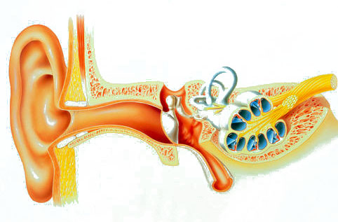
The middle ear consists of cavities and canals communicating with each other: the tympanic cavity, the auditory (Eustachian) tube, the passage to the antrum, the antrum and the cells of the mastoid process (Fig.). The boundary between the outer and middle ear is the eardrum (see).

Rice. 1. Lateral wall of the tympanic cavity. Rice. 2. Medial wall of the tympanic cavity. Rice. 3. Section of the head, carried out along the axis of the auditory tube (lower part of the section): 1 - ostium tympanicum tubae audltivae; 2 - tegmen tympani; 3 - membrane tympani; 4 - manubrium mallei; 5 - recessus epitympanicus; 6 -caput mallei; 7 -incus; 8 - cellulae mastoldeae; 9 - chorda tympani; 10 - n. facialis; 11 - a. carotis int.; 12 - canalis caroticus; 13 - tuba auditiva (pars ossea); 14 - prominentia canalis semicircularis lat.; 15 - prominentia canalis facialis; 16 - a. petrosus major; 17 - m. tensor tympani; 18 - promontorium; 19 - plexus tympanicus; 20 - steps; 21- fossula fenestrae cochleae; 22 - eminentia pyramidalis; 23 - sinus sigmoides; 24 - cavum tympani; 25 - entrance to meatus acustlcus ext.; 26 - auricula; 27 - meatus acustlcus ext.; 28 - a. et v. temporales superficiales; 29 - glandula parotis; 30 - articulatio temporomandibularis; 31 - ostium pharyngeum tubae auditivae; 32 - pharynx; 33 - cartilago tubae auditivae; 34 - pars cartilaginea tubae auditivae; 35 - n. mandibularis; 36 - a. meningea media; 37 - m. pterygoideus lat.; 38 - in. temporalis.

The middle ear consists of the tympanic cavity, the eustachian tube and the mastoid air cells.
Between the outer and inner ear is the tympanic cavity. Its volume is about 2 cm3. It is lined with mucous membrane, filled with air and contains a number of important elements. Inside the tympanic cavity there are three auditory ossicles: the malleus, the incus and the stirrup, so named for their resemblance to the indicated objects (Fig. 3). The auditory ossicles are connected to each other by movable joints. The hammer is the beginning of this chain; it is woven into the eardrum. The anvil occupies a middle position and is located between the malleus and stapes. The stirrup is the final link in the chain of auditory ossicles. On the inside of the tympanic cavity there are two windows: one is round, leading into the cochlea, covered by a secondary membrane (unlike the already described tympanic membrane), the other is oval, into which a stirrup is inserted, as if in a frame. The average weight of the malleus is 30 mg, the incus is 27 mg, and the stapes is 2.5 mg. The malleus has a head, a neck, a short process and a handle. The handle of the hammer is woven into the eardrum. The head of the malleus is connected to the incus joint. Both of these bones are suspended by ligaments from the walls of the tympanic cavity and can move in response to vibrations of the eardrum. When examining the tympanic membrane, a short process and the handle of the malleus are visible through it.

Rice. 3. Auditory ossicles.
1 - anvil body; 2 - short process of the incus; 3 - long process of the anvil; 4 - rear leg of the stirrup; 5 - foot plate of the stirrup; 6 - hammer handle; 7 - anterior process; 8 - neck of the malleus; 9 - head of the hammer; 10 - malleus-incus joint.
The anvil has a body, short and long processes. With the help of the latter, it is connected to the stirrup. The stirrup has a head, a neck, two legs and a main plate. The handle of the malleus is woven into the eardrum, and the footplate of the stapes is inserted into the oval window, thereby forming a chain of auditory ossicles. Sound vibrations travel from the eardrum to the chain of auditory ossicles, which form a lever mechanism.
There are six walls in the tympanic cavity; The outer wall of the tympanic cavity is mainly the eardrum. But since the tympanic cavity extends upward and downward beyond the tympanic membrane, bone elements, in addition to the tympanic membrane, also participate in the formation of its outer wall.
The upper wall - the roof of the tympanic cavity (tegmen tympani) - separates the middle ear from the cranial cavity (middle cranial fossa) and is a thin bone plate. The inferior wall, or floor of the tympanic cavity, is located slightly below the edge of the eardrum. Below it is the bulb of the jugular vein (bulbus venae jugularis).
The posterior wall borders the pneumatic system of the mastoid process (antrum and cells of the mastoid process). The descending part of the facial nerve passes through the posterior wall of the tympanic cavity, from which the auricular chord (chorda tympani) arises here.
The anterior wall in its upper part is occupied by the mouth of the Eustachian tube, connecting the tympanic cavity with the nasopharynx (see Fig. 1). The lower section of this wall is a thin bone plate that separates the tympanic cavity from the ascending segment of the internal carotid artery.
The inner wall of the tympanic cavity simultaneously forms the outer wall of the inner ear. Between the oval and round windows there is a protrusion on it - a cape (promontorium), corresponding to the main curl of the cochlea. On this wall of the tympanic cavity above the oval window there are two elevations: one corresponds to the facial nerve canal passing here directly above the oval window, and the second corresponds to the protrusion of the horizontal semicircular canal, which lies above the facial nerve canal.
There are two muscles in the tympanic cavity: the stapedius muscle and the tensor tympani muscle. The first is attached to the head of the stapes and is innervated by the facial nerve, the second is attached to the handle of the malleus and is innervated by a branch of the trigeminal nerve.
The Eustachian tube connects the tympanic cavity with the nasopharynx cavity. In the unified International Anatomical Nomenclature, approved in 1960 at the VII International Congress of Anatomists, the name “Eustachian tube” is replaced by the term “auditory tube” (tuba anditiva). The eustachian tube has bony and cartilaginous parts. It is covered with a mucous membrane lined with ciliated columnar epithelium. The cilia of the epithelium move towards the nasopharynx. The length of the pipe is about 3.5 cm. In children, the pipe is shorter and wider than in adults. In a calm state, the tube is closed, since its walls in the narrowest place (at the place where the bone part of the tube transitions into the cartilaginous part) are adjacent to each other. When swallowing movements, the tube opens and air enters the tympanic cavity.
The mastoid process of the temporal bone is located behind the auricle and external auditory canal.
The outer surface of the mastoid process consists of compact bone tissue and ends at the bottom with an apex. The mastoid process consists of a large number of air (pneumatic) cells, separated from each other by bony septa. Often there are mastoid processes, the so-called diploetic ones, when their basis is spongy bone, and the number of air cells is insignificant. In some people, especially those suffering from chronic suppurative disease of the middle ear, the mastoid process consists of dense bone and does not contain air cells. These are the so-called sclerotic mastoid processes.
The central part of the mastoid process is a cave - the antrum. It is a large air cell that communicates with the tympanic cavity and with other air cells of the mastoid process. The upper wall, or roof of the cave, separates it from the middle cranial fossa. In newborns, the mastoid process is absent (not yet developed). It usually develops in the 2nd year of life. However, the antrum is also present in newborns; it is located above the ear canal, very superficially (at a depth of 2-4 mm) and subsequently moves posteriorly and downward.
The upper border of the mastoid process is the temporal line - a protrusion in the form of a roller, which is like a continuation of the zygomatic process. In most cases, the floor of the middle cranial fossa is located at the level of this line. On the inner surface of the mastoid process, which faces the posterior cranial fossa, there is a grooved depression in which the sigmoid sinus is located, which drains venous blood from the brain to the bulb of the jugular vein.
The middle ear is supplied with arterial blood mainly from the external and to a lesser extent from the internal carotid arteries. The innervation of the middle ear is carried out by the branches of the glossopharyngeal, facial and sympathetic nerves.
Anyone who looks deeper into the ear to see how our hearing organ works will be disappointed. The most interesting structures of this apparatus are hidden deep inside the skull, behind the bone wall. You can get to these structures only by opening the skull, removing the brain, and then also breaking open the bone wall itself. If you are lucky or if you know how to do it masterfully, then an amazing structure will appear before your eyes - the inner ear. At first glance, it resembles a small snail, like the ones you might find in a pond.
It may look unassuming, but upon closer examination it turns out to be a very complex device, reminiscent of the most ingenious human inventions. When sounds reach us, they enter the funnel of the auricle (which we usually call the ear). Through the external auditory canal they reach the eardrum and cause it to vibrate. The eardrum is connected to three miniature bones that vibrate behind it. One of these bones is connected by something like a piston to a snail-like structure. The vibration of the eardrum causes this piston to move back and forth. As a result, a special jelly-like substance moves back and forth inside the snail. The movements of this substance are perceived by nerve cells, which send signals to the brain, and the brain interprets these signals as sound. The next time you listen to music, just imagine all the pandemonium that is happening in your head.
This entire system has three parts: the outer, middle and inner ear. The outer ear is that part of the hearing organ that is visible from the outside. The middle ear is made up of three miniature bones. Finally, the inner ear is made up of sensory nerve cells, a jelly-like substance, and the tissues that surround them. By considering these three components separately, we can understand our hearing organs, their origin and development.
Our ear consists of three parts: the outer, middle and inner ear. The oldest of them is the inner ear. It controls nerve impulses sent from the ear to the brain.
The auricle, which we usually call the ear, was given to our ancestors in the course of evolution relatively recently. You can verify this by visiting a zoo or aquarium. Which sharks, bony fishes, amphibians and reptiles have ears? This structure is characteristic only of mammals. In some amphibians and reptiles, the outer ear is clearly visible, but they do not have an auricle, and the outer ear usually looks like a membrane, like the one stretched over a drum.
The subtle and deep connection that exists between us and fish (both cartilaginous, sharks and rays, and bony ones) will only be revealed to us when we consider the structures located deep in the ears. At first glance, it may seem strange to look for connections between humans and sharks in the ears, especially since sharks do not have them. But they are there, and we will find them. Let's start with the auditory ossicles.
Middle ear - three auditory ossicles
Mammals are special creatures. Hair and mammary glands distinguish us mammals from all other living organisms. But many may be surprised to learn that the structures located deep in the ear are also important distinguishing features of mammals. No other animal has bones like those in our middle ear: mammals have three of these bones, while amphibians and reptiles have only one. But fish don’t have these bones at all. How then did the bones of our middle ear arise?
A little anatomy: let me remind you that these three bones are called the malleus, incus and stirrup. As already mentioned, they develop from the gill arches: the malleus and incus - from the first arch, and the stirrup - from the second. This is where our story begins.
In 1837, German anatomist Carl Reichert studied embryos of mammals and reptiles to understand how the skull is formed. He traced the development of gill arch structures in different species to understand where they end up in the skulls of different animals. The result of lengthy research was a very strange conclusion: two of the three auditory ossicles of mammals correspond to fragments of the lower jaw of reptiles. Reichert couldn't believe his eyes! Describing this discovery in his monograph, he did not hide his surprise and delight. When he comes to compare the auditory ossicles and jaw bones, the usual dry style of 19th-century anatomical descriptions gives way to a much more emotional style, showing how amazed Reichert was by this discovery. From the results he obtained, an inevitable conclusion followed: the same gill arch that forms part of the jaw in reptiles forms the auditory ossicles in mammals. Reichert put forward the thesis, which he himself found difficult to believe, that the structures of the middle ear of mammals correspond to the structures of the jaw of reptiles. The situation will look more complicated if we remember that Reichert came to this conclusion more than twenty years earlier than Darwin’s position about a single family tree of all living things was announced (this happened in 1859). What is the point of saying that different structures in two different groups of animals "correspond" to each other, without a concept of evolution?
Much later, in 1910 and 1912, another German anatomist, Ernst Gaupp, continued Reichert's work and published the results of his exhaustive studies on the embryology of the mammalian auditory organs. Gaupp provided more details, and, given the time in which he worked, was able to interpret Reichert's discovery within the framework of ideas about evolution. Here are the conclusions he came to: the three bones of the middle ear demonstrate a connection between reptiles and mammals. The single ossicle of the middle ear of reptiles corresponds to the stapes of mammals - both develop from the second branchial arch. But the truly stunning discovery was not this, but the fact that the other two bones of the mammalian middle ear - the malleus and the incus - developed from ossicles located at the back of the jaw in reptiles. If this is true, then the fossils should show how the ossicles passed from the jaw to the middle ear during the rise of mammals. But Gaupp, unfortunately, studied only modern animals and was not ready to fully appreciate the role that fossils could play in his theory.
Since the forties of the 19th century, fossil remains of animals of a previously unknown group began to be mined in South Africa and Russia. Many well-preserved finds were discovered - entire skeletons of creatures the size of a dog. Soon after these skeletons were discovered, many of their specimens were packed into boxes and sent to Richard Owen in London for identification and study. Owen discovered that these creatures had a striking mixture of characteristics from different animals. Some of their skeletal structures resembled reptiles. At the same time, others, especially the teeth, were more like those of mammals. Moreover, these were not just isolated finds. In many localities, these mammal-like reptiles were the most abundant fossils. They were not only numerous, but also quite diverse. After Owen's research, such reptiles were discovered in other areas of the Earth, in several layers of rocks corresponding to different periods of earth's history. These finds formed an excellent transitional series leading from reptiles to mammals.
Until 1913, embryologists and paleontologists worked in isolation from each other. But this year was significant in that the American paleontologist William King Gregory, an employee of the American Museum of Natural History in New York, drew attention to the connection between the embryos that Gaupp studied and fossils discovered in Africa. The most "reptilian" of all mammal-like reptiles had only one bone in the middle ear, and its jaw, like other reptiles, consisted of several bones. But as Gregory studied a series of increasingly mammalian-like reptiles, Gregory discovered something quite remarkable—something that would have deeply astonished Reichert had he lived: a successive series of shapes that clearly showed that the bones of the back of the jaw in mammal-like reptiles were gradually decreased and shifted until, finally, in their descendants, mammals, they took their place in the middle ear. The malleus and incus actually developed from the jaw bones! What Reichert discovered in embryos had long ago lain in the ground in fossil form, awaiting its discoverer.
Why did mammals need to have three bones in the middle ear? The system of these three bones allows us to hear sounds of a higher frequency than those animals that have only one bone in the middle ear are able to hear. The emergence of mammals was associated with the development not only of bite, which we discussed in the fourth chapter, but also of more acute hearing. Moreover, what helped mammals improve their hearing was not the appearance of new bones, but the adaptation of old ones to perform new functions. Bones that originally served to help reptiles bite now help mammals hear.
This, it turns out, is where the hammer and the anvil came from. But where, in turn, did the stirrup come from?
If I just showed you how an adult and a shark work, you would never guess that this tiny bone in the depths of the human ear corresponds to the large cartilage in the upper jaw of a marine predator. However, by studying the development of humans and sharks, we are convinced that this is exactly the case. The stapes is a modified skeletal structure of the second branchial arch similar to that of a shark's cartilage, which is called the pendulum, or hyomandibular. But the pendant is not the bone of the middle ear, because sharks do not have ears. In our aquatic relatives - cartilaginous and bony fish - this structure connects the upper jaw with the skull. Despite the obvious difference in the structure and functions of the stapes and pendulum, their relationship is manifested not only in their similar origin, but also in the fact that they are served by the same nerves. The main nerve leading to both these structures is the nerve of the second arch, that is, the facial nerve. So, we have before us a case where two completely different skeletal structures have a similar origin during embryonic development and a similar innervation system. How can this be explained?
Once again we should turn to fossils. If we trace the changes in the pendant from cartilaginous fishes to such creatures as Tiktaalik, and further to amphibians, we are convinced that it gradually decreases and finally separates from the upper jaw and becomes part of the organ of hearing. At the same time, the name of this structure also changes: when it is large and supports the jaw, it is called the dewlap, and when it is small and participates in the work of the ear, it is called the stapes. The transition from pendant to stirrup occurred when the fish came to land. To hear in water, you need completely different organs than on land. The small size and position of the stirrup perfectly allow it to capture small vibrations occurring in the air. And this structure arose due to modifications in the structure of the upper jaw.

We can trace the origin of our auditory ossicles from the skeletal structures of the first and second branchial arches. The history of the malleus and incus (left) is shown from ancient reptiles, and the history of the stapes (right) is shown from even more ancient cartilaginous fish.
Our middle ear stores traces of two major changes in the history of life on Earth. The appearance of the stapes - its development from the suspension of the upper jaw - was caused by the transition of fish to life on land. In turn, the malleus and incus arose during the transformation of ancient reptiles, in which these structures were part of the lower jaw, into mammals, for whom they help to hear.
Let's look deeper into the ear - into the inner ear.
Inner ear - movement of jelly and vibration of hairs
Imagine that we enter the ear canal, pass through the eardrum, past the three bones of the middle ear and find ourselves deep inside the skull. This is where the inner ear is located - tubes and cavities filled with a jelly-like substance. In humans, as in other mammals, this structure resembles a snail with a curled shell. Her characteristic appearance immediately catches the eye when we dissect bodies in anatomy classes.
Different parts of the inner ear perform different functions. One of them is for hearing, the other is to tell us how our head is tilted, and the third is for us to feel how the movement of our head is speeding up or slowing down. All of these functions are carried out in the inner ear in a fairly similar way.
All parts of the inner ear are filled with a jelly-like substance that can change its position. Special nerve cells send their endings to this substance. When this substance moves, flowing inside the cavities, the hairs at the ends of the nerve cells bend as if by the wind. When they bend, nerve cells send electrical impulses to the brain, and the brain receives information about sounds and the position and acceleration of the head.

Every time we tilt our heads, tiny pebbles move out of place in the inner ear, lying on the shell of the cavity filled with a jelly-like substance. The flowing substance affects the nerve endings inside this cavity, and the nerves send impulses to the brain telling it that the head is tilted.
To understand the principle of operation of the structure that allows us to feel the position of the head in space, imagine a Christmas toy - a hemisphere filled with liquid in which “snowflakes” float. This hemisphere is made of plastic, and it is filled with a viscous liquid, in which, if you shake it, a blizzard of plastic snowflakes begins. Now imagine the same hemisphere, only made not of a solid, but of an elastic substance. If you sharply tilt it, the liquid in it will move, and then the “snowflakes” will settle, but not to the bottom, but to the side. This is exactly what happens in our inner ear, only in a greatly reduced form, when we tilt our head. In the inner ear there is a cavity with a jelly-like substance into which nerve endings emerge. The flow of this substance allows us to feel what position our head is in: when the head tilts, the substance flows to the appropriate side, and impulses are sent to the brain.
Additional sensitivity is given to this system by tiny pebbles lying on the elastic shell of the cavity. When we tilt our heads, the pebbles rolling in the liquid medium press on the shell and increase the movement of the jelly-like substance enclosed in this shell. Due to this, the entire system becomes even more sensitive and allows us to perceive even small changes in the position of the head. As soon as we tilt our heads, tiny pebbles are already rolling around inside our skull.
You can imagine how difficult it is to live in space. Our senses are configured to work under the constant influence of Earth's gravity, and not in low-Earth orbit, where the Earth's gravity is compensated by the movement of the spacecraft and is not felt at all. An unprepared person in such conditions becomes ill, because the eyes do not allow one to understand where is up and where is down, and the sensitive structures of the inner ear are completely confused. This is why space sickness is a serious problem for those who work on orbital vehicles.
We perceive acceleration due to another structure of the inner ear, connected to the other two. It consists of three semicircular tubes, also filled with a jelly-like substance. Whenever we accelerate or brake, the substance inside these tubes shifts, tilting the nerve endings and causing impulses to travel to the brain.

Whenever we speed up or slow down, it causes the jelly-like substance in the semicircular tubes of the inner ear to flow. The movements of this substance cause nerve impulses sent to the brain.
Our entire system for perceiving the position and acceleration of the body is connected with the eye muscles. Eye movement is controlled by six small muscles attached to the walls of the eyeball. Their contraction allows you to move your eyes up, down, left and right. We can voluntarily move our eyes, contracting these muscles in a certain way when we want to look in some direction, but their most unusual property is the ability to work involuntarily. They control our eyes all the time, even when we don't think about it at all.
To assess the sensitivity of the connection between these muscles and the eyes, move your head this way and that way without taking your eyes off this page. Moving your head, look intently at the same point.
What happens? The head moves, but the position of the eyes remains almost unchanged. Such movements are so familiar to us that we perceive them as something simple, self-evident, but in reality they are extremely complex. Each of the six muscles that control each eye responds sensitively to any movement of the head. Sensitive structures located inside the head, which will be discussed below, continuously record the direction and speed of its movements. From these structures signals go to the brain, which in response to them sends other signals that cause contractions of the eye muscles. Remember this the next time you stare at something while moving your head. This complex system can sometimes malfunction, which can tell a lot about what problems in the body’s functioning are caused.
To understand the connections between the eyes and the inner ear, the easiest way is to cause various disruptions to these connections and see what effect they produce. One of the most common ways to cause such disorders is through excessive alcohol consumption. When we drink a lot of ethyl alcohol, we say and do stupid things because alcohol weakens our internal limiters. And if we drink not just a lot, but a lot, we also start to feel dizzy. Such dizziness often foreshadows a difficult morning - we are in for a hangover, the symptoms of which will be new dizziness, nausea and headache.
When we drink too much, we have a lot of ethyl alcohol in our blood, but the alcohol does not immediately enter the substance that fills the cavities and tubes of the inner ear. Only some time later it leaks from the bloodstream into various organs and ends up in the jelly-like substance of the inner ear. Alcohol is lighter than this substance, so the result is about the same as pouring a little alcohol into a glass of olive oil. This creates random swirls in the oil, and the same thing happens in our inner ear. These chaotic turbulences cause chaos in the body of an intemperate person. The hairs at the ends of the sensory cells vibrate, and the brain thinks that the body is in motion. But it doesn't move - it rests on the floor or on the bar counter. The brain is deceived.
Vision is also not left out. The brain thinks that the body is rotating, and it sends corresponding signals to the eye muscles. The eyes begin to move to one side (usually to the right) when we try to keep them on something by moving our head. If you open the eye of a dead drunk person, you can see characteristic twitching, the so-called nystagmus. This symptom is well known to police officers, who often test drivers stopped for careless driving for it.
With a severe hangover, something different happens. The next day after drinking, the liver had already removed alcohol from the blood. She does this surprisingly quickly and even too quickly, because alcohol still remains in the cavities and tubes of the inner ear. It gradually leaks from the inner ear back into the bloodstream and in the process again agitates the jelly-like substance. If you take the same dead-drunk person whose eyes twitched involuntarily in the evening, and examine him during a hangover, the next morning, you may find that his eyes twitch again, only in a different direction.
We owe all this to our distant ancestors - fish. If you've ever fished for trout, you've probably encountered the workings of the organ from which our inner ear apparently originates. Fishermen are well aware that trout stay only in certain areas of the riverbed - usually where they can be especially successful in obtaining food for themselves while avoiding predators. These are often shaded areas where the current creates eddies. Large fish are especially willing to hide behind large stones or fallen trunks. Trout, like all fish, has a mechanism that allows it to sense the speed and direction of movement of the surrounding water, much like the mechanism of our senses of touch.
In the skin and bones of fish there are small sensitive structures that run in rows along the body from head to tail - the so-called lateral line organ. These structures form small tufts from which miniature hair-like projections emerge. The outgrowths of each bundle protrude into a cavity filled with a jelly-like substance. Let's remember once again the Christmas toy - a hemisphere filled with a viscous liquid. The cavities of the lateral line organ also resemble such a toy, only equipped with sensitive hairs looking inward. When water flows around the body of a fish, it presses on the walls of these cavities, forcing the substance filling them to move and tilting the hair-like outgrowths of nerve cells. These cells, like the sensory cells in our inner ear, send impulses to the brain that enable the fish to sense the movement of the water around it. Both sharks and bony fish can sense the direction of water movement, and some sharks even sense small turbulence in the surrounding water, caused, for example, by other fish swimming by. We used a system very similar to this one, where we looked intently at one point, moving our heads, and saw disruptions in its operation when we opened our eyes to a drunk person. If our ancestors, common with sharks and trout, had used some other jelly-like substance in the lateral line organs, in which turbulence would not have arisen when alcohol was added, we would never have become dizzy from drinking alcoholic beverages.
It is likely that our inner ear and the fish's lateral line organ are variants of the same structure. Both of these organs are formed during development from the same embryonic tissue and are very similar in internal structure. But which came first, the lateral line or the inner ear? We do not have clear data on this matter. If we look at some of the oldest head-bearing fossils, which lived about 500 million years ago, we see small pits in their dense protective coverings, which leads us to assume that they already had a lateral line organ. Unfortunately, we know nothing about the inner ear of these fossils because we have no specimens that preserve this part of the head. Until we have new data, we are left with an alternative: either the inner ear developed from the lateral line organ, or, conversely, the lateral line developed from the inner ear. In any case, this is an example of a principle that we have already observed in other structures of the body: organs often arise to perform one function, and then are rebuilt to perform a completely different one - or many others.
Our inner ear has grown larger than that of fish. Like all mammals, the part of the inner ear responsible for hearing is very large and curled, like a snail. In more primitive organisms, such as amphibians and reptiles, the inner ear is simpler and not curled like a snail. Obviously, our ancestors - ancient mammals - developed a new, more effective hearing organ than their reptilian ancestors had. The same applies to structures that allow you to feel acceleration. In our inner ear there are three tubes (semicircular canals) responsible for sensing acceleration. They are located in three planes, lying at right angles to each other, and this allows us to feel how we move in three-dimensional space. The oldest known vertebrate to possess such canals, the hagfish-like jawless one, had only one canal in each ear. Later organisms already had two such channels. And finally, most modern fish, like other vertebrates, have three semicircular canals, like us.
As we have seen, our inner ear has a long history, dating back to the earliest vertebrates, even before the appearance of fish. It is noteworthy that the neurons (nerve cells) whose endings are embedded in a jelly-like substance in our inner ear are even older than the inner ear itself.
These cells, the so-called hair-like cells, have characteristics not found in other neurons. The hair-like outgrowths of each of these cells, including one long “hair” and several short ones, and these cells themselves, both in our inner ear and in the lateral line fish organ, are strictly oriented. Recently, a search has been made for such cells in other animals, and they were found not only in organisms that do not have such developed sensory organs as we do, but also in organisms that do not even have a head. These cells are found in lancelets, which we met in the fifth chapter. They have no ears, no eyes, no skull.
Therefore, hair cells appeared long before our ears arose, and initially performed other functions.
Of course, all this is written in our genes. If a mutation occurs in a person or mouse that turns off a gene Pax 2, a full inner ear does not develop.

A primitive version of one of the structures of our inner ear can be found under the skin of fish. Small cavities of the lateral line organ are located along the entire body, from head to tail. Changes in the flow of surrounding water deform these cavities, and the sensory cells located in them send information about these changes to the brain.
Gene Pax 2 works in the embryo in the area where ears are formed, and likely sets off a chain reaction of genes turning on and off that leads to the formation of our inner ear. If we look for this gene in more primitive animals, we will find that it works in the head of the embryo, and also, imagine, in the rudiments of the lateral line organ. The same genes are responsible for dizziness in drunk people and the feeling of water in fish, suggesting that these different feelings have a common history.
Jellyfish and the origin of eyes and ears
Similar to the gene responsible for eye development Pax 6, which we have already discussed, Pax 2, in turn, is one of the main genes necessary for ear development. Interestingly, these two genes are quite similar. This suggests that eyes and ears may come from the same ancient structures.
Here we need to talk about box jellyfish. Those who regularly swim in the sea off the coast of Australia are well aware of them, because these jellyfish have an unusually strong poison. They differ from most jellyfish in that they have eyes - more than twenty of them. Most of these eyes are simple pits scattered in the integument. But several eyes are surprisingly similar to ours: they have something like a cornea and even a lens, as well as an innervation system similar to ours.
Jellyfish have neither Pax 6, neither Pax 2 - these genes arose later than jellyfish. But with box jellyfish we find something quite remarkable. The gene that is responsible for the formation of their eyes is not a gene Pax 6, nor the genome Pax 2, but is like a mosaic mixture both of these genes. In other words, this gene looks like a primitive version of the genes Pax 6 And Pax 2 characteristic of other animals.
The most important genes that control the development of our eyes and ears, in more primitive organisms - jellyfish - correspond to a single gene. You may ask: "So what?" But this is a pretty important conclusion. The ancient connection we discovered between ear and eye genes helps us understand much of what modern doctors face in their practice: many of the human birth defects affect on both of these organs- both before our eyes and ears. And it all reflects our deep connection with creatures like the poisonous sea jellyfish.
An important element of the human body are the auditory ossicles. These miniature formations play almost the main role in the process of sound perception. Without them, it is impossible to imagine the transmission of wave vibrations and vibrations, so it is important to protect them from diseases. These bones themselves have an interesting structure. This, as well as the principle of their operation, should be discussed in more detail.
Types of auditory ossicles and their location
In the cavity of the middle ear, sound vibrations are perceived and subsequently transmitted to the internal part of the organ. All this becomes possible thanks to the presence of special bone formations.
The bones are covered with a layer of epithelium, so they do not injure the eardrum.
They are combined into a single group - the auditory ossicles. To understand the principle of their operation, you need to know what these elements are called:
- hammer;
- anvil;
- stapes.
Despite their tiny size, the role of each is simply invaluable. They got their names due to their special shape, reminiscent of a hammer, anvil and stirrup, respectively. Let's look at what exactly each auditory bone serves for next.
As for location, the ossicles are located in the middle ear cavity. By fastening with muscle formations, they adjoin the eardrum and exit into the window of the vestibule. The latter opens the passage from the middle ear to the inner ear.

All three bones form an integral system. They are connected to each other using joints, and their shape ensures perfect joining. The following connections can be distinguished:
- in the body of the incus there is an articular fossa that connects to the malleus, or more precisely, to its head;
- the lenticular process on the long stalk of the incus connects to the head of the stapes.
- the posterior and anterior legs of the stirrup bone are united by its base.
As a result, two articular joints are formed, and the extreme elements are connected to the muscles. The tensor tympani muscle grips the handle of the malleus. With its help it is set in motion. Its antagonist muscle, which connects to the posterior leg of the stapes, regulates pressure on the base of the bone in the window of the vestibule.
Functions performed
Next, you need to find out what role the auditory ossicles play in the process of sound perception. Their adequate operation is necessary for the full transmission of sound signals. At the slightest deviation from the norm, conductive hearing loss occurs.
Two main tasks of these elements should be highlighted:
- bone conduction of sound waves and vibrations;
- mechanical transmission of external signals.

When sound waves enter the ear, vibrations of the eardrum occur. This is possible due to muscle contraction and bone movement. To prevent damage to the middle ear cavity, control over the reaction of mobile elements is partially carried out at the reflex level. Muscle contraction keeps the bones from oscillating excessively.
Due to the fact that the handle of the hammer is quite long, when the muscle is tense, a lever effect occurs. As a result, even small sound signals cause an appropriate reaction. The auricular ligament of the malleus, incus and stapes transmits the signal to the vestibule of the inner ear. Further, the leading role in transmitting information belongs to sensors and nerve endings.
Relationship with other elements
The auditory ossicles are closely connected to each other using articular nodes. In addition, they are connected to other elements, forming a continuous chain of sound transmission systems. Communication with previous and subsequent links is carried out using muscles.
The first direction is the eardrum and the muscle that tenses it. A thin membrane forms a ligament due to the process of a muscle connected to the handle of the malleus. Reflex contractions protect the membrane from rupture during sudden loud sounds. However, excessive loads can not only damage such a sensitive membrane, but also displace the bone itself.

The second direction is the exit of the base of the stapes into the oval window. The stapedius muscle holds its pedicle and relieves pressure on the window of the vestibule. It is in this part that the signal is transmitted to the next level. From the ossicles of the middle ear, impulses pass to the inner ear, where the signal is converted and subsequently transmitted along the auditory nerve to the brain.
Thus, the bones act as a connecting link in the system of receiving, transmitting and processing sound information. If the middle ear cavity is subject to changes due to pathologies, injuries or diseases, the functioning of the elements may be impaired. It is important to prevent displacement, blocking and deformation of fragile bones. In some cases, otosurgery and prosthetics come to the rescue.
The ear is a paired organ located deep in the temporal bone. The structure of the human ear allows it to receive mechanical vibrations in the air, transmit them through internal media, transform them and transmit them to the brain.
The most important functions of the ear include analysis of body position and coordination of movements.
The anatomical structure of the human ear is conventionally divided into three sections:
- external;
- average;
- internal.
Ear shell
It consists of cartilage up to 1 mm thick, above which there are layers of perichondrium and skin. The earlobe is devoid of cartilage and consists of adipose tissue covered with skin. The shell is concave, along the edge there is a roll - a curl.
Inside it there is an antihelix, separated from the helix by an elongated depression - a rook. From the antihelix to the ear canal there is a depression called the auricle cavity. The tragus protrudes in front of the ear canal.
auditory canal
Reflecting from the folds of the concha of the ear, the sound moves into the auditory ear 2.5 cm in length, with a diameter of 0.9 cm. The basis of the ear canal in the initial section is cartilage. It resembles the shape of a gutter, open upward. In the cartilaginous section there are santorium fissures bordering the salivary gland.
The initial cartilaginous section of the ear canal passes into the bone section. The passage is curved in a horizontal direction; to examine the ear, the shell is pulled back and up. For children - back and down.
The ear canal is lined with skin containing sebaceous and sulfur glands. Sulfur glands are modified sebaceous glands that produce. It is removed by chewing due to vibrations of the walls of the ear canal.
It ends with the eardrum, blindly closing the auditory canal, bordering:
- with the joint of the lower jaw, when chewing, the movement is transmitted to the cartilaginous part of the passage;
- with cells of the mastoid process, facial nerve;
- with the salivary gland.
 The membrane between the outer ear and the middle ear is an oval translucent fibrous plate, measuring 10 mm in length, 8-9 mm in width, 0.1 mm in thickness. The membrane area is about 60 mm 2.
The plane of the membrane is located obliquely to the axis of the ear canal at an angle, drawn funnel-shaped into the cavity. The maximum tension of the membrane is in the center. Behind the eardrum is the middle ear cavity.
There are:
- middle ear cavity (tympanum);
- auditory tube (Eustachian tube);
- auditory ossicles.
Tympanic cavity
The cavity is located in the temporal bone, its volume is 1 cm 3. It houses the auditory ossicles, articulated with the eardrum.
The mastoid process, consisting of air cells, is located above the cavity. It houses a cave - an air cell that serves in the anatomy of the human ear as the most characteristic landmark when performing any operations on the ear.
Eustachian tube
 The formation is 3.5 cm long, with a lumen diameter of up to 2 mm. Its upper mouth is located in the tympanic cavity, the lower pharyngeal mouth opens in the nasopharynx at the level of the hard palate.
The auditory tube consists of two sections, separated by its narrowest point - the isthmus. A bony part extends from the tympanic cavity, and below the isthmus there is a membranous-cartilaginous part.
The walls of the tube in the cartilaginous section are normally closed, opening slightly during chewing, swallowing, and yawning. The expansion of the lumen of the tube is provided by two muscles associated with the velum palatine. The mucous membrane is lined with epithelium, the cilia of which move towards the pharyngeal mouth, providing the drainage function of the pipe.
 The smallest bones in human anatomy, the auditory ossicles of the ear, are designed to conduct sound vibrations. In the middle ear there is a chain: malleus, stirrup, incus.
The malleus is attached to the tympanic membrane, its head articulates with the incus. The incus process is connected to the stapes, which is attached at its base to the window of the vestibule, located on the labyrinthine wall between the middle and inner ear.

The structure is a labyrinth consisting of a bone capsule and a membranous formation that follows the shape of the capsule.
In the bone labyrinth there are:
- vestibule;
- snail;
- 3 semicircular canals.
Snail
The bone formation is a three-dimensional spiral of 2.5 turns around the bone rod. The width of the base of the cochlear cone is 9 mm, the height is 5 mm, the length of the bone spiral is 32 mm. A spiral plate extends from the bone rod into the labyrinth, which divides the bone labyrinth into two channels.
At the base of the spiral lamina are the auditory neurons of the spiral ganglion. The bony labyrinth contains perilymph and a membranous labyrinth filled with endolymph. The membranous labyrinth is suspended in the bony labyrinth using cords.
Perilymph and endolymph are functionally connected.
- Perilymph – its ionic composition is close to blood plasma;
- endolymph - similar to intracellular fluid.
 Violation of this balance leads to increased pressure in the labyrinth.
The cochlea is an organ in which physical vibrations of the perilymph fluid are converted into electrical impulses from the nerve endings of the cranial centers, which are transmitted to the auditory nerve and the brain. At the top of the cochlea there is an auditory analyzer - the organ of Corti.
vestibule
The most ancient anatomically middle part of the inner ear is the cavity bordering the scala cochlea through a spherical sac and semicircular canals. On the wall of the vestibule leading into the tympanic cavity, there are two windows - an oval window, covered by the stapes, and a round window, which represents the secondary eardrum.
Features of the structure of the semicircular canals
All three mutually perpendicular bony semicircular canals have a similar structure: they consist of an expanded and simple pedicle. Inside the bones there are membranous canals that repeat their shape. The semicircular canals and vestibular sacs make up the vestibular apparatus and are responsible for balance, coordination, and determining the position of the body in space.
In a newborn, the organ is not formed and differs from an adult in a number of structural features.
Auricle
- The shell is soft;
- the lobe and curl are weakly expressed and are formed by the age of 4 years.
auditory canal
- The bone part is not developed;
- the walls of the passage are located almost closely;
- The drum membrane lies almost horizontally.
- Almost the same size as an adult;
- In children, the eardrum is thicker than in adults;
- covered with mucous membrane.
Tympanic cavity
 In the upper part of the cavity there is an open gap, through which, in acute otitis media, the infection can penetrate into the brain, causing the phenomenon of meningism. In an adult, this gap closes.
The mastoid process in children is not developed; it is a cavity (atrium). The development of the appendage begins at the age of 2 years and ends by 6 years.
Eustachian tube
In children, the auditory tube is wider, shorter than in adults, and located horizontally.
The complex paired organ receives sound vibrations of 16 Hz - 20,000 Hz. Injuries and infectious diseases reduce the sensitivity threshold and lead to gradual hearing loss. Advances in medicine in the treatment of ear diseases and hearing aids make it possible to restore hearing in the most difficult cases of hearing loss.
Video about the structure of the auditory analyzer
middle ear, auris mebia , includes a mucous membrane-lined and air-filled tympanic cavity (about 1 cm3 in volume) and an auditory (Eustachian) tube. The middle ear cavity communicates with the mastoid cave and through it with the mastoid cells located in the thickness of the mastoid process.
tympanic cavity,cdvitas tympani [ cavitas tympanicaj, located in the thickness of the pyramid of the temporal bone, between the external auditory canal laterally and the bony labyrinth of the inner ear medially. The tympanic cavity, in which 6 walls are distinguished, is compared in shape to a tambourine placed on its edge and tilted outward.
1. Upper tegmental wall,paries tagmentlis, formed by a thin plate of bone substance (tegmen tympani), separating the tympanic cavity from the cranial cavity. 2. Bottom jugular wall,paries juguldris, corresponds to the lower wall of the pyramid in the place where the jugular fossa is located. 3. Medial labyrinth wall,paries labyrinthicus, complexly arranged, separates the tympanic cavity from the bony labyrinth of the inner ear. On this wall there is a protruding towards the tympanic cavity cape,promontorium. Above the promontory and somewhat posteriorly there is an oval vestibule window,fenestra vestii- buli, leading to the vestibule of the bony labyrinth; it is covered by the base of the stirrup. Somewhat higher than the oval window and behind it there is a transverse facial canal projection(walls of the facial nerve canal), prominentia candlis facidlis. Behind and below the cape is snail window,fenestra cochleae, closed secondary tympanic membrane,membrdna tympani secundd- ria, separating the tympanic cavity from the scala tympani. 4. Rear mastoid wall,paries mastoideus, at the bottom has pyramidal elevation,eminentia pyramidlis, inside which it begins stapedius muscle,m. stapedius. In the upper part of the posterior wall, the tympanic cavity continues into mastoid cave,dntrum mastoideum, into which the mastoid cells of the process of the same name also open. 5. Front sleepy wall,paries cardticus, in its lower part it separates the tympanic cavity from the carotid canal, in which the internal carotid artery passes. In the upper part of the wall there is an opening of the auditory tube, which connects the tympanic cavity with the nasopharynx. 6. Lateral membranous wallparies membrandceus, formed by the eardrum and the surrounding parts of the temporal bone.
In the tympanic cavity there are three auditory ossicles covered with mucous membrane, as well as ligaments and muscles.
auditory ossicles,ossicula auditus [ auditoria], miniature in size, connecting with each other, they form a chain that continues from the eardrum to the end of the vestibule, which opens into the inner ear. In accordance with their shape, the bones received the names: hammer, anvil, stirrup (Fig. 211). Hammer, malleus, has a rounded head,cdput mallei, which turns into a long one hammer handle,manubrium mallei, with two processes: lateral and anterior,processus laterlis et anterior. Anvil, incus, consists of a body corpus incudis, with an articular fossa for articulation with the head of the malleus and two legs: one short leg,crus breve, another - long,crus longum, with a thickening at the end. This thickening is lenticular process,pro cessus lenticuldris, for connection to the head of the stirrup. S t r e m, stages, has a head cdput stapedis, two legs - front and back,crus anterius el crus posterius, connected by base of the stirrup,basis stapedis, inserted into the window of the vestibule. The hammer, with its handle, is fused along its entire length with the eardrum so that the end of the handle corresponds to the navel on the outer side of the eardrum. The head of the malleus is connected to the body of the incus by means of a joint and forms incus-malleus joint,articulatio in- cudomallearls, and the anvil, in turn, connects with the head of the stapes with its lenticular process, forming incus-stapedial joint,articulatio incudostapedia [ incudo- stapedialisj. The joints are strengthened by miniature ligaments.
With the help of a chain movable in the joints, consisting of three auditory ossicles, vibrations of the eardrum, resulting from the impact of a sound wave on it, are transmitted to the window of the vestibule, in which the base of the stapes is movably fixed with the help of annular ligament of the stapes,lig. anuldre stapedius [ stapediale]. Two muscles attached to the auditory ossicles regulate the movements of the ossicles and protect them from excessive vibrations during strong sounds. The tensor tympani musclem. tensor tympani, lies in the semi-canal of the muscular-tubal canal of the same name, and its thin and long tendon is attached to the initial part of the handle of the malleus. This muscle, pulling the handle of the hammer, strains the eardrum. stapedius muscle,m. stapedius, starting in the pyramidal eminence, it is attached by a thin tendon to the posterior leg of the stapes, near its head. When the stapedius muscle contracts, the pressure of the base of the stapes inserted into the window of the vestibule is weakened.
Auditory (Eustachian) tube,tuba auditiva [ auditorial, with an average length of 35 mm and a width of 2 mm, it serves to bring air from the pharynx into the tympanic cavity and maintain pressure in the cavity equal to the external one, which is important for the normal operation of the sound-conducting apparatus (tympanic membrane and auditory ossicles). The auditory tube consists of bone part,pars ossea, And cartilaginous part(elastic cartilage), pars cartilaginea. Clearance of the pipe at the junction - isthmus of the auditory tube,isthmus tubae auditivae / auditoriaej, tapers to 1 mm. The upper bony part of the tube is located in the hemicanal of the same name of the myotubal canal of the temporal bone and opens on the anterior wall of the tympanic cavity tympanic opening of the auditory tube,ostium tympanicum tubae auditivae [ auditoriaej. The lower cartilaginous part, which accounts for 2 /z the length of the tube, has the appearance of a groove, open at the bottom, formed by the medial and lateral cartilaginous plates and the membranous plate connecting them. At the point where the auditory tube opens on the side wall of the nasopharynx pharyngeal opening of the auditory tube,ostium pharyngeum tubae auditivae /" auditor iaeJ, the medial (posterior) plate of the elastic cartilage of the tube thickens and protrudes into the pharyngeal cavity in the form roller,torus tubdrius. The longitudinal axis of the auditory tube from its pharyngeal opening is directed upward and laterally, forming an angle of 40-45° with the horizontal and sagittal planes.
The tensor muscle and the levator palatine muscle originate from the cartilaginous part of the auditory tube. When they contract, the cartilage of the tube and its membranous plate,lamina membrandcea, are retracted, the pipe channel expands and air from the pharynx enters the tympanic cavity. The mucous membrane of the tube forms longitudinal folds and is covered with ciliated epithelium, the movements of the cilia are directed towards the pharynx. The mucous membrane of the auditory tube contains many mucous glands, gldndulae tubdriae, lymphoid tissue, which forms a cluster near the tubal ridge and around the pharyngeal opening of the auditory tube - the tubal tonsil (see “Organs of hematopoiesis and the immune system”)
