Uterine blood flow is increased during pregnancy. Disturbance of uteroplacental blood flow during pregnancy
During the gestation process, the mother's body is closely connected with the fetus through placental structures that provide full fetal development, and also deliver nutrition and oxygen, produce hormonal components and remove metabolic products. In general, the placenta is responsible for everything critical processes during the gestation period. Sometimes, for various reasons, a woman develops blood flow problems during pregnancy. Such conditions are incredibly dangerous for pregnancy; they can provoke fetal pathologies and pregnancy complications.
Scheduled ultrasound allows you to recognize any abnormalities in time
During gestation, a close connection is established between the child and the mother, which is also called the fetoplacental system. It consists of several departments.
- The central role is given to the placenta, which grows with villi into the uterine wall and through them nourishes the fetus in the uterus necessary substances. Moreover, maternal and fetal blood do not mix, since there is a hematoplacental barrier through which the blood is filtered, returning back to the woman.
- Part of the fetoplacental system is arterial network uterine body. Before conception, these arteries are spasmed and twisted in the form of a spiral, but already from the first month of pregnancy muscle layer, which provides a spasmodic state, resolves, and by the fourth month of gestation, the arteries are transformed into special trunks that carry blood to the placental structures, fully participating in the blood supply. This phenomenon was specifically provided by nature for better intrauterine nutrition of the child. If they start uterine bleeding, That vascular walls will no longer be able to contract, which is fraught with heavy blood loss, even death.
- Another part of the blood supply system between mother and fetus are the umbilical vessels, which form another route of blood supply. A vein and two arterial canals pass through the umbilical cord, which connect the fetus to the placenta. If blood flow disturbances occur in this part, the fetus is subject to severe damage.
Causes of uterine blood flow disturbances
Placental insufficiency can be caused by various reasons. The main thing is that such a condition pathologically affects such placental functions as metabolic and trophic, endocrine, transport, etc. In such conditions, material exchange processes between the fetal and maternal organism are seriously disrupted, which leads to serious consequences.

If you have diabetes, you should check your blood sugar levels regularly
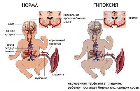
Intrauterine complications can cause disruption of the blood supply. infectious lesions, pneumonia or fetal hypoxia, hypertensive disorders, etc. Also, normal blood supply is disrupted by pathologies such as asthma, heart disease pathological conditions such as low blood pressure, activity defects, etc. Often the reasons pathological disorders blood circulation is caused by neuroendocrine diseases such as hyperthyroidism or diabetes, hypothyroidism, as well as hypothalamic or adrenal pathologies. Blood supply disturbances are provoked by kidney failure or pyelonephritis.
Also, problems with blood supply can be caused by anemia or blood clots, which is actually the norm for similar pathologies. Quite often women suffer from exacerbations various kinds infectious pathologies, which also lead to placental changes. A variety of infectious agents cause inflammatory lesions, which in the first weeks of gestation can cause spontaneous abortion, and in later various abnormalities in fetal intrauterine development. No less dangerous are the various uterine pathologies such as fibroids, endometriosis, hypoplasia or malformations, as well as pathological changes in the myometrial layer.
Moms over 35, those pregnant for the first time and those with large fibroids are at risk of encountering blood flow disorders. Also, placental or breech presentation, gestational processes and multiple pregnancy, the presence of Rh conflict, abortions in the past and a tendency to unhealthy habits, social and everyday dissatisfaction and other factors.
Types of fetoplacental insufficiency
First of all, placental insufficiency is classified into chronic and acute forms. Acute pathology can occur at any stage and even during delivery. IN placental tissues Meanwhile, gas exchange is disrupted, leading to acute fetal hypoxia or the death of the baby. Often similar phenomenon occurs against the background of premature placental infarction or detachment, bleeding or thrombosis of blood vessels.
Chronic forms of fetoplacental insufficiency are diagnosed much more often and occur mainly during the second trimester of gestation, although they are detected only in the third trimester. On the surface of the villi, which grow into the uterine wall, fibrin begins to be deposited, which interferes with the normal course of metabolic processes. As a result, premature placental aging begins.
Chronic insufficiency of fetoplacental blood supply is divided into the following types:
- Critical. With this form, serious functional and morphological changes, which cannot be influenced in any way, so fetal death becomes inevitable.
- Subcompensated failure. With such a violation, the female body cannot cope with disturbances in placental activity, so fetal development is delayed, which leads to complications during gestation and fetal development.
- Decompensated - when compensation mechanisms lose the ability to deal with pathological placental changes, therefore the norm of indicators during the development of pregnancy is violated, the fetus begins to suffer from hypoxia, cardiac dysfunction, developmental delays, etc. The likelihood of intrauterine fetal death is high.
- Compensated deficiency is considered the most favorable of all of the above, since the fetus continues to develop without suffering from various abnormalities. Female body with such a deficiency, he is able to adapt and compensate the child for the deviations that have occurred. If a woman gets correct treatment, then such deviations will not affect the health of the baby and the timing of delivery.
Degrees of hemodynamic deviations

Breathing exercises are useful for the expectant mother and baby
In addition to the types of disorders described above, there are various fetoplacental degrees during pregnancy. At elementary degrees pathological abnormalities, the fetus remains in in good condition, blood flow deviations are harmless and affect only the uteroplacental area. It is important to detect such violations in a timely manner; if the patient does not receive necessary assistance, then within a month pathological abnormalities worsen, becoming more serious.
Blood flow disorders of the 1st degree are conventionally divided into two types: deviations of degree 1A and 1B. In the latter case, the blood flow between the placenta and uterine tissues is normal, but there are deviations in the placental-fetal circulation. In approximately 80% of cases, the fetus with such disorders develops developmental delay. Deviations of blood flow of 1A degree are characterized by disturbances between the placental and uterine blood circulation, while in the placental-fetal blood flow the norm of indicators is observed. About 90% of cases similar violations accompanied by a child's developmental delay.
In the second degree, pathologies are observed serious violations in the bloodstream of the fetal vessels and uterine body. This stage is usually short-lived, lasting about a week, and quickly progresses to the next stage of disturbances. The third degree of hemodynamic disturbances is characterized by critical disturbances in the fetal blood supply or its complete absence. You can only try to cure stage 1B pathology; in other cases, deviations are irreversible and often require premature birth.
Signs of pathology
The clinical picture of blood flow disorders is determined by their severity. Compensated disorders usually occur latently and are revealed only when ultrasound diagnostics. Decompensated and sharp forms fetoplacental insufficiency is usually accompanied by changes motor activity fetus, which is either minimized or becomes excessively pronounced. Normally, the fetus should move at least ten times per day.
Sometimes such deviations are accompanied by insufficient growth of the pregnant woman’s tummy, polyhydramnios or oligohydramnios, severe gestosis or hyperedema, sudden weight gain or pressure surges, and the appearance of protein compounds in urine. Most dangerous manifestation insufficiency placental circulation Uterine bleeding is considered, which usually occurs against the background of placental abruption. In a situation like this meaningful help a woman can only get it from specialists, so she needs to call an ambulance.
Why is a blood flow disorder dangerous?
During pregnancy, problems with blood flow are dangerous, because even a slight circulatory disorder reduces the amount of nutrition and oxygen supplied to the fetus. With prolonged fasting of this kind, complications such as:

When identifying initial stage There are no particularly dangerous risks for the baby; with age, the child will catch up with the development of his peers. If more than severe degrees violations, the prognosis is unfavorable, such pregnancies usually end in a frozen fetus or the appearance of a child with various anomalies, disabilities and other ailments.
Diagnosis of disorders
If the development of placental blood flow disorders is suspected, the patient undergoes comprehensive examination. The leading role in diagnosis is given to Doppler ultrasound in combination with ultrasound examination. Such techniques make it possible to promptly identify pathological blood flow disorders and determine the degree of complications caused by them. Usually Doppler testing is prescribed for premature placental aging, deficiency or excessive content amniotic fluid, congenital genetic pathologies or fetal defects, with hypoxic manifestations or intrauterine retention fetal development.
How to normalize blood supply to the uterus
The first stage of blood flow fetoplacental abnormalities responds most positively to treatment. In the second degree, therapy is practically ineffective, and in the third degree it is necessary emergency birth through surgical intervention. During therapy, it is necessary to influence all parts of the blood flow structures. For minor microcirculatory disorders, to improve blood flow, patients are advised to take Hofitol, which belongs to the category homeopathic remedies. If the treatment does not provide the desired effect, then resort to medications such as Actovegin or Pentoxipharma.
To expand the vascular passages, No-Shpa or Drotaverine is used, and for thrombophlebitis, Curantil is indicated. Improves blood flow and reduces uterine muscle tone Magnesium B6 and magnesium infusion. And for antioxidant purposes, it is recommended to take ascorbic acid and vitamin E.
In order not to treat blood flow disorders or their consequences, it is necessary to take preventive measures in advance to prevent these conditions. The mother must exclude all risk factors that provoke the development of fetoplacental insufficiency. Mommy needs to control her body weight, go to more fresh air and undergo all planned procedures on time, diagnostic studies, laboratory tests And gynecological examinations. This is the only way to detect deviations in time and prevent their further development.
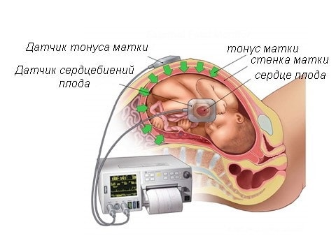
During pregnancy it is very important to constant surveillance over the state of the organisms of the mother and fetus and their performance of vital functions. One of the most significant research is an analysis of blood flow in the arteries of the uterus, the woman’s umbilical cord, as well as in the aorta and cerebral vessels child.
Among the main causes of perinatal mortality and morbidity, violation of uterine blood flow(utero-placental and fetal-placental).
Blood flow in the placenta
The placenta, in which the fetus is located, supplies it with nutrition and oxygen from the mother’s blood and removes metabolic products child's body. It is this organ that unites two complex vascular systems - the maternal one, which connects the vessels of the uterus and the placenta, and the fetal one, which passes into the umbilical arteries and leading to the fetus.
The above circulatory systems separates the membrane, which does not allow the blood of the mother and child to mix. The placenta acts as a kind of barrier, resistant to many viruses and harmful substances.
In some cases, completely various reasons Placental insufficiency may develop, which inevitably affects the performance of the trophic, metabolic, transport, endocrine and other vital functions of the placenta. In this condition, the metabolism between the body of mother and child deteriorates significantly, which is fraught with consequences.
Causes of uterine blood flow disorders
Poor blood circulation in the uterus can be caused by increased pressure, pneumonia, intrauterine infection and insufficient supply of oxygen to the fetal body ().
To diagnose the blood flow system in obstetric practice three-dimensional is applied ultrasound examination(Doppler), with the help of which the vessels are visible in the so-called 3D (three-dimensional) image. With this modern diagnostic method there was a prospect of diagnosing retroplacental bleeding and assessing cardiac malformations by monitoring blood flow. This method is indispensable, since with its help you can see defects even in the smallest vessels that form the microvasculature, monitor the development and formation of intraplacental hemodynamics, and also control the amount of oxygen and nutrients, which must enter the fetal body. New opportunities for early detection have opened up obstetric complications, and if correction or treatment is started without wasting time, then circulatory disorders and further associated pathologies can be practically avoided.
Hemodynamic disorders during pregnancy
Hemodynamic disorders are divided into three degrees of severity:
First degree includes two subspecies:
- 1A is a violation of uteroplacental blood flow, which is the mildest. Fetal-placental circulation is preserved. In most cases, this problem is caused by intrauterine infection;
- 1B - uteroplacental blood flow is preserved, while pathologies occur in the fetoplacental blood flow.
Second degree characterized by disturbances of both blood flow systems, but does not involve cardinal changes.
Third degree is that disruption of the uteroplacental circulation leads to defects in blood circulation at the utero-fetal level.
In the first degree of violations, due to timely detection and adequate treatment, cases of fetal death can be avoided. Perinatal mortality in the second degree is 13.3%, in the third - 46.7%. During this time, it was found that correction of placental insufficiency in patients with third-degree hemodynamic impairment was ineffective. In this case, perinatal mortality during conservative birth was 50%, while it helps to avoid losses. To the ward intensive care 35.5% of newborns fall into the first degree, 45.5% into the second, and 88.2% into the third.
Prevention of blood flow disorders during pregnancy
Every woman who wants to give birth to a child must remember that the mother’s condition is completely transmitted to the unborn baby. Therefore, in order for the fetus to develop without complications, she needs to make up her diet from food containing a maximum of vitamins, micro- and macroelements, as well as rich required quantity carbohydrates, proteins and fats. If a pregnant woman is not bothered by swelling, then fluid intake should be at least 1-1.5 liters.
It is important to monitor changes in body weight, since by the end of pregnancy the weight gain should not exceed 10 kg.
There are risk groups that need to be used drug prevention, which promotes the interaction of the body systems of the fetus and mother and prevents dysfunction of the uteroplacental circulation.
Timely corrected methods of labor management will help to significantly reduce perinatal morbidity and mortality. drug therapy. But high risk the appearance of severe neurological complications still not excluded.
Especially for Elena Zhirko
After a woman finds out about her pregnancy, she must realize that now the body belongs not only to her, but also to her unborn child. Hormonal surges and complete restructuring of the pelvic organs quite often result in a disruption in the blood supply to the fetus. In this article we will talk about impaired blood flow during pregnancy, what it entails, what symptoms are inherent, what therapy can be used and how to bear a healthy child.
How blood flow changes during pregnancy
First, let's figure out how everything works in mothers' tummies. During pregnancy for transmission useful microelements The placenta is responsible for oxygen supply to the baby. It is the same unifier through which the pregnant woman’s vascular system connects with vascular system fruit, becoming common. Any disturbances in the functioning of the placenta affect the condition of the baby, so it is necessary to monitor blood flow during pregnancy. Diagnosis is made through research - Doppler. Let's talk about it in more detail below.
What is a blood flow disorder during pregnancy?
Impaired blood flow can appear at any stage of pregnancy. A pregnant woman is diagnosed with placental insufficiency. This is one of the most common complications during pregnancy associated with dysfunction of the placenta. This pathology occurs in two forms:
- The acute form appears suddenly and is most often a consequence of placental abruption. May cause disruption of gas exchange in this body and, as a consequence, oxygen starvation in the fetus.
- Chronic, it is also called premature aging placenta. Most often detected in the third trimester of pregnancy. Divided into the following types:
- compensated - considered minimally dangerous, since with this form the child continues normal physiological development. In the mother’s body they “turn on” defense mechanisms, which compensate for impaired blood flow;
- decompensated - the mother’s body cannot cope with the problem, pathological changes in the placenta. The first ones appear oxygen starvation fetus, leading to developmental delays, possible intrauterine death of the baby;
- subcompensated - with this form, the condition of the fetus worsens, it lags significantly behind in development;
- critical - with this form of deficiency, the death of the child is inevitable.
Diagnosis of pathology
It was previously said that during pregnancy, blood flow disorders can be diagnosed using Doppler ultrasound. It is an ultrasound examination that can detect any pathological abnormalities in blood flow. When diagnosed, a pregnant woman takes a horizontal position on her back or side. The specialist conducts the examination using the transabdominal method. Usually Doppler testing is prescribed twice:
- at 20–22 weeks, to ensure that there are no abnormalities in the development of the fetus;
- at 32 weeks.
Degrees of blood flow disturbances in pregnant women
Conventionally, the blood flow system during pregnancy can be divided into two subsystems:
- woman (uterus) – placenta;
- placenta - baby.
In medicine, there are standards for Doppler readings. They are used from the second trimester. Doctors compare the diagnostic data obtained with the norms and identify the degree of blood flow impairment during pregnancy.
I degree
During diagnosis, the presence of deviations in one of two forms is recorded:
- I-a degree - disruption of blood flow occurs in the area of the pregnant woman - placenta (uteroplacental blood flow);
- I-b degree - pathological changes are observed in the placenta-child subsystem.
Impaired blood flow during first-degree pregnancy does not affect the condition of the unborn baby and is easily amenable to medical correction.
II degree
In the second degree, both subsystems are affected. Over the course of 7–12 days, this condition threatens to develop into III degree, which could end tragically.
III degree
A critical point at which the baby's blood supply may be completely absent or reversed. If within 72 hours, with reverse blood flow, it is not possible to stabilize the condition, then a diagnosis is made artificial childbirth, or premature termination of pregnancy.
What are the dangers of impaired blood flow during pregnancy?
Complications and dangers that can develop due to impaired blood flow include:
- placental abruption;
- hypoxia;
- fetal hypotrophy;
- developmental pathologies;
- intrauterine death.
In grade I, if the fetus does not suffer from hypoxia, then the woman is allowed to give birth on her own. In other cases, delivery is carried out through C-section.
Why do blood flow disorders occur during pregnancy?
There are many reasons that contribute to impaired blood flow during pregnancy. Let's consider the most common factors that provoke blood flow disorders.
- Diseases of the uterus: bicornuate uterus, endometriosis, uterine hypoplasia, presence of fibroids, etc.
- Maternal health problems: renal failure, diabetes mellitus, hypotension, pyelonephritis, disease endocrine system, bronchial asthma etc.
- Unfavorable gestation conditions: Rh-conflict, multiple births, gestosis, malpresentation of the fetus, etc.
- External factors: drinking alcohol during pregnancy, smoking, constantly being in a nervous environment, first birth (and the woman is over 35), poor (limited) maternal nutrition.
Symptoms of pathology
At the first degree of manifestation of the pathology, the symptoms do not manifest themselves in any way, therefore expectant mother finds out about the problem by visiting the next scheduled ultrasound. If the pathology occurs in an acute or decompensated form, then changes in the activity (movement, movement) of the fetus can be noticed. Such conditions are characterized by changes that are too strong movements with subsidence.
In addition to behavioral changes in the baby in the stomach, there may be:
- slow growth of the mother’s belly;
- late toxicosis;
- increased swelling;
- low or high water levels are diagnosed.
Treatment methods
If the disease occurs in mild form(first degree), then the doctor may prescribe medications that improve blood circulation.
The dynamics of the fetal condition are carried out; weekly, until the indicators normalize, the pregnant woman undergoes Doppler measurements and checks the fetal heartbeat. If the indicators stabilize, the woman will continue to bear the child. In case of deterioration, it is recommended to have a cesarean section (if the pregnancy is more than 25–28 weeks).
In the second degree, the pregnant woman is hospitalized and treated under the strict supervision of medical staff. If the condition worsens, an unscheduled operation is performed.
As for the third degree, it cannot be treated, since the development of the fetus begins irreversible changes. Therefore, in order not to risk the child’s life, doctors insist on an immediate cesarean section.
In conclusion about prevention
Prevention activities should be aimed at creating conditions for healthy growth and intrauterine development child. To do this, a woman must:
- watch your diet;
- rest more often;
- regularly spend time in the fresh air;
- give up bad habits;
- minimize emotional stress.
The main thing is to regularly visit your doctor and follow his recommendations.
During the period of bearing a child, the body of the expectant mother is subjected to strong hormonal changes. In this regard, it is very important to constantly monitor the woman’s health and the condition of the fetus. IN medical practice very often there is a violation of blood flow in women expecting a new addition to the family. The appearance of an additional circle of blood circulation in the mother’s body requires frequent examination from a specialist. After all, if blood flow is impaired during pregnancy, then there is a risk of fetal death, and different dates its gestation.
Blood flow during pregnancy: normal
Many women, especially those who are carrying their first child, are not aware of the existence of such a study as Doppler. It consists of ultrasound diagnostics, which is able to assess the intensity of blood flow in different vessels. This study is mainly carried out in the third trimester of pregnancy. But in some cases they resort to it even after the twentieth week of bearing a child. Doppler is considered serious research, which allows you to diagnose vascular pathology in the uterus and placenta, in the brain and carotid arteries and fetal aorta. By comparing the obtained figures and the norms of blood flow during pregnancy, the specialist determines whether the child in the mother’s womb suffers from lack of oxygen or not.
There are approved Doppler standards starting from the second trimester of pregnancy. These are the norms of the vascular resistance index of the uterus, umbilical cord, aorta and cerebral artery fetus Doctors recommend not trying to decipher the results yourself. There is a certain formula for accurately calculating the vascular resistance index - this procedure should only be done by a doctor.
Impaired blood flow during pregnancy: degrees
For many expectant mothers, this diagnosis causes panic and confusion. Should you be nervous? Can this pathology have any consequences for the child? What are the degrees of this disease? Let's try to find answers to these questions.
There are three degrees of disturbances in blood circulation through the blood vessels during pregnancy. The first is characterized by impaired blood flow, which does not reach critical values (in the umbilical cord and artery). In this case, a positive state of fetal hemodynamics is observed. In both ventricles of his heart, there is a decrease in the index of diastolic function, as well as an increase in the maximum speed of blood flow through all the heart valves. The first degree of the disease is divided into 1-a, in which only uteroplacental blood flow is impaired, and 1-b degree, in which defective fetal-placental blood flow is observed.
In the second degree, fetal hemodynamics are disrupted. In 50% of cases it decreases maximum speed blood flow through all heart valves. It should be noted that in the left sections this phenomenon is less pronounced. Disturbances in blood flow are observed both in the fetus and in uterine arteries. The second degree often turns into the third, and in a very short period.
The third degree signals critical condition blood supply to the fetus. At this stage, a deeper restructuring occurs intracardiac hemodynamics. It is directly related to the centralization of blood circulation. Fetal hypoxia cannot be ruled out. It is also possible to reduce diastolic blood flow in the aorta, until it disappears. There is simultaneous inadequate blood movement in the aorta and carotid artery.
What are the consequences of impaired blood flow during pregnancy: consequences
This pathology leads to placental insufficiency, which is observed in 25% of pregnant women.
It is known that the placenta is the main organ during the gestation of the unborn baby, with the help of which its breathing and nutrition occurs, as well as the excretion of waste products. It is in the placenta that two systems of blood vessels converge, between which there is a membrane that acts as a kind of barrier between the body of the child and the mother. Thanks to the membrane, the blood of the mother and the unborn child does not mix. The placenta is also a protective shield against viruses and bacteria. She performs immune function, providing protection to the fetus.
With placental insufficiency, the uteroplacental and fetal-placental blood flow is disrupted, and the placenta itself is not fully matured. Due to these changes, the unborn child is not admitted to sufficient quantity useful substances and oxygen. For this reason, its development and growth slows down, and existing pregnancy complications worsen.
Naturally, due to the fact that blood flow is low during pregnancy, such serious changes can even lead to the death of the fetus. But this happens in in rare cases. Often this pathology is detected at the initial stage and can be successfully treated.
Disturbance of uteroplacental blood flow
IN medical terminology violation of uteroplacental blood circulation is designated degree 1a. The occurrence of this pathology indicates dangerous complication pregnancy. It usually occurs in the later stages.
Inadequate blood flow occurs between the uterus and placenta. Similar condition contributes to a significant deterioration of metabolism between the body of a woman and the fetus. Naturally, this condition leads to certain consequences.
There are reasons that provoke the development of this condition. These include an increase blood pressure mother, diabetes mellitus, pneumonia and kidney disease in a pregnant woman, as well as the presence of infection in the fetus itself. It should be noted that timely detection possible groups the risk is serious preventative measure. Therefore, it is important for a pregnant woman to monitor her health and, even with minor ailments, seek medical help.
What are the dangers of poor blood flow in a child?
In a single functional system mother-placenta-fetus defective fetal-placental blood flow leads to placental insufficiency. After all, the placenta supplies the unborn baby with nutrition and oxygen. It is she who is the connecting link that unites two complex systems - maternal and fetal. When such a pathology occurs, a disturbance in the child’s blood flow is observed. It should be noted that inadequate blood movement in the vessels of any degree leads to malnutrition of the fetus. Its condition also depends on the stage of blood flow disturbance. Naturally, the third degree signals the critical condition of the child.
In case early detection For this pathology, the doctor must determine the need for treatment in a hospital or at home. It all depends on the specific case and stage of pregnancy.
In medical terminology, a violation of fetal-placental blood flow is designated degree 1b.
How to treat blood flow problems during pregnancy
For the treatment of inadequate blood flow during pregnancy, it is used various drugs, helping to increase the resistance of the fetal brain to hypoxia, improve blood microcirculation and reduce blood clotting. If necessary, the doctor prescribes antibacterial and antiviral drugs, as well as immunomodulators.
A good remedy that improves cerebral circulation, the work of the heart and metabolism in a child during hypoxia, is the drug Instenon. It is used in combination with other drugs.
The expectant mother is also credited with using Actovegin, a drug that helps increase the resistance of fetal tissues and brain to hypoxia. It also improves metabolism, stimulates cell renewal of the unborn baby, and improves blood circulation in the complex system - mother-placenta-fetus. As a result of treatment with Actovegin, blood flow indicators improve, and active growth child in the mother's womb.
It is known that with placental insufficiency, as a rule, there is chronic disorder blood clotting. In this regard, experts recommend the use of drugs that prevent the formation of blood clots in blood vessels (for example, Curantil).
In the case of the 3rd (most complex) degree of blood flow disturbance, specialists cause premature birth.
Especially for -Ksenia Manevich
During pregnancy, the expectant mother should be observed by a gynecologist so that the fetus develops correctly. During the functioning of the body, disturbances may occur that affect the formation of the fetus; the doctor helps the woman avoid unpleasant consequences. An additional circle of blood circulation appears in the mother’s body, and disturbances in this area can cause the death of the baby during pregnancy.
The circulatory system connecting the uterus, placenta and baby has different functional value: supplies the baby with nutrition and oxygen, removes fetal metabolic products. The placenta is an obstacle to viruses that can enter through the mother's blood. If the blood supply to the placenta is disrupted, this leads to placental insufficiency and impaired functionality of the placenta.
Why is blood flow disrupted?
There are reasons for this:
- Due to high blood pressure.
- At the expense of .
- For pneumonia.
- With hypoxia.
- Due to thrombosis.
- Presence of gynecological disorders.
- Due to miscarriages, abortions.
Violations are of the following types:
- Uteroplacental.
- Placental.
- Feto-placental.
Diagnostic methods
Do they exist? To answer this question, it is necessary to identify blood flow disorders. Doppler ultrasound and ultrasound should be performed. Blood flow testing is carried out in different blood vessels mother and child.
What does the doctor pay attention to when making a diagnosis: thin placenta, the presence of infections, disturbances in the amniotic fluid.
Using Dopplerometry, three degrees of blood flow disturbance can be determined:
1st degree – mild, divided into categories:
1B - fetoplacental - uteroplacental blood flow is preserved;
2nd degree – both blood flow systems are disrupted.
3rd degree – circulatory disorders in a critical stage.
Doppler measurements are carried out at any stage of pregnancy, especially when there is a suspicion of circulatory problems.
In addition, they use laboratory methods blood tests of pregnant women.
Therapeutic methods
Treatment of circulatory disorders is carried out comprehensively so that there are no complications. If a pregnant woman falls into a risk category (abortion, gynecological diseases), is carried out preventive treatment, the expectant mother is constantly monitored. Treatment methods are determined based on the degree of circulatory impairment.
The use of drugs that can reduce the tone in the uterus and improve blood circulation, for example, Magne-B6, depends on the individual state of the mother’s body. No-shpa may also be prescribed to dilate blood vessels. In addition, therapy methods include drugs that help blood clotting, for example, Curantil.
A woman whose blood flow is impaired, mandatory at 36 weeks they are placed on inpatient treatment to carry out prenatal diagnostics. Natural childbirth pregnant women with 1st degree blood flow impairment are carried out under special supervision. For grades 2 and 3 disorders, a caesarean section is performed.
Prevention to reduce the risk of blood flow disorders:
- Proper nutrition.
- Relief from stress.
- Walking in the fresh air.
- Vitamins.
Impaired blood flow must be treated under the supervision of a physician. If a pregnant woman behaves incorrectly and is not treated, this may cause premature birth or complications of fetal development.
